1. Carcinogenesis vol.31 no.6 pp.1110–1115, 2010
doi:10.1093/carcin/bgp300
Advance Access publication December 3, 2009
Hypomethylation and genome instability in the germline of exposed parents and their
progeny is associated with altered miRNA expression
Jody N.Filkowskiy
, Yaroslav Ilnytskyyy
, Jan Tamminga,
Igor Koturbash, Andrey Golubov, Tetyana Bagnyukova1
,
Igor P.Pogribny1
and Olga KovalchukÃ
Department of Biological Sciences, University of Lethbridge, 4401 University
of Lethbridge, Alberta, Canada T1K 3M4 and 1
Division of Biochemical
Toxicology, National Center for Toxicological Research, Jefferson, AR 72079,
USA
Ã
To whom correspondence should be addressed. Tel: þ1 403 394 3916;
Fax: þ1 403 329 2242;
Email: olga.kovalchuk@uleth.ca
Recent studies suggest that transgenerational genome instability
may be epigenetic in nature and mediated via altered DNA meth-
ylation and microRNAome. Here, we investigated the nature and
mechanisms underlying the disruption of DNA methylation and
microRNA expression status in the germline and progeny of ex-
posed parents. We have found that paternal irradiation leads to
upregulation of the miR-29 family in the exposed male germline,
which causes decreased expression of de novo methyltransferase,
DNA methyltransferase 3a, and profound hypomethylation of long
interspersed nuclear elements 1 (LINE1) and short interspersed
nuclear elements B2 (SINE B2). Epigenetic changes in the male
germline further resulted in deleterious effects in the somatic thy-
mus tissue from the progeny of exposed animals, including hypo-
methylation of LINE1 and SINE B2. Hypomethylation of LINE1
and SINE B2 in the thymus tissue from the progeny was associated
with a significant decrease in the levels of lymphoid-specific helicase
(LSH) that is crucial for the maintenance of methylation and
silencing of repetitive elements. Furthermore, we noted a significant
upregulation of miR-468 that targets LSH and leads to its de-
creased expression in thymus in the progeny of exposed parents.
We suggest that miR-468-mediated suppression of LSH leads to
aberrant methylation of LINE1 and SINE B2. In summary, altered
microRNAome and hypomethylation of retroelements constitute
deleterious effects that may significantly influence genome stability
of the parental germline and consequently cause genome instability
in the progeny.
Introduction
Paternal exposure to genotoxic agents such as ionizing radiation (1–5),
environmental toxicants (6,7) and chemotherapeutic drugs (8,9) poses
a great threat to the progeny of exposed parents by inducing trans-
generational genome instability. While the occurrence of transgenera-
tional genome instability, especially radiation-induced genome
instability, has been well documented, the mechanisms by which it
arises remain elusive.
It has been suggested that transgenerational genome instability may
be epigenetic by its nature (2,10–12). Epigenetic changes are
alterations in gene expression that include DNA methylation,
histone modification and RNA-associated silencing (13–15). Epige-
netic changes also include the best-known and most studied epige-
netic mechanism—DNA methylation, the covalent addition of
a methyl group to the cytosine residue at CpG sequences that affects
gene expression and genome stability (15). Amongst short RNAs,
microRNAs (miRNAs) deserve special attention. MiRNAs are evolu-
tionally conserved, small, single-stranded RNA molecules that are
recognized as major regulators of gene expression, cellular functions
and genome stability (16,17).
To fully understand transgenerational genome instability, it is im-
portant to define what happens in the germline of exposed parents and
in the progeny. In our previous study, we demonstrated that paternal
X-ray irradiation leads to a significant accumulation of DNA damage,
loss of global methylation and altered global expression of miRNAs in
the paternal germline (18,19). We found that it also influences global
DNA methylation in bone marrow, thymus and thymus in unexposed
offspring (19,20).
Materials and methods
Model
In this study, we utilized an in vivo murine model to analyze the role of
epigenetic alterations in transgenerational radiation effects. The murine
model is widely used, well characterized and generally accepted for studies
of radiation-induced changes and transgenerational effects (21–24). Mice
(mature 60-day-old male C57BL/6J animals) were randomly assigned to
different treatment groups. Handling and care of animals were performed
in accordance with the recommendations of the Canadian Council for Animal
Care and Use. The procedures were approved by the University of Lethbridge
Animal Welfare Committee. Animals were housed in a virus-free facility and
given food and water ad libitum. The exposed cohort (10 animals) received
2.5 Gy (3 Gy/min) of X-rays (90 kV, 5 mA) to the whole body. In our previous
studies, this dose led to significant deleterious effects in the progeny (20). For
the irradiation procedure, animals were placed in small (10 Â 5 Â 6 cm)
plastic vented containers. These containers limit the movement of animals
and insure the dose uniformity. Control mice (10 animals) were sham treated.
For sham treatment, containers with animals were placed into the irradiator
machine, but X-rays were not turned on. Four days (96 h) after exposure,
mice were humanly killed, and testes were sampled and processed for further
analysis.
To analyze the effects of exposure on the progeny, 4 days after irradiation
control (10 mice) and exposed (10 mice) animals were mated with unexposed
females. Both sets of progeny were killed 6 months after birth. The thymus
tissues were extracted, immediately frozen and stored at À80°C until the
analysis.
miRNA microarray expression analysis
Total RNA was extracted from mouse testes and thymus tissues using TRIzol
Reagent (Invitrogen, Burlington, Ontario, Canada) according to the manufac-
turer’s instructions. Tissues from two animals per group (paternal germline
analysis) or eight animals per group (progeny analysis) were used for the
analysis. The miRNA microarray analysis was performed by LC Sciences
(Houston, TX) and was confirmed by quantitative real-time polymerase chain
reaction (PCR) as described before (25,26). Quantitative real-time-PCR was
conducted using tissues of six animals per group (paternal germline analysis)
or eight animals per group (progeny analysis).
Immunohistochemistry
Paraffin embedding and sectioning were conducted at Histoprobe Consulting
(Surray, British Columbia, Canada). The sections were stained with hematox-
ylin and eosin for the histopathological examination. Following the patholog-
ical examination, the tissues were assembled into tissue microarrays with
2.5 mm cores by Pantomics (www.pantomics.com; Richmond, CA). Immuno-
histochemical staining was conducted using the antibodies against Dicer
(Santa Cruz Biotechnology, Santa Cruz, CA) in accordance with the manufac-
turer’s recommendations, as described previously (27).
Luciferase reporter assay for targeting lymphoid-specific helicase-3#-
untranslated region
For the luciferase reporter experiments, a 3#-untranslated region (UTR) seg-
ment of lymphoid-specific helicase (Lsh) gene corresponding to a region of 366
nt (from 2642 nt through 3008 nt of the total transcript) for Lsh (Acc.
# NM_008234) was amplified by PCR from mouse genomic DNA and cloned
Abbreviations: DNMT, DNA methyltransferase; LINE1, long interspersed
nuclear elements 1; LSH, lymphoid-specific helicase; miRNA, microRNA;
PCR, polymerase chain reaction; SINE B2, short interspersed nuclear elements
B2; UTR, untranslated region.
y
These authors contributed equally to this work.
Ó The Author 2009. Published by Oxford University Press. All rights reserved. For Permissions, please email: journals.permissions@oxfordjournals.org 1110
byguestonJune16,2012http://carcin.oxfordjournals.org/Downloadedfrom
2. into the pGL3-control vector (Promega, Madison, WI). In parallel, the
miR-468 seed sequence-binding region in the 3#-UTR segment (5#-tcagttatg-
3#) of the LSH gene was mutated to 5#-gagagggga-3#. The HEK293 cells were
transfected with the firefly luciferase UTR-report vector, control Renilla lucif-
erase pRL-TK vector (Promega), transfection controls and precursor miR-468
using lipofectamine 2000 reagent according to the manufacturer’s protocol
(Invitrogen, Carlsbad, CA), as described previously. Twenty-four hours after
transfection, cells were lysed with a 1Â passive lysis buffer and the activity of
both renilla and firefly luciferases was assayed using the dual-luciferase re-
porter assay system (Promega) according to the manufacturer’s instructions, as
described previously (28,29).
To check the suppression of the native mouse LSH protein by miR-468,
mouse NIH3T3 cells were transfected with miR-468 precursors (100 nM).
Twenty-four hours following transfection, the cellular levels of LSH were
detected by immunofluorescence or western immunoblotting using anti-
LSH antibodies (Santa Cruz Biotechnology) according to manufacturer’s
instructions.
Western immunoblotting
Western immunoblotting was conducted as described previously, using anti-
bodies against LSH (1:500; Abcam, Cambridge, MA), DNA methyltransferase
(DNMT)3a (1:500; Santa Cruz Biotechnology) and actin (loading control)
(1:2000; Abcam).
DNA extraction and DNA methylation analysis
DNA was extracted from thymus using a QIAGEN (Valencia, CA) DNAeasy
kit (Qiagen), according to the manufacturer’s instructions.
Methylation analysis of long interspersed nuclear elements 1 (LINE1) and
short interspersed nuclear elements B2 (SINE B2) retrotransposons was
determined by the methylation-sensitive McrBC-qPCR assay as described
previously (28). Genomic DNA (1 lg) was digested overnight with the
methylation-specific restriction enzyme McrBC (New England Biolabs,
Ipswich, MA) and then analyzed by quantitative PCR using primers de-
scribed in Martens et al. (28). Two-step quantitative PCR was performed
using a SYBRÒ GreenERTM
SuperMix (Invitrogen, Carlsbad, CA) for
iCycler (Bio-Rad, Hercules, CA) with 40 cycles of 45 s at 95°C and 90 s
at 58°C. After the final cycle, melting curve analysis of all samples was
conducted within the range of 55–95°C. All reactions were run in triplicate.
The threshold cycle (Ct) is defined as the fractional cycle number that passes
the fixed threshold. Ct values for each repetitive element were converted into
the absolute amount of input DNA using the absolute standard curve method.
An increased amount of input DNA after digestion with McrBC is indicative
of hypomethylation, whereas a decreased amount of input DNA is indicative
of hypermethylation.
Global DNA methylation was analyzed using a well-established HpaII-
based cytosine extension assay as described previously (19,20).
Statistical analysis
Statistical analysis was performed using MS Excel 2007 and JMP5 software
packages.
Results
Altered miRNA expression leads to decreased levels of de novo
methyltransferases in the germline of exposed male mice
In the present study, we investigated the exact nature and mechanisms
underlying the disruption of microRNAome and DNA methylation
status in the germline of exposed fathers and in the radiation target
thymus tissue of their 6-month-old progeny that was conceived 4 days
after paternal exposure to 2.5 Gy of X-rays.
The paternal radiation exposure resulted in profound miRNA
changes in the germline of exposed fathers (18), particularly in a sig-
nificant upregulation of miR-29a and 29b (Figure 1A). These changes
were confirmed by real-time PCR. The miR-29 family influences the
de novo DNMTs, DNMT3a and DNMT3b. Fabbri et al. (31) showed
that miR-29 expression could directly affect methylation patterns.
Here, we noted that miR-29a and miR-29b upregulation was paral-
leled by a significant downregulation of DNMT3a in the testes of
exposed animals (Figure 1B). These data are in good agreement with
a significant loss of global DNA methylation and altered miRNAome
changes in the exposed mouse testes tissues, previously reported by
our group (18). Interestingly, irradiation did not cause any significant
changes in the levels of maintenance DNMT1. Furthermore, it did not
affect the cellular levels of LSH, a chromatin remodeling protein that
is thought to be important for the maintenance of genome-wide DNA
methylation (supplementary Figure 1 is available at Carcinogenesis
Online). Thus, the main changes were seen only in the levels of
de novo methyltransferases.
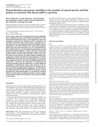
Fig. 1. Radiation exposure alters miRNA expression and methylation of
LINE1 and SINE B2 retrotransposons in male germline. We utilized an
in vivo murine model to analyze the role of epigenetic alterations in
transgenerational radiation effects. The murine model is widely used, well
characterized and generally accepted for studies of radiation-induced
changes and transgenerational effects (21–24). Mice (mature 60-day-old
male C57BL/6 animals) were randomly assigned to different treatment
groups. The exposed cohort (10 animals) received 2.5 Gy (3 Gy/min) of
X-rays (90 kV, 5 mA) to the whole body. In our previous studies, this dose
led to significant deleterious effects in the progeny (20). Control mice (10
animals) were sham treated. Four days (96 h) after exposure, mice were
humanly killed, and testes were sampled and processed for further
analysis. (A). Altered expression of miR-29 family in germline of exposed
male animals. Global miRNA expression was previously determined by
the LC Sciences miRNA microarray platform (18). Levels of miR-29a and
miR-29b were confirmed by quantitative real-time PCR using specific
primers (Ambion) as described (26). (B). Decreased levels of DNMT3a in
germline of exposed male mice. Lysates from testes tissue were subjected
to immunoblotting using antibodies against DNMT3a. Protein levels
relative to those of control animals are shown as the mean ± SD,
Ã
significant, 95% confidence limit, P , 0.05, Student’s t-test.
Representative western blots. Each experiment included pooled lysates
from two animals for each cohort, with equal representation of each
animal. Western blots were repeated at least three times to ensure the
reproducibility and robustness of the results. (C). Hypomethylation of
LINE1 and SINE B2 in the germline of exposed animals as determined by
methylation-sensitive McrBC-based quantitative real-time PCR analysis.
The methylation-sensitive McrBC endonuclease is a restriction enzyme
that digests only methylated DNA sequences (30), n 5 10; Ã
P , 0.05,
Student’s t-test. CT, control animals; EX, exposed animals, n 5 10;
Ã
P , 0.05, Student’s t-test.
Association of altered miRNA expression with hypomethylation and genome instability
1111
byguestonJune16,2012http://carcin.oxfordjournals.org/Downloadedfrom
3. Loss of DNA methylation of transposable elements in the germline of
exposed mice
It is well known that DNMT3a partakes in methylation and silencing
of transposable elements and safeguarding genome stability. There-
fore, we determined the DNA methylation status of LINE1 and SINE
B2, two of the most prevalent classes of repetitive sequences that
compose .20% of the mouse genome (32). The status of LINE1
and SINE B2 methylation was determined by methylation-sensitive
McrBC quantitative PCR analysis (30). The methylation-sensitive
McrBC endonuclease is a restriction enzyme that digests only
methylated DNA sequences but does not cleave unmethylated DNA
(33). Importantly, we found a significant loss of LINE1 methylation and
a tendency of SINE B2 hypomethylation upon radiation exposure in the
male germline (Figure 1C). This is a very interesting finding because
hypomethylation of certain repetitive elements, including LINE1, is
considered to be a hallmark of genomic instability (34,35).
Paternal exposed leads to altered LINE1 and SINE B2 methylation in
the offspring
Genome instability in the paternal germline may exert a negative
influence on fertilized eggs and therefore cause deleterious effects
in the offspring (36). Our next step was to investigate the extent of
inherited epigenetic changes in the thymus tissue of unexposed prog-
eny of exposed male mice. Thymus is an important lymphoid hema-
topoietic organ that is a target of radiation carcinogenesis.
Furthermore, in our previous studies, we observed a loss of global
genome-wide methylation in the thymus tissue of the progeny of
exposed parents (20). One of the main functions of DNA methylation
in normal mammalian somatic tissues is the suppression of transpos-
able repetitive elements (37,38). With this in mind, we determined the
DNA methylation status of LINEs and SINEs, particularly LINE1 and
SINE B2, in thymus tissues of the progeny of exposed parents.
Figure 2A demonstrates a profound level of hypomethylation of
LINE1 and SINE B2 sequences in the thymus tissue of the progeny
of exposed male mice as evidenced by a significantly (P , 0.05)
greater recovery of LINE1 and SINE B2 PCR products (by 3.8 and
2.2 times, respectively) after pretreatment of DNA with McrBC en-
donuclease. This may be indicative of genomic instability in the
thymus tissue of the progeny (34,35).
Decreased levels of LSH in the progeny of exposed parents
In order to determine whether or not loss of DNA methylation in the
thymus of the offspring of irradiated male mice is associated with the
dysregulated function of the DNA methylation machinery, we mea-
sured the protein levels of DNMTs. Surprisingly, we did not detect
changes in protein levels of either the maintenance DNMT1 or the
de novo DNMT3a and DNMT3b (data not shown). However, we
detected a substantial decrease in the levels of the LSH protein in
the thymus tissue of the offspring of exposed parents as compared
with controls (Figure 2B). LSH, a member of the SNF2 family of
chromatin remodeling proteins, is thought to be crucial for the main-
tenance of genome-wide CpG methylation (39), especially for meth-
ylation and silencing of repetitive elements such as LINEs and SINEs
(40,41). Recently, it has been demonstrated that LSH is directly in-
volved in the control of de novo methylation of DNA (42), and LSH
deficiency leads to aberrant upregulation of retroviral repetitive ele-
ments in the genome, abnormal mitosis with amplified centrosomes
and genomic instability (43). Therefore, LINE1 and SINE B2 hypo-
methylation in the thymus tissue of the progeny of exposed mice may
be related to the decreased levels of LSH.
LSH expression is mediated by a miRNA
To further explore molecular mechanisms underlying hypomethylation
of repetitive elements in the progeny of irradiated male mice driven by
LSH downregulation, we studied the regulation of LSH expression
mediated by short noncoding RNAs. It has been demonstrated that
miRNAs participate in the regulation of a variety of cellular processes
in mammals, including DNA methylation (31,44–46). In total, miRNAs
control the activity of $30% of all protein-coding genes (47), including
proteins that control methylation of DNA (31). Additionally, the results
of recent studies have shown that deficiency of Dicer, an RNAse III
family nuclease that generates miRNAs, is associated with decreased
DNA methylation (48). Indeed, Figure 3A demonstrates a decrease in
Dicer expression in the thymus tissue of the offspring.
Next, we analyzed miRNA expression profiles using miRNA
microarrays in the thymus tissue of the progeny of control and exposed
parents. Cluster analysis revealed that preconceptional paternal
exposure led to significant changes in miRNA expressions in the thymus
tissue of the offspring. We identified 25 miRNA genes (17 upregulated
and 8 downregulated) that were differentially expressed (P , 0.05) in
the progeny of exposed animals as compared with controls (Figure 3B).
Interestingly, we did not detect changes in the expression of two
miRNA families, miR-29 and miR-290, that regulate levels of DNMTs
(31,48,49). However, computational analysis revealed that miR-468,
which was upregulated in the progeny of exposed parents, targets
Fig. 2. Paternal radiation exposure causes hypomethylation of LINE1 and
SINE B2 retrotransposons and alters cellular levels of the methylation
regulator LSH. To analyze the effects of exposure on the progeny, 4 days after
irradiation control and exposed animals were mated with unexposed females.
Both sets of progeny were killed 6 months after birth. The thymus tissues were
extracted, immediately frozen and stored at À80°C until the analysis.
(A). Hypomethylation of LINE1 and SINE B2 in the progeny of exposed
parents as determined by methylation-sensitive McrBC-based quantitative
real-time PCR analysis. The methylation-sensitive McrBC endonuclease is
a restriction enzyme that digests only methylated DNA sequences (30),
n 5 10; Ã
P , 0.05, Student’s t-test. (B). Paternal radiation exposure
decreases LSH protein levels in the thymus of the progeny in vivo. Lysates
from thymus tissue were subjected to immunoblotting using antibodies against
LSH. Representative blots from four independent experiments are shown; each
experiment included pooled lysates from four animals for each exposure
condition with equal representation of each animal.
J.N.Filkowski et al.
1112
byguestonJune16,2012http://carcin.oxfordjournals.org/Downloadedfrom
4. murine LSH (Figure 4A). This prediction was further confirmed by the
comprehensive microRNA.org data resource (51). Therefore, to exam-
ine whether or not LSH is indeed functionally targeted by miR-468,
a segment of Lsh-3#-UTR containing the miR-468 complementary re-
gion was cloned into the 3#-UTR of the luciferase reporter system.
Additionally, we have mutated the miR-468 seed-interacting sequence
in the Lsh-3#-UTR. The resulting reporter vectors were then transfected
into HEK293 cells together with transfection controls and miR-468.
The luciferase reporter construct that did not contain the Lsh UTR was
used as a negative control. Figure 4B shows that miR-468 inhibited the
luciferase activity of the construct containing the Lsh-3#-UTR segment
(Figure 4B). There was no change in the luciferase reporter activity if
cells were co-transfected with a negative control (scrambled oligonu-
cleotides) or with a vector harboring mutated Lsh-3#-UTR. No lucifer-
ase expression changes were observed if cells were transfected with the
plasmid lacking a Lsh-3#-UTR fragment (Figure 4B).
To further confirm that miR-468 indeed affects the protein levels of
LSH in mouse NIH3T3 cells, these cells were transfected with miR-
468, and the level of LSH was determined by immunocytochemistry
and western immunoblotting 24 h after transfection. Transfection of
NIH3T3 cells with miR-468 resulted in a decrease of LSH levels
(Figure 5A). Therefore, we propose that miR-468-mediated suppres-
sion of LSH leads to aberrant methylation of LINE1 and SINE B2 in
the thymus tissue of the progeny of exposed parents.
The mechanistic link between miR-468-mediated LSH downregu-
lation and the DNA hypomethylation was confirmed by transfection
of mouse NIH3T3 cells with miR-468 or scrambled oligonucleotides.
Cells were harvested 48 h later and the levels of global DNA meth-
ylation were analyzed. Interestingly, we noted a 9% decrease global
genomic DNA methylation in the miR-468-transfected cells that was
statistically significant at 90% confidence.
Furthermore, transfection of NIH3T3 cells with miR-468 led to
a significant (P , 0.05) 24% loss of methylation of SINE elements.
Methylation levels of LINE1 elements also tended to decrease 48 h
after transfection of the cells with miR-468 precursors (Figure 5).
Discussion
Approximately 5% of human live births today have a birth defect,
a de novo genetic disease or chromosomal abnormality (52). Even
having this knowledge, we still have little understanding of the mech-
anisms of genome instability. Recent studies suggest that transgenera-
tional genome instability may be epigenetic in nature and mediated
via altered DNA methylation and microRNAome. To fully understand
transgenerational genome instability, it is important to define what
happens in the germline of exposed parents and in the progeny.
Here, we for the first time analyzed the nature and mechanisms
underlying the disruption of DNA methylation and miRNA expres-
sion status in the germline and progeny of exposed parents. We dem-
onstrate that paternal irradiation leads to upregulation of the miR-29
family in the exposed male germline, which causes decreased
Fig. 3. Paternal radiation exposure results in microRNAome deregulation in the thymus tissue of the unexposed progeny. (A) Decreased levels of Dicer in the
thymus tissue of the progeny of exposed animals as determined by immunohistochemistry (IHC). IHC was conducted as described before (27). Levels of Dicer-
positive cells per field of view are shown, mean values ± SEM, n 5 10 fields per animals, 40 cells per field, Ã
P , 0.05, Student’s t-test. (B). Hierarchical clustering
of differentially expressed miRNA genes in the thymus tissue of the progeny of control and exposed mice. Total RNA was extracted from the thymus tissue using
TRIzol Reagent (Invitrogen, Burlington, Ontario, Canada). miRNA microarray analysis was performed by LC Sciences (Houston, TX). Red denotes high
expression levels, whereas green depicts low expression levels.
Fig. 4. mir-468 targets LSH. (A) Complementary site for miR-468 in the
3#-UTR of Lsh1. (B) A dose-dependent inhibition of Lsh expression in the
luciferase assay. A 3#-UTR segment of the Lsh gene corresponding to
a region of 366 nts (from 2642 nt through 3008 nt of the total transcript) of
Lsh (Acc. # NM_008234) was amplified by PCR from mouse genomic DNA
and cloned into the pGL3-control vector (Promega, Madison, WI). HEK293
cells were transfected with the firefly luciferase UTR-report vector, the
control Renilla luciferase pRL-TK vector (Promega), transfection controls
either with precursor miR-468 or with miRNAs that do not have binding sites
within the 3#-UTR of Lsh (Ambion) as described previously (26). Twenty-
four hours after transfection, cells were lysed, and the activity of both renilla
and firefly luciferases was assayed using the dual-luciferase reporter assay
system (Promega) as described previously (26,50). The graph depicts an
inhibition of Lsh expression in the luciferase assay after transfection of
HEK293 cells with miR-468. RLU, relative luminescence units; Ã
P , 0.05,
Student’s t-test.
Association of altered miRNA expression with hypomethylation and genome instability
1113
byguestonJune16,2012http://carcin.oxfordjournals.org/Downloadedfrom
5. expression of de novo methyltransferase DNMT3a (31) and profound
hypomethylation of transposable LINE1 and SINE B2 sequences.
Therefore, radiation-induced hypomethylation in the male germline
may be explained, at least in part, by the radiation-induced miR-29
changes. In our previous studies, we have shown that radiation-
induced deregulation of another important miRNA—miR-709—also
partakes in regulation DNA methylation in the male germline (18).
Importantly, epigenetic DNA methylation and microRNAome
changes observed in the male germline led to deleterious molecular
effects in the somatic thymus tissue from the progeny of exposed
animals, including hypomethylation of LINE1 and SINE B2. Hypo-
methylation of LINE1 and SINE B2 in the thymus tissue from the
progeny was associated with a significant decrease in the levels of
LSH. LSH previously reported to be crucial for the maintenance of
methylation and silencing of repetitive elements (39–41,43,53). LSH
cooperates with DNMTs in setting DNA methylation patterns and
silencing (54). Therefore, even though we have not seen any changes
in the levels of DNMTs in the thymus tissue of the progeny of exposed
parents, the changes observed in the levels of LSH may have possibly
affected the functioning of DNMTs. The role of DNMT–LSH inter-
actions in the germline and transgenerational effects has still to be
analyzed in detail. Additionally, the tissue-specific roles of LSH and
DNMTs need to be further analyzed.
Furthermore, our results demonstrate for the first time that pater-
nal irradiation leads to microRNAome changes in the progeny. Spe-
cifically, we noted a significant upregulation of miR-468 that targets
LSH and leads to its decreased expression in thymus tissue of the
progeny of exposed parents. We suggest that miR-468-mediated
suppression of LSH leads to aberrant methylation of LINE1 and
SINE B2 in thymus of the progeny of exposed parents. Transfection
of mouse fibroblast NIH3T3 cells with miR-468 led to a significant
decrease in the levels of Lsh and resulted in decreased global geno-
mic DNA methylation levels. Mammalian genomes have high levels
of DNA methylation at repetitive satellite DNA sequences, retro-
and DNA transposons. Repetitive sequences are the first targets of
DNA methylation loss caused by the altered levels of the methyla-
tion machinery (30,40,53,55). This regulation may be specifically
required when the cells that were exposed to a genotoxic stressor,
such as radiation, and for their descendants. Radiation induces DNA
damage, which is known to cause significant DNA hypomethylation.
In normal cells, after DNA is repaired, DNA methylation machinery
will most probably relatively quickly restore the methylation pat-
terns, Yet, if miR-468 is overexpressed and LSH is suppressed such
a restoration will not be as efficient due to the lack of LSH and will
take longer time. Further studies are needed to analyze the roles of
mir-468-mediated LSH changes in normal conditions and upon
stress exposure.
Altered microRNAome levels, global DNA hypomethylation and
reactivation of retroelements all constitute deleterious effects that
may significantly influence genome stability and therefore lead to
carcinogenesis. Importantly, changes were observed in radiation
carcinogenesis of the target organ (thymus). Biological repercus-
sions of these molecular and cellular changes and their etiological
role in transgeneration carcinogenesis need to be further analyzed in
detail. Additionally, further studies are needed to address epigenetic
and miRNAome changes in the germline of progeny of exposed
parents.
Epigenetic changes in the progeny were linked to male parent
exposure. The paternal genome is extremely sensitive to exposure
to genotoxic agents (1,4,5,56). Therefore, our data agree with the
previous reports on radiation induction of transgenerational genomic
instability upon paternal exposure. The epigenetic alterations
observed may be associated with the transmission of altered DNA
methylation, reactivation of transposons and DNA damage in parental
sperm cells. After fertilization, mammalian genomes undergo marked
methylation reprogramming in order to establish correct parent-of-
origin developmental programs (57,58). The epigenetic changes
observed in sperm cells may interfere with post-fertilization epige-
netic reprogramming, thus affecting fertilized eggs and leading to
subsequent deleterious changes in the embryo (36). Interestingly,
some studies reported that transgenerational genome instability is
more pronounced in mice than in humans. Our data may offer a plau-
sible explanation for an apparent discrepancy in the magnitude of
transgenerational effects in mice and humans. Detailed analysis of
miRNA and genome databases revealed that miR-468 is a mouse-
specific miRNA. While the 3#-UTR of human and mouse Lsh genes
are 98% identical and the miR-468-binding sites are preserved, func-
tional miR-468 does not exists in a human genome. The lack of miR-
468 may explain, at least in part, why transgenerational genome
instability is less pronounced in humans. Further in depth studies
are needed to understand the roles of miRNAs in germline and trans-
generational effects. Therefore, this study may serve as a future road-
map for analyzing the role of methylation and microRNAome in
transgenerational genomic instability.
Supplementary material
Supplementary Figure 1 can be found at http://carcin.oxfordjournals.org/
Funding
Alberta Cancer Board operating grant (22180) to O.K. J.F. was a re-
cipient of the Alberta Heritage Foundation for Medical Research and
the National Science and Engineering Research Council Graduate
Scholarships.
Fig. 5. miR-468 reduces cellular levels of LSH and affects DNA methylation.
(A) Transfection of mouse NIH3T3 cells with miR-468 effectively reduces
cellular levels of LSH. Levels of LSH were detected by immunofluorescence
and by western immunoblotting using anti-LSH antibodies. Red indicates LSH
and blue indicates nuclear 4#,6-diamidino-2-phenylindole stain. (B) Altered
methylation of LINE1 and SINE B2 as determined by methylation-sensitive
McrBC-based real-time PCR analysis in NIH3T3 cells 48 h after transfection
with miR-468, Ã
P , 0.05, Student’s t-test.
J.N.Filkowski et al.
1114
byguestonJune16,2012http://carcin.oxfordjournals.org/Downloadedfrom
6. Acknowledgements
We thank James Meservy for technical assistance. We are grateful to Valentina
Titova for proofreading this manuscript. Note: The views expressed in this paper
do not necessarily represent those of the US Food and Drug Administration.
Conflict of Interest Statement: None declared.
References
1.Barber,R.C. et al. (2006) The offspring of irradiated parents, are they sta-
ble? Mutat. Res., 598, 50–60.
2.Dubrova,Y.E. (2003) Radiation-induced transgenerational instability. On-
cogene, 22, 7087–7093.
3.Niwa,O. (2006) Radiation induced dynamic mutations and transgeneration-
al effects. J. Radiat. Res. (Tokyo), 47 (suppl. B), B25–B30.
4.Morgan,W.F. (2003) Non-targeted and delayed effects of exposure to ion-
izing radiation: II. Radiation-induced genomic instability and bystander
effects in vivo, clastogenic factors and transgenerational effects. Radiat.
Res., 159, 581–596.
5.Morgan,W.F. (2003) Non-targeted and delayed effects of exposure to ion-
izing radiation: I. Radiation-induced genomic instability and bystander
effects in vitro. Radiat. Res., 159, 567–580.
6.Dubrova,Y.E. et al. (2008) Paternal exposure to ethylnitrosourea results in
transgenerational genomic instability in mice. Environ. Mol. Mutagen., 49,
308–311.
7.Nomura,T. (2008) Transgenerational effects from exposure to environmen-
tal toxic substances. Mutat. Res., 659, 185–193.
8.Glen,C.D. et al. (2008) Single-molecule PCR analysis of germ line muta-
tion induction by anticancer drugs in mice. Cancer Res., 68, 3630–3636.
9.Hales,B.F. et al. (2005) Impact of paternal exposure to chemotherapy on
offspring in the rat. J. Natl Cancer Inst. Monogr., 34, 28–31.
10.Dubrova,Y.E. (2003) Long-term genetic effects of radiation exposure.
Mutat. Res., 544, 433–439.
11.Dubrova,Y.E. et al. (2000) Transgenerational mutation by radiation.
Nature, 405, 37.
12.Baulch,J.E. et al. (2005) Gamma irradiation of Type B spermatogonia leads
to heritable genomic instability in four generations of mice. Mutagenesis,
20, 337–43.
13.Egger,G. et al. (2004) Epigenetics in human disease and prospects for
epigenetic therapy. Nature, 429, 457–463.
14.Feinberg,A.P. (2008) Epigenetics at the epicenter of modern medicine.
JAMA, 299, 1345–1350.
15.Jaenisch,R. et al. (2003) Epigenetic regulation of gene expression: how the
genome integrates intrinsic and environmental signals. Nat. Genet., 33
(suppl.), 245–254.
16.Huppi,K. et al. (2008) The identification of microRNAs in a genomically
unstable region of human chromosome 8q24. Mol. Cancer Res., 6,
212–221.
17.Erson,A.E. et al. (2008) MicroRNAs in development and disease. Clin.
Genet., 74, 296–306.
18.Tamminga,J. et al. (2008) DNA damage-induced upregulation of miR-709
in the germline downregulates BORIS to counteract aberrant DNA hypo-
methylation. Cell Cycle, 7, 3731–3736.
19.Tamminga,J. et al. (2008) Paternal cranial irradiation induces distant by-
stander DNA damage in the germline and leads to epigenetic alterations in
the offspring. Cell Cycle, 7, 1238–1245.
20.Koturbash,I. et al. (2006) Epigenetic dysregulation underlies radiation-
induced transgenerational genome instability in vivo. Int. J. Radiat. Oncol.
Biol. Phys., 66, 327–330.
21.Boulton,E. et al. (2002) Myeloid, B and T lymphoid and mixed lineage
thymic lymphomas in the irradiated mouse. Carcinogenesis, 23, 1079–1085.
22.Barber,R.C. et al. (2006) Radiation-induced transgenerational alterations in
genome stability and DNA damage. Oncogene, 25, 7336–7342.
23.Yauk,C.L. et al. (2002) A novel single molecule analysis of spontaneous
and radiation-induced mutation at a mouse tandem repeat locus. Mutat.
Res., 500, 147–156.
24.Utsuyama,M. et al. (2003) Radiation-induced-thymic lymphoma occurs in
young, but not in old mice. Exp. Mol. Pathol., 74, 319–325.
25.Ilnytskyy,Y. et al. (2009) Radiation-induced bystander effects in vivo are
epigenetically regulated in a tissue-specific manner. Environ. Mol. Muta-
gen., 50, 105–113.
26.Ilnytskyy,Y. et al. (2008) Altered microRNA expression patterns in irradi-
ated hematopoietic tissues suggest a sex-specific protective mechanism.
Biochem. Biophys. Res. Commun., 377, 41–45.
27.Koturbash,I. et al. (2008) Sex-specific microRNAome deregulation in the
shielded bystander spleen of cranially exposed mice. Cell Cycle, 7,
1658–1667.
28.Chang,J. et al. (2008) Liver-specific microRNA miR-122 enhances the
replication of hepatitis C virus in nonhepatic cells. J. Virol., 82, 8215–8223.
29.Koh,T.C. et al. (2009) Identification and expression analysis of miRNAs
during batch culture of HEK-293 cells. J. Biotechnol., 140, 149–155.
30.Martens,J.H. et al. (2005) The profile of repeat-associated histone lysine
methylation states in the mouse epigenome. EMBO J., 24, 800–812.
31.Fabbri,M. et al. (2007) MicroRNA-29 family reverts aberrant methylation
in lung cancer by targeting DNA methyltransferases 3A and 3B. Proc. Natl
Acad. Sci. USA, 104, 15805–15810.
32.Waterston,R.H. et al. (2002) Initial sequencing and comparative analysis of
the mouse genome. Nature, 420, 520–562.
33.Sutherland,E. et al. (1992) McrBC: a multisubunit GTP-dependent restric-
tion endonuclease. J. Mol. Biol., 225, 327–348.
34.Roman-Gomez,J. et al. (2005) Promoter hypomethylation of the LINE-1
retrotransposable elements activates sense/antisense transcription and
marks the progression of chronic myeloid leukemia. Oncogene, 24,
7213–7223.
35.Howard,G. et al. (2008) Activation and transposition of endogenous retro-
viral elements in hypomethylation induced tumors in mice. Oncogene, 27,
404–408.
36.Aitken,R.J. et al. (2007) Origins and consequences of DNA damage in male
germ cells. Reprod. Biomed. Online, 14, 727–733.
37.Yoder,J.A. et al. (1997) Cytosine methylation and the ecology of intra-
genomic parasites. Trends Genet., 13, 335–340.
38.Miranda,T.B. et al. (2007) DNA methylation: the nuts and bolts of repres-
sion. J. Cell. Physiol., 213, 384–390.
39.Dennis,K. et al. (2001) Lsh, a member of the SNF2 family, is required for
genome-wide methylation. Genes Dev., 15, 2940–2944.
40.Huang,J. et al. (2004) Lsh, an epigenetic guardian of repetitive elements.
Nucleic Acids Res., 32, 5019–5028.
41.Yan,Q. et al. (2003) Lsh, a modulator of CpG methylation, is crucial for
normal histone methylation. EMBO J., 22, 5154–5162.
42.Zhu,H. et al. (2006) Lsh is involved in de novo methylation of DNA. EMBO
J., 25, 335–345.
43.Muegge,K. (2005) Lsh, a guardian of heterochromatin at repeat elements.
Biochem. Cell Biol., 83, 548–554.
44.Bartel,D.P. (2004) MicroRNAs: genomics, biogenesis, mechanism, and
function. Cell, 116, 281–297.
45.Sevignani,C. et al. (2006) Mammalian microRNAs: a small world for fine-
tuning gene expression. Mamm. Genome, 17, 189–202.
46.Hobert,O. (2008) Gene regulation by transcription factors and microRNAs.
Science, 319, 1785–1786.
47.Filipowicz,W. et al. (2008) Mechanisms of post-transcriptional regulation
by microRNAs: are the answers in sight? Nat. Rev. Genet., 9, 102–114.
48.Benetti,R. et al. (2008) A mammalian microRNA cluster controls DNA
methylation and telomere recombination via Rbl2-dependent regulation of
DNA methyltransferases. Nat. Struct. Mol. Biol., 15, 998.
49.Sinkkonen,L. et al. (2008) MicroRNAs control de novo DNA methylation
through regulation of transcriptional repressors in mouse embryonic stem
cells. Nat. Struct. Mol. Biol., 15, 259–67.
50.Kovalchuk,O. et al. (2008) Involvement of microRNA-451 in resistance of
the MCF-7 breast cancer cells to chemotherapeutic drug doxorubicin. Mol.
Cancer Ther., 7, 2152–2159.
51.Betel,D. et al. (2008) The microRNA.org resource: targets and expression.
Nucleic Acids Res., 36, D149–D53.
52.Wyrobek,A.J. et al. (2007) Assessing human germ-cell mutagenesis in the
Postgenome Era: a celebration of the legacy of William Lawson (Bill)
Russell. Environ. Mol. Mutagen., 48, 71–95.
53.Fan,T. et al. (2008) DNA hypomethylation caused by Lsh deletion pro-
motes erythroleukemia development. Epigenetics, 3, 134–142.
54.Myant,K. et al. (2008) LSH cooperates with DNA methyltransferases to
repress transcription. Mol. Cell. Biol., 28, 215–226.
55.Rollins,R.A. et al. (2006) Large-scale structure of genomic methylation
patterns. Genome Res., 16, 157–163.
56.Cordier,S. (2008) Evidence for a role of paternal exposures in developmen-
tal toxicity. Basic Clin. Pharmacol. Toxicol., 102, 176–181.
57.Anway,M.D. et al. (2005) Epigenetic transgenerational actions of endo-
crine disruptors and male fertility. Science, 308, 1466–1469.
58.Mayer,W. et al. (2000) Demethylation of the zygotic paternal genome.
Nature, 403, 501–502.
Received April 22, 2009; revised November 13, 2009;
accepted November 23, 2009
Association of altered miRNA expression with hypomethylation and genome instability
1115
byguestonJune16,2012http://carcin.oxfordjournals.org/Downloadedfrom