General dentistry an evaluation and adjustment method article
1. Journal of the Academy of General Dentistry
August, 2013
An evaluation and adjustment method for natural proximal contacts of crowns using diamond dental strips: a case report
Daniel S. Kim, DDS, FAGD n John A. Rothchild, DDS, MAGD n Kyu-Won Suh, DDS, MSD, DSO
The best way to adjust proximal contacts of newly fabricated indirect restorations has been a long- standing unresolved issue in dentistry. Excessively tight contacts cause incomplete seating of indirect restorations and intrusion of adjacent teeth, which leads to patient discomfort, hypersensitivity, and recurrent dental caries at the crown margins. When seating indirect restorations, interproximal relief should be restored as it exists in natural dentition.
This article presents an innovative method of crown seating using diamond strips. This simple, consistent method makes it easier for clinicians to provide comfortable and long-lasting restorations with minimal time and effort. Laboratory technicians utilize diamond strips to provide properly fitting indirect restorations that require minimal adjustment upon clinical delivery. Diamond strips also allow for accurate determination of heavier proximal contacts, allowing dentists to adjust the proximal contact properly in the patients’ mouths.
Clinically, restoring natural proximal contacts is a critical factor to the success of indirect restorations. Using this method standardizes proper proximal contact adjustments of laboratory-fabricated indirect restorations between dental labs and dental offices. The method also helps to limit or eliminate the lingering proximal contact issue between clinicians and laboratory technicians.
Received: February 19, 2012
Revised: May 7, 2012
Accepted: June 13, 2012
Key words: proximal contact, interproximal relief, contact adjustment, diamond dental strips, crown seating
Restoring natural proximal contacts is critical to the success of indirect restorations. This restoration directly affects a dentist’s ability to achieve complete marginal seating, maintain existing occlusion, and provide patient comfort. Inadequately restored proximal contacts may cause food impaction by open contact, or may cause incomplete marginal seating of indirect restorations by overly tight contact.1 Dental school curriculums offer surprisingly little instruction on fixing and adjusting tight proximal contacts, and there is little written research on this subject.
2. In the authors’ experience, crowns sometimes must be refabricated due to improper proximal contacts of the final restoration after chairside adjustment. This occurs when the restoration is too tight on one side and opens up on the opposite side. This report presents a clinically proven method for determining and adjusting proximal contact strength by using diamond dental strips. Ideal proximal contacts can be achieved accurately with minimal time and effort, while enhancing patient comfort and functionality immediately upon definitive cementation.
Proximal contacts in the literature
The term proximal contact area refers to the area of proximal contour height on the mesial or distal surface of a tooth that touches its adjacent tooth in the same arch.2 The variants that affect proximal contact are location, tooth type, chewing, and time of day.3 If a patient has a nocturnal parafunctional habit of clenching or grinding, the interproximal contact would be significantly tighter in the morning upon wakening. Proximal contact location is in fact a physiological entity of multifactorial origin. Dorfer et al measured proximal contact strength with a calibrated metal strip (0.05 mm thick), and reported that the strength varied between teeth, arches, and function.4
In natural dentition, small spaces occur between natural teeth at rest, and floss does not properly penetrate these spaces.3,5-7 A 1987 study by Boice et al reported that a shim stock passed through 90% of natural contacts, regardless of the sex or age of the patient.6 When restoring natural proximal contacts, the clinician should not intentionally build the contact so tight that it would prevent a 0.0127 mm shim stock from passing through the contact.6,8 Although there is an association between open contacts and food impaction, open contacts can occur in interproximal sites where contacts are tight, marginal ridges are uneven, when the patient has a lack of adequate escape grooves, and/or prominent opposing cusps.9 A properly restored proximal contact should offer passive contact or microscopic clearance that relieves pressure between the proximal contact surfaces of indirect restorations and adjacent teeth. This relief of pressure is referred to as interproximal relief.10 Interproximal space is restored accurately by proper restoration of both the deflecting contours and the occlusal anatomy of the teeth, which prevents food impaction. Proper restoration of interproximal relief allows for complete marginal seating of an indirect restoration, and prevents occlusal interferences.10 The periodontal ligaments allow for sufficient minor tooth movement during proximal contact adjustment.
Materials and methods
To fabricate an indirect restoration, it is necessary to abrade the proximal contact surfaces of adjacent teeth on working models to compensate for proper proximal contacts. It also is necessary to have a predictable and consistent method for making crowns, with proper measurements of contact tightness.
Dental laboratory technicians use their own techniques to abrade adjacent proximal contacts. These techniques can vary from crown to crown, from technician to technician, and from laboratory to laboratory.
Due to these inconsistencies, the final restoration almost always requires a dentist to perform proximal contact adjustment, which may require extra time for a restoration; this chair time increases if a dentist
3. is placing several restorations. The present study was performed to develop a consistent method for abrading proximal contacts, and to ensure that the laboratory-processed crowns will fulfill dentists’ expectations every time.
A section of a stone die with a dowel pin attached was obtained from a used stone model (Heraeus Kulzer), and it was used as a specimen. The section of the stone die block was ground to a rectangular shape (6 mm x 6 mm x 3 mm), with one side rounded to resemble an adjacent proximal contact of a working model. An electronic caliper (Starrett 797, The L.S. Starrett Company) was used to measure the thickness of the block after each abrasion with diamond strips.
The specimen was abraded on the surface of its rounded end by passing the diamond strip once. Using the caliper, the thickness of the specimen was measured (in μ). This procedure was repeated 10 times, and the difference in thickness calculated between each measurement; the differences between reductions (DBRs) were the amount of abrasion. The abrasive surface of the diamond strip was air blown after each use for accuracy. This procedure was repeated in 4 sets, and the median number was chosen from each set of 10 DBRs.
Three different strips were used for this experiment: 15 μ grit, 17 μ grit, and 30 μ grit. (All strips mentioned in this article are manufactured by ContacEZ Company.). The median score for the 17 μ diamond strip was a 0.01 mm reduction per pass, as was the average for the 4 sets. The 17 μ grit, 0.09 mm diamond strip was chosen to abrade the stone die because it consistently abraded 0.01 mm with each pass when controlled finger pressure was applied. The travel distance of the strip movement was 16 mm.
Laboratory procedures
Abrade the proximal surfaces of adjacent teeth (stone) on the working model by passing a 17 μ grit diamond strip 6 times (in the authors’ experience, using a strip versus a knife produces more consistent, accurate results), reducing the thickness by 0.06 mm. Construct the laboratory fabricated crown to fit into the space between the 2 adjacent teeth on the stone model. When the crown is made, place it on the working model and pass a thin (0.06 mm) single-sided diamond strip through the interproximal spaces, facing the abrasive surface of the strip toward the bisque-baked crown, until the strip encounters light resistance. Now the crown is ready for glazing.
Adjust the proximal contacts of the glazed crown on the working stone model by passing an ultrafine 0.05 mm diamond strip until the strip encounters light resistance, meaning, the distance between the crown and proximal teeth is 0.01 mm larger than the normal 0.1 mm lateral movement of teeth in natural dentition. Thus, this crown will be seated on the abutment completely with slight resistance.
At this point, the crown fabrication is complete and ready to send to the dental office. Do not rotary polish the proximal contact surfaces of the crown. This procedure eliminates guesswork about the accuracy of a crown’s proximal contacts. The clinical seating of the crown will be consistent and accurate.
4. Evaluation and adjustment
To determine and adjust the proximal contact prior to cementation, place the crown on the abutment tooth and press it to the prepared margin. If there is slight resistance to seating the crown against the prepared margin due to proximal contacts, proceed with the following protocol.
First, lift the crown slightly and insert a 0.05 mm ultrafine or a 0.06 mm fine diamond strip into the interproximal space, with the abrasive side facing the crown. Allow the crown to sit on the prepared tooth, but do not press down yet; instead, simply hold the crown in place and start passing the diamond strip in a buccolingual direction to check interproximal pressure against the strip. Repeat this procedure in the other interproximal space. The diamond strip may move in one direction at the beginning because of the orientation of the diamond particles.
When more pressure is detected on one side than the other, pass the 0.06 mm diamond strip 5-6 times through the side in a buccolingual direction with more pressure. Repeat this procedure until there are equal amounts of light resistance in both the mesial and distal interproximal spaces, indicating that ideal proximal contact for the crown has been attained and that the crown is ready for cementation. If the proximal contact is too heavy, move the crown back to the working model and adjust the side of the closed margin of the proximal contact of the crown by using the 17 μ grit diamond strip with the abrasive side facing the crown. Next, using an explorer, remove the excess resin cement around the crown. Pass an ultrathin (0.04 mm) serrated strip (Serrated Dental Strip I) back and forth gently and buccolingually, to cut and clean out any trapped and remaining resin cement in the interproximal spaces. Pass a single-sided, ultrafine (0.05 mm) diamond strip through the interproximal spaces to polish the contact surfaces of restorations, confirm interproximal relief, and restore a natural finish. Dental floss should pass through the interproximal space with firm resistance (that is, snapping in and out). At this point, the crown seating is considered to be complete with proper proximal contacts.
Case report
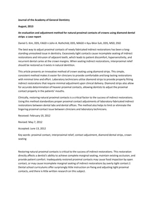
A 55-year-old man presented with dental decay under an existing gold crown on Tooth No. 19. A porcelain crown was recommended and the patient consented. The tooth was prepared for the crown and an impression was taken and sent to the dental laboratory for fabrication. On the working model, a diamond strip (LAB Stone Strip) was used to abrade the proximal contacts of the adjacent teeth 6 times (Fig. 1). As shown in the experiment in Materials and Methods, each 16 mm passing of a strip with 17 μ diamond particles reduces the proximal surface of the adjacent stone teeth 0.01 mm in thickness. A porcelain crown (Ceramco, DENTSPLY International) was fabricated (via the conventional laboratory method) to fit the crown into the space between the 2 adjacent teeth (No. 18 and 20).
5. Fig. 1. The proximal surfaces of tooth No. 18 on the working stone model are abraded with a 17 μ grit diamond strip.
When the crown was made for tooth No. 19, a thin (0.06 mm) diamond strip (LAB Bisque Strip) was passed through the interproximal space until light resistance occurred and the crown was ready for glazing.
After glazing, the proximal contacts of the glazed crown were adjusted on the working stone model by passing an ultrafine (0.05 mm) diamond strip (until light resistance was felt against the strip. Rotary polishing was not used on the proximal contact surfaces of the crown. Upon completion of fabrication, the crown was sent to the dental office.
In the dental office
The provisional crown (Luxatemp, DMG America) was removed and the porcelain crown was placed on the abutment and seated almost to the margin, indicating that the proximal contact was slightly heavy and needed adjustment. The crown was lifted slightly, and a thin diamond strip (Black Diamond Strip) was inserted into the distal interproximal space with the abrasive side facing the porcelain crown. The strip passed buccolingually through the distal interproximal space a few times (Fig. 2). This procedure was repeated in the mesial interproximal space, revealing additional pressure on the strip, which indicated a heavier proximal contact on the mesial side of the crown. The strip was passed, starting at the mesial interproximal space. The process was repeated until there was equal light bilateral resistance. A periapical radiograph was taken to confirm the complete marginal seating of the crown. Now the crown was ready for cementation.
Fig. 2. A thin diamond strip is passed in a buccolingual direction through the interproximal space between the fabricated crown for tooth No. 19 and the adjacent teeth.
The crown was cemented with glass ionomer cement (GC Fuji II, GC America, Inc.). The crown was seated with finger pressure and excess cement was removed with an explorer. The remaining excess cement was cut, cleaned, and removed using an ultrathin serrated dental strip (White Strip, 0.04 mm). Dental floss (Reach, Johnson & Johnson) was used to check the interproximal spaces. The floss snapped in and out of the spaces firmly. A periapical radiograph was taken to confirm complete marginal seating of the crown and no residual cement left in the interproximal space (Fig. 3). After the interproximal spaces were cleaned, an ultrafine (0.05 mm) diamond strip was used to polish the interproximal surfaces and restore a natural finish. The ultrafine strip confirmed interproximal relief and proper proximal contacts.10
6. Fig. 3. A radiograph shows complete marginal seating for the crown.
Using a round diamond point, minimal occlusal adjustment was performed and polished using rubber points (Shofu Dental Corporation). At that point, crown seating was complete. The patient reported no pressure from the new crown and said that he felt as comfortable as if it was his own tooth.
Discussion
Excessive tight proximal contacts can be multifactorial and may include parafunctional habits such as bruxing and/or clenching, occlusal discrepancies, crowding, malposition of teeth, and iatrogenic dentistry. Tight proximal contacts can cause not only patient discomfort, but also migration of teeth with consequent crowding and/or repositioning of teeth.10 When replacing existing dental restorations or restoring virgin teeth, proximal contacts must be restored to appropriate pressure/tightness. There is no true definition of appropriate pressure or tightness for a physiological proximal contact, other than the fact that there is contact between 2 teeth. Typically, appropriate contacts are confirmed by a snapping sound between the teeth when flossing with waxed or unwaxed floss (0.05 mm thick when passing interproximal contacts), by using a shim stock as a guide, or by placing very thin articulation paper or film (0.05 mm) between the contacts. Because lateral mobility of teeth in normal natural dentition is 0.1 mm, the microscopic thickness of the medium can be used to detect and adjust the tightness of a crown’s proximal contacts. The goal is to achieve ideal contact, which is passive and without pressure, thus providing the patient with immediate comfort and functionality after crown seating.10
Crowns fabricated in the dental laboratory often have heavy bilateral proximal contact. The dentist usually needs to adjust both contacts but first must determine which of the 2 is heavier. An incorrect assessment can result in weak proximal contact on one side, with the heavy proximal contact intact on the opposite side. In such cases, it may be necessary for the laboratory to add porcelain to close the open contact, or to fabricate a whole new crown.
Traditionally, proximal contacts of indirect restorations are adjusted when a dentist holds a restoration between their fingers, using an indicating medium (such as articulating paper), and reducing the heavy contact with rotary instruments. Small restorations are difficult to hold and to see, and can get caught in the folds of gloves or end up on the floor. In addition, incremental adjustments require multiple trials, which can be tedious and time consuming for the clinicians and uncomfortable for the patients.
7. By contrast, the method described in this article results in consistent placement, as well as patient comfort and functionality. In addition, the complete marginal seating minimizes the need for occlusal adjustment and prevents adjacent teeth movement and extrusion due to excessive interproximal pressure. In mature dentition, the proximal contact should be an area rather than a point.10 In the authors’ experience, most proximal contact surfaces are curved. They become concave or convex by aging due to lateral jaw movement during chewing. The method described in this article also allows dentists to determine easily which proximal contact is heavier and to adjust it properly and immediately in the patient’s mouth. Interproximal relief is confirmed easily by using an ultrathin diamond strip (0.04 mm). Ideal proximal contact of restorations can be achieved accurately with minimal time and effort, enhancing patient comfort and functionality immediately upon definitive cementation.
Summary
When seating indirect restorations, interproximal relief should be restored as it exists in natural dentition. This method of proximal contact adjustment uses diamond dental strips to standardize the proximal contact adjustment method of indirect restorations. Thanks to diamond dental strips, laboratory technicians can provide clinicians with consistent indirect restorations that fit properly, and dentists can restore natural proximal contacts and achieve patient comfort and functionality immediately after definitive cementation, with minimal time and effort.
Author information
Dr. Kim is in private practice in Vancouver, Washington. Dr. Rothchild is in private practice in Durango, Colorado; an associate professor, Dental Medicine Emeritus, Capital University of Integrative Medicine, Washington, DC; and an assistant professor, Department of Surgery, Rush University Medical School, Chicago. Dr. Suh is professor emeritus, College of Medicine, Korea University, Seoul, South Korea.
Acknowledgments
The authors would like to thank Carmen Spulber, CDA, for the photographs used in this article, and Ki Duk Ki from Cascade Dental Laboratory for his cooperation in following the laboratory procedures as instructed by the authors.
Disclaimer
Dr. Kim is the inventor and founder of ContacEZ Restorative Strip System and has a financial interest in the ContacEZ Company. He received no compensation for writing this article. Drs. Rothchild and Suh have no financial interest in any of the companies mentioned in this article and received no compensation for writing this article.
References
1. Howard WW, Moller RC, eds. Atlas of Operative Dentistry. 3rd ed. St. Louis: C.V. Mosby; 1981:127.
2. Sluder TB. Clinical dental anatomy, histology, physiology, and occlusion. In: Studevant CM, ed. The Art and Science of Operative Dentistry. 2nd ed. New York: Mc- Graw-Hill;1985:20.
8. 3. DuBois LM, Niles SM, Boice PA. The magnitude of interproximal spaces between adjacent teeth. Am J Dent. 1993;6(6):315-317.
4. Dorfer CE, von Bethlenfalvy ER, Staehle HJ, Ploch T. Factors influencing proximal dental contact strengths. Eur J Oral Sci. 2000;108(5):368-377.
5. Lindquist JT. A study of the intra-arch relationships in normal human dentitions [master’s thesis]. Indianapolis: Indiana University School of Dentistry;1951.
6. Boice PA, Niles SM, DuBois LM. Evaluation of proximal contacts with shim stock. J Oral Rehabil. 1987;14(1): 91-94
7. Kasahara K, Miura H, Kuritama M, Kato H, Hasegawa S. Observations of interproximal contact relations during clenching. Int J Prosthodont. 2000;13(4):289-294.
8. Campagni WV. The final touch in the delivery of a fixed prosthesis. J Calif Dent Assoc. 1984;12(2):21- 29.
9. Newell DH, John V, Kim SJ. A technique of occlusal adjustment for food impaction in the presence of tight proximal contacts. Oper Dent. 2002;27(1):95-100.
10. Kim DS, Suh KW. A proximal contact adjustment and interproximal relief method. J Prosthet Dent. 2007; 97(4):244-245.
Manufacturers
ContacEZ Company, Vancouver, WA
360.694.1000, www.contacez.com
DENTSPLY International, York, PA
800.243.1942, www.dentsply.com
DMG America, Englewood, NJ
800.662.6383, www.dmg-america.com
GC America, Inc., Alsip, IL
800.323.7063, www.gcamerica.com
Heraeus Kulzer, South Bend, IN
800.435.1785, www.heraeus-dental-us.com
Johnson & Johnson, Skillman, NJ
800.690.1826, www.jnj.com
The L.S. Starrett Company, Athol, MA
978.249.3551, www.starrett.com
Shofu Dental Corporation, San Marcos, CA
800.827.4638, www.shofu.com