1. Skeletal Muscle Contraction
OVERVIEW:
Filaments of actin &
myosin “slide” past each
other when triggered
SLIDING-FILAMENT
THEORY
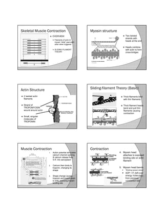
Myosin structure
Two twisted
strands with
heads at the end
Heads combine
with actin to form
cross-bridges
Actin Structure
2 twisted actin
filaments
Strand of
TROPOMYOSIN
wound around actin
Small, singular
molecules of
TROPONIN
Sliding-filament Theory (Basic)
Thick filaments bind
with thin filaments
Thick filament heads
bend and pull thin
filaments causing
contraction
Muscle Contraction
1. Action potential stimulates
calcium channel opening
& calcium release from
S.R. into sarcoplasm
2. Calcium then binds to
troponin, changing its
shape
3. Shape change causes
troponin and tropomyosin
to shift, exposing myosin
binding site
Contraction
4. Myosin head
attaches to exposed
binding site on actin
filament
5. Myosin head bends
“Working (power) stroke”
ADP + Pi (left-over
energy molecules)
from previous
contraction lost
2. Contraction
6. New ATP molecule binds to myosin,
causing it to change shape and release
it from actin
Contraction
7. ATPase hydrolyzes
(splits) ATP into ADP
& Pi giving off energy
8. Returns myosin head
to “high-energy”
configuration for next
cycle
Contraction
9. Calcium pumps in
S.R. reabsorb ions
10. Troponin returns to
normal position
11. Acetylcholinesterase
(AChE) destroys ACh