2. 110 P. Danielson and A. Scott
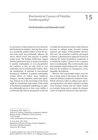
Fig. 15.1 Evidence of intratendinous ACh production.
Tenocytes of patellar tendon tissue have been shown to
harbor enzymes related to acetylcholine (ACh) production
in tendinopathy patients. The ACh synthesizing enzyme
choline acetyltransferase (ChAT), as well as its mRNA,
have been found at intracellular locations. Furthermore,
vesicular acetylcholine transporter (VAChT) – an enzyme
that shuffles ACh from an intracellular site of synthesis
into vesicles – has also been detected inside tenocytes as
shown by this picture. Immunohistochemical staining
(immunofluorescence method, TRITC) show specific
immunoreactions inside the tenocytes, some indicated
with arrows
Fig. 15.2 Evidence of intratendinous catecholamine pro-
duction. In-situ hybridization method shows reactions for
tyrosine hydroxylase (TH) mRNA within some tenocytes
(filled arrow) in patellar tendon tissue from a patient with
tendinopathy. Other tenocytes (unfilled arrows) are nega-
tive in this regard. TH is the rate-limiting enzyme in the
synthesis of catecholamines
3. 11115 Biochemical Causes of Patellar Tendinopathy?
Ach
Catecholamines
Glutamate
mAChR
AR
NMDAR
Fig. 15.3 The biochemical model for patellar tendinopa-
thy. Schematic figure of patellar tendon tissue, showing
the possible roles of biochemical mediators. The micro-
scopic milieu is depicted in the frame to the right. Afferent
sensory nerve fibers, here seen to the left in close associa-
tion with a blood vessel, express muscarinic acetylcholine
receptors (mAChR), N-methyl-d-aspartate receptors
(NMDA R), and adrenergic receptors (AR). The sensory
nerves are hereby susceptible to stimulation by the neu-
rotransmitters acetylcholine (ACh) and glutamate, as well
as by catecholamines. All these substances might thus
theoretically affect pain signalling from the tendon. The
adrenergic receptors might be influenced by cate-
cholamines produced by neighboring efferent sympathetic
nerves (1) “sympathetically maintained pain”). However,
the mAChRs, the NMDA Rs, and the adrenergic receptors
on the sensory nerves might also be stimulated by acetyl-
choline (ACh), glutamate, and catecholamines, respec-
tively, which are produced by the tenocytes themselves (2)
since these principal tendon cells have been shown to
express biosynthetic enzymes for the substances in ques-
tion when tendinopathy occurs. This phenomenon has
been noted for the morphologically disfigured tenocytes
that are frequently seen in tendinosis tissue (upper teno-
cyte in picture). Such tenocytes lack the slender, spindle-
shaped, appearance of normal tenocytes (lower tenocyte
in picture). The efferent sympathetic nerves are further-
more likely to affect blood vessel regulation, via stimula-
tion of adrenergic receptors in the blood vessel walls (3).
Such receptors, alongside mAChRs in the blood vessel
walls, are moreover expected to be stimulated by circulat-
ing catecholamines and ACh, respectively (4). A third
possible source of catecholamines and ACh affecting
blood vessel regulation is the tenocytes of the tendon tis-
sue (5). The tenocytes, in addition to producing the signal
substances in question, express adrenergic receptors and
mAChRs, making them receptive to catecholaminergic
and cholinergic effects (proliferation, changes in collagen
production, and/or degeneration/apoptosis). The receptors
on the tenocytes might, in the case of adrenergic recep-
tors, be influenced by signal substances (catecholamines)
produced by efferent nerves (6), or by signal substances
(ACh and catecholamines) produced by the tenocytes
themselves. In the latter case, autocrine (7) as well as
paracrine (8) loops are suggested to occur. In summary,
receptors on sensory nerves, blood vessels, and tenocytes
in patellar tendons, might be affected by substances from
efferent nerves (green arrows), the blood circulation (red
arrows), and/or the tendon tissue itself (purple arrows)
Copyright with artist: Gustav Andersson