Optimization of 5-HT2Abinding site to fit Serotonin and other tryptamineclass agonists
1. M334 15/11-2010
Dias 1
Optimization of 5-HT2A binding site to fit Serotonin and other tryptamine class agonists.
Martin Strøm Jørgensen and Özlem Karabudak
Introduction
Choice of compounds
Methods
Results
Discussion
Conclusion
Serotonin, or 5-hydroxytryptamine (5-HT), is
a very effective endogenous ligand for the A
family of G-protein coupled receptors.
5-HT2A is a serotonergic receptor which
induces the secretion of different hormones
e.g. renin, ACTH, corticosterone after its
activation (Ísberg V. et al 2010). The aim is
to re-shape the active binding site of a given
5-HT2A model, by moving the amino acid
side chains, in order to fit serotonin.
To fulfill the aim of project we have used
following structure and receptor models.
•As a starting point, we have used serotonin
which was presented in the 5-HT1A receptor
model in the article (Kitson SL. 2007).
•We used the 5-HT2A receptor model from
the Ísberg’s article. This model was
generated by homology modeling based on
the β2-adrenergic receptor and the
G-protein-bound opsin crystal structure.
•To validate the re-shaped binding site,196
known ligands from 3 different agonist
classes and several antagonists were docked
into the new binding site.
•Blast alignment tool was used to find out the
corresponding binding site residues in 5-HT1A
and 5-HT2A.
•Manual docking of serotonin was carried out
by moving amino acid site chains on the
5-HT2A receptor. Molecular dynamics were run
after each manual docking using distance
constraints to optimize the binding pose.
•Minimization was applied to find the local
minimum conformation
•Site map was used to evaluate the interactions
obtained from molecular dynamics and
consider the possibility for further interactions.
•Automated docking with Glide was
performed to test the re-shaped binding site.
5-HT1A 5-HT2A
Asp-166 Asp-155
Ser-198 Ser-239
Thr-199 Ser-242
Table1. Corresponding residues
The blast alignment revealed 3 corresponding
residues involved in the binding of serotonin.
(Table 1)
Manual docking and site map showed 5
potential hydrogen bonds in the binding site
(Figure 2). Efforts to improve the hydrophobic
interactions in the binding site was
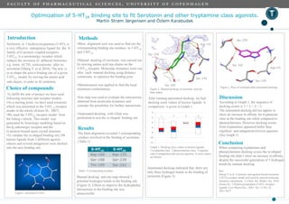
unsuccessful.Figure1. Serotonin (5-HT)
Figure 2. Manual docking of serotonin and site
map output
Ser -239
Ser -242
Ser -159
Asp-155
Automated docking indicated that there are
only three hydrogen bonds in the binding of
serotonin (Figure 3)
After running automated docking we had
docking score values of known ligands. A
comparison is given in Graph 1.
Graph 1. Docking score values of known ligands.
1.tryptamine class 2.phenetylamine class 3.ergoline
class 4.antagonists and inverse agonists. (Lower values
are better)
Figure 3. Pose of serotonin after automated docking
According to Graph 1, the sequence of
docking scores is 1 ≈ 2 > 4 > 3.
The automated docking did not appear to
show an increase in affinity for tryptamine
class at the binding site when compared to
phenetylamines. However docking scores
from tryptamines appeared better than
ergolines’ and antagonists/inverse agonists.
(See Graph 1)
Ser -239
Ser -242
Ser -159
Asp-155
When comparing tryptamines and
phenetylamines docking scores the re-shaped
binding site didn’t show an increase in affinity,
despite the successful generation of 5 hydrogen
bonds by manual docking.
Ref:
Ísberg V. et al. G protein- and agonist-bound serotonin
5-HT2a receptor model activated by steered molecular
dynamics simulations. J. Chem. Inf. Model. Oct. 2010
Kitson SL. 5-Hydroxytryptamine (5-HT) receptor
ligands. Curr Pharm Des., 2007, Vol. 13 No. 25,
2621-2637
-12
-10
-8
-6
-4
-2
0
1 2 3 4