Case record...Myxopapillary ependymoma
•
3 likes•974 views

Case record...Myxopapillary ependymoma http://yassermetwally.com http://yassermetwally.net

Recommended
More Related Content
More from Professor Yasser Metwally
More from Professor Yasser Metwally (20)
Recently uploaded
Recently uploaded (20)
Case record...Myxopapillary ependymoma
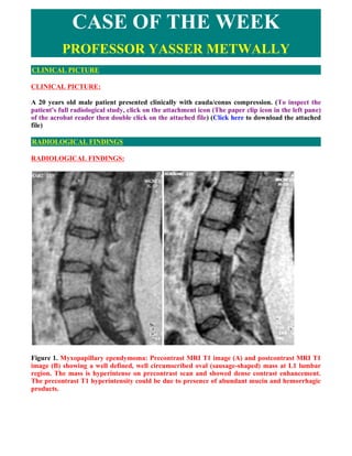
- 1. CASE OF THE WEEK PROFESSOR YASSER METWALLY CLINICAL PICTURE CLINICAL PICTURE: A 20 years old male patient presented clinically with cauda/conus compression. (To inspect the patient's full radiological study, click on the attachment icon (The paper clip icon in the left pane) of the acrobat reader then double click on the attached file) (Click here to download the attached file) RADIOLOGICAL FINDINGS RADIOLOGICAL FINDINGS: Figure 1. Myxopapillary ependymoma: Precontrast MRI T1 image (A) and postcontrast MRI T1 image (B) showing a well defined, well circumscribed oval (sausage-shaped) mass at L1 lumbar region. The mass is hyperintense on precontrast scan and showed dense contrast enhancement. The precontrast T1 hyperintensity could be due to presence of abundant mucin and hemorrhagic products.
- 2. Figure 2. MRI T2 image showing the lumbar myxopapillary ependymoma as a well defined, well circumscribed oval (sausage-shaped) mass at L1 lumbar region. The ependymoma is hypointense relative to the CSF. The patient was operated upon, the tumor was removed and histopathological findings revealed a myxopapillary ependymoma Histologic Findings in ependymomas Ependymoma pathology includes cellular, papillary, and clear cell variants as well as anaplastic ependymomas, myxopapillary ependymomas, and subependymomas. Histologically, ependymomas are characterized by ependymal pseudorosettes with glial fibrillary acidic protein (GFAP)-positive processes tapering toward blood vessels. Myxopapillary ependymomas are located at the cauda equina and conus, while subependymoma and anaplastic ependymomas are described at intramedullary locations. Figure 3. This is a myxopapillary ependymoma, which is typically found arising in the filum terminale of the spinal cord. Note the cells around papillations that have a myxoid connective tissue core. Surgical removal is made easier if this tumor has not grown around nerve roots of the cauda equina. A variety of histological ependymoma subtypes may be encountered. The cellular ependymoma is
- 3. the most common, but epithelial, tanycytic (fibrillar), subependymoma, myxopapillary, or mixed examples also occur. Histological differentiation from astrocytoma may be difficult, but the presence of perivascular pseudorosettes or true rosettes establishes the diagnosis. Most spinal ependymomas are histologically benign, although necrosis and intratumoral hemorrhage are frequent. Although unencapsulated, these glial-derived tumors are usually well circumscribed and do not infiltrate adjacent spinal cord tissue. Recent attempts to correlate the expression of MIB-1 antigen with malignancy of ependymomas have been confounded by tumor heterogeneity. Myxopapillary ependymoma histology consists of a papillary arrangement of cuboidal or columnar tumor cells surrounding a vascularized core of hyalinized and poorly cellular connective tissue. Cellular type The tumours are highly cellular and composed of polygonal cells and little supporting stroma. Two architectural features are found Cellular Ependymal tubules (ependymal rosettes) :- Composed of concentric arrangement of cilia ted ependymal cells around a genuine cavity Cellular Perivascular pseudorosettes :- Perivascular arrangement of ependymal cells Cellular forming pseud rosettes Papillary type (common spinally) Papillary The papillary ependymomas:- The ependymal cells rests upon glial fibrillary stromas Papillary Myxopapillary ependymomas:- The connective tissue stroma is the seat Papillary myxomatous degeneration DIAGNOSIS: DIAGNOSIS: MYXOPAPILLARY LUMBAR EPENDYMOMA DISCUSSION DISCUSSION: Spinal ependymomas most commonly occur as intramedullary tumors throughout the spinal axis. In the lumbosacral region, ependymomas are most commonly associated with the conus medullaris and cauda equina, but can also occur extradurally in the sacrum, presacral tissues, or subcutaneous tissues over the sacrum. These two tumor locations produce different management concerns. Intradural ependymomas, especially those in the lumbosacral region, are now recognized for their potential to spread throughout the central nervous system (CNS), whereas extradural tumors elicit more concern for their association with extraneural metastases. In this discussion the author has reviewed the literature regarding both of these distinct tumors and have summarized recommendations for the management of intra- and extradural lumbosacral ependymomas. For both tumors, it appears that gross-total resection is the treatment of choice when feasible. The role of radiation therapy has not been adequately studied for either tumor location, but most clinicians use this modality in patients with subtotal resection of intradural ependymomas, local recurrence, or CNS dissemination. Data supporting the use of
- 4. radiation therapy for extradural ependymomas are lacking. There does not appear to be a significant role for chemotherapy in either tumor location. Despite the risk for local recurrence and CNS dissemination, the prognosis for intradural lumbosacral ependymomas is good, with a greater than 90% 10-year patient survival in most series. The prognosis for extradural ependymomas does not appear to be as good. Much depends on extradural tumor location, however; the outlook is better for dorsal sacral tumors than presacral tumors. Spinal ependymomas are very rare tumors, with approximately 227 intradural spinal ependymomas diagnosed each year in the US. Of the intradural ependymomas, approximately 50% are intramedullary, above the conus, and the remaining 50% occur in association with the terminal filum, cauda equina, and conus medullaris.[3,6,22] In the lumbosacral region, ependymomas account for 90% of intradural tumors.[10] In addition to occurring intradurally, ependymomas can also sometimes occur extradurally around or in the sacrum. Spinal ependymomas are most common in the fourth decade of life and are more common in male patients.[5,33,37] Sonneland, et al.,[32] reported a 1.7:1.0 male/female ratio and a mean age at onset of 36.4 years (range 6–82 years) in the largest series (77 patients) of myxopapillary ependymomas reported in the literature. All of the tumors were intradural in this series, with the majority occurring in the lumbosacral region. Bimodal peaks have been suggested for extraspinal ependymomas in the sacral region, with one peak at less than 8 years of age and one in the fourth decade of life.[20,21] Others have suggested that extraspinal ependymomas occur in younger patients compared with intraspinal ependymomas.[1,15,17,21,25] There appears to be no significant sex predominance for extradural ependymomas.[15,21,25] OVERVIEW Tumor Locations and Features Spinal ependymomas can originate from either the ependymal lining of the central canal of the spinal cord, ependymal cell clusters in the terminal filum, or from ependymal rests left during embryonic development. Intramedullary tumors above the lumbosacral region almost uniformly arise from the ependymal lining of the spinal cord. These intradural, intramedullary ependymomas are predominantly the cellular ependymoma histiotype.[6,22] In the lumbosacral region, the majority of ependymomas arise from the intradural terminal filum. These are characteristically well-encapsulated, sausage-shaped tumors with the nerve roots of the cauda equina draped over the surface of the lesion or enveloped by it (Fig. 1). Myxopapillary ependymomas comprise the majority of ependymomas that arise in this area, but papillary and cellular histiotypes can also occur here.[3–5,9,10,13,22]
- 5. Figure 1. Sagital MR image with Gd contrast demonstrating an intradural ependymoma at the level of the cauda equina. Extradural ependymomas are very rare and are known to occur in four perisacral locations, as follows: 1) the extradural spinal canal in association with the dural part of the terminal filum; 2) the bone substance of the sacrum; 3) the pelvic cavity anterior to the sacrum; and 4) the subcutaneous tissues dorsal to the sacrum.[10,14,17,20,21,30] Because of the erosive nature of these tumors, it may be impossible to differentiate between one that starts within the bone of the sacrum and one that starts in the extradural spinal canal and erodes into the sacrum. Based on the number of case reports in the literature, the posterior subcutaneous location is the most common one for extradural ependymomas, followed by the presacral region.[10,11,25] Intraspinal extradural sacral ependymomas arise from ependymal cell remnants in the extradural dural part of the terminal filum. The other extradural ependymomas likely arise from ependymal rests that are present at the time of birth. Bale[2] reported finding ependymal rests in the dorsal sacral subcutaneous tissues in necropsy samples obtained in 10 of 15 infants. Others have reported finding ependymal rests in tissue obtained during pilonidal cyst resection.[25] One theory is that the ependymal cell rests arise from the coccygeal medullary vestige, a remnant of the dural part of the terminal filum that involutes during embryonic development. Others hypothesize that these ependymal rests are heterotopias that occur as a result of incomplete closure of the neural arch. [12,16,17,20] Presacral ependymomas are thought to arise from ependymal cell rests or in association with cauda equina nerve roots that have exited the sacral neural foramen. Histologically, all reported cases of extradural ependymomas have been myxopapillary types. [8,10,16,17,25,34] Tumor Symptoms
- 6. Back pain and occasionally radicular pain are the most common presenting symptoms for intradural ependymomas arising from the terminal filum in the lumbosacral region. Motor or sensory abnormalities and bladder dysfunction may also develop in association with these lesions involving the cauda equina.[5,24] Extraspinal ependymomas can present with a variety of symptoms caused by local mass effect. Intrasacral and intraspinal extradural tumors present most commonly with local pain caused by erosion into the sacrum, but can also present with radicular or neurological symptoms caused by neural element compression.[2,24] Dorsal subcutaneous tumors typically present as a growing, asymptomatic mass in the intergluteal fold or buttocks. Occasionally there can be pain or tenderness caused by local mass effect, and very rarely neurological symptoms are reported. [1,9,17,21,25] Presacral pelvic masses can grow to be very large, because of significant tolerance for masses in this area. With sufficient size, presacral masses eventually cause bowel or bladder dysfunction from local mass effect or neural involvement (Fig. 2).[2,10,21] Some patients may have a palpable mass on rectal examination. Rarely, presacral tumors cause pain in a sciatic nerve distribution or local pain from erosion into the sacrum.[2,20,21,34] Figure 2. Left: Sagittal noncontrast MR image demonstrating a presacral ependymoma and an enlarged bladder due to urinary retention. Right: Axial noncontrast MR image demonstrating a presacral ependymoma. Preoperative Evaluation All patients with these tumors require a complete neurological examination and MR imaging to delineate local disease. Intradural ependymomas can disseminate through CSF pathways throughout the CNS, but are not likely to metastasize outside it,[21] although rare cases of this phenomenon have been reported.[27,35] Therefore, we recommend preoperative screening MR imaging of the entire CNS to rule out tumor dissemination. Screening of other organ systems for metastases is not needed for intradural tumors. Preoperative plain anteroposterior and lateral x- ray films of the lumbosacral spine may also be valuable for comparison if instability or deformity develop after laminectomy surgery.[23] Extradural tumors, in contrast, are not likely to disseminate within the CNS, but are at significant risk of metastasizing to other organ systems, such as lymph, bone, lung, and liver. Use of MR or computerized tomography studies is sufficient to evaluate local disease and we see no need to screen the entire CNS. In addition, plain x-ray films or computerized tomography scans may be warranted in extradural ependymomas to provide further detail about bone involvement. We recommend paying particular attention to general examination for lymphadenopathy, especially in the inguinal lymph nodes,[15] and would also obtain chest x-ray films, liver function tests, and alkaline phosphatase levels preoperatively. Presacral tumors warrant a special workup that likely requires an abdominal/ pelvic surgical specialist. Other studies, such as barium enema, intravenous pyelogram, cystoscopy, and pelvic examination may need to be performed for
- 7. presacral tumors.[21] TREATMENT OPTIONS There are no prospective, randomized studies in which the management options of surgery alone, surgery followed by radiation, radiation alone, or chemotherapy are examined. Surgery Alone The surgical goal with all ependymomas, whether they are intra- or extradural, is gross-total resection when feasible.[16] Gross-total resection provides a possibility for cure without the definite need for adjuvant therapy. For intradural ependymomas in this location, gross-total resection is the most frequently cited factor influencing prognosis in terms of local recurrence and patient survival.[5,28,33] Some authors report higher recurrence rates for tumors removed in a piecemeal fashion, even though gross-total resection was achieved.[33,35] Therefore, when possible, en bloc resection is preferred over piecemeal removal. Other factors, such as duration of clinical symptoms and tumor involvement with conus medullaris or cauda equina, have also been cited (Fig. 3).[5] Figure 3. Intraoperative photograph of an intradural ependymoma in the lumbar thecal sac. Obviously, when the conus medullaris and cauda equina are involved with the tumor, achieving gross-total resection becomes more difficult. In fact, Celli, et al.,[5] found that gross-total resection could be obtained in only 43% of cases when the conus medullaris or cauda were involved with the tumor. They also noted that, despite gross-total resection at the time of surgery, tumors involving the conus medullaris or cauda equina were more likely to recur. Sonneland, et al.,[33] reported gross-total resection in 59% of a series of 77 patients with predominantly terminal filum/conus medullaris/cauda equina lesions. For posterior extradural lesions without sacral involvement, wide local excision for gross-total resection is advocated.[10,20] Plastic surgery assistance may be needed for complex skin closure
- 8. techniques required after resection of large masses. Some authors have recommended coccygectomy in addition to local excision as a means of reducing the risk of recurrence.[15] The surgical approach for presacral lesions depends on the degree of sacral involvement. Isolated presacral tumors with minimal to no sacral involvement may be radically resected via just an anterior approach. When the sacrum is involved, combined anterior–posterior approaches, either in the same sitting or during different operations, may be needed.[10,20] Figure 4. Lumbar myxopapillary ependymoma Resection Followed by Radiation The role of radiation for lumbosacral ependymomas has not been adequately studied to warrant firm conclusions. There appears to be a consensus for radiation therapy in cases of subtotal resections of intradural tumors, although the role for radiation in cases of gross-total resection and extradural ependymomas is controversial.[3,5,7,10,11,16,28,29,31,33,35]
- 9. Figure 5. Myxopapillary ependymoma. Notice hemorrhagic zones and cystic degeneration. Sonneland, et al.,[33] based on their large series of patients, recommended local radiation therapy in cases of subtotal or piecemeal gross-total resection. Nevertheless, in reviewing their data, there appears to be no trend toward improved survival or recurrence rates with radiation therapy. Ross, et al.,[28] in a review of the 131 cases in the literature, concluded that although no differences in outcome after radiation have been shown for gross-total and subtotal resection, results in individual cases show that long-term tumor control can be achieved with this modality. Waldron, et al.,[35] in a large series of spinal ependymomas, performed multivariate analysis in patients who received radiation after surgery. They evaluated the following variables: age, sex, preoperative symptom duration, preoperative functional status, tumor location, and histological grade. They found that only histological grade was statistically significant in regard to predicting recurrence-free survival and cause-specific survival. They reported a cause-specific actuarial survival at 5 and 10 years of 86 and 81%, respectively. Nevertheless, their results may not correspond completely with lumbosacral ependymomas because in their group of 59 patients approximately half of the tumors were above the level of the conus medullaris. They note that in patients with well-differentiated tumors, like those typically found in the lumbosacral region, there was a 97% 5-year, cause-specific survival. Although these survival rates with radiation appear to be excellent, when we compare these figures with overall survival rates for lumbosacral ependymomas there appears to be no significant difference. Waldron, et al.,[35] reported no cases of radiation myelopathy in the 59 spinal ependymomas treated, and others report very low complication rates with spinal irradiation in this area.[31] Therefore, although they are not scientifically proven, the potential benefits of radiation therapy appear to outweigh the risks and can be justified in cases of subtotal tumor resection. Radiation Therapy After Biopsy Sampling
- 10. Waldron, et al.,[35] report on five patients who received radiation therapy after biopsy sampling in which a diagnosis of ependymoma was made. This subgroup was included in a larger series of 59 patients who received radiation after surgery (biopsy or resection), and it is difficult to discern from the authors´ data the tumor location, histological findings, and results in these five patients. Waldron, et al., report that three of these five patients were alive at the last follow-up review. Others have used radiation therapy alone after biopsy procedures for lumbosacral ependymomas, with apparently very good long-term results, although the number of cases in each series is small. [32,36] Radiation Therapy for Recurrence or Metastases For intradural ependymomas, radiation therapy is often used in cases of local recurrence and disseminated disease within the CNS. Intradural myxopapillary ependymomas have been shown to be radiosensitive, and long-term tumor control can be achieved with this adjunctive therapy in most cases of local recurrence or CNS metastases.[7,28] Extradural ependymomas have not shown as promising results as intradural tumors. These extradural tumors are rare; therefore, sufficient data are lacking to draw conclusions on the effectiveness of radiotherapy. Most cases of local recurrence are treated with surgery and not radiation. With the limited data available, prolonged survival has not been observed in patients who receive radiotherapy for extradural ependymomas. Metastatic disease to other organ systems typically does not respond to radiation therapy or chemotherapy.[15] Chemotherapy This modality has been restricted primarily to patients with recurrent disease that has been refractory to resection and radiation. A number of chemotherapeutic agents have been tried in small series of recurrent spinal ependymomas, but no studies have shown compelling evidence to indicate that chemotherapy may be a primary treatment for these tumors.[6,10,33,34] Individual case reports[18] describe control of myxopapillary ependymoma with chemotherapy, but sufficient data are lacking to draw any significant conclusions regarding this regimen. PROGNOSIS Most ependymomas occurring in the lumbosacral region are myxopapillary on histological examination. Even though intraspinal myxopapillary ependymomas are considered benign, they are known to recur locally and can disseminate through CSF pathways. Even with gross-total resection, recurrence rates of 4 to 29% have been reported.[3–5] Although described by Davis and Barnard[9] in 1985, the potential for dissemination of these lesions throughout the CNS has only recently been truly appreciated.[26,27,38] Disseminated disease can arise many years after initial tumor presentation.[9,26] Despite the significant risk of local recurrence and CSF dissemination, the overall prognosis for lumbosacral intradural myxopapillary ependymomas appears to be very good. Sonneland, et al., [33] reported long-term survival in approximately 95% of patients in their series of 77 patients. Mork and Loken,[22] in another large series of spinal ependymomas, reported a 10-year survival rate of 94% for patients with lumbosacral intradural ependymomas and noted that the myxopapillary histiotype had a favorable prognosis. The few patients who die as a result of primary disease typically do so after a prolonged course with multiple recurrences. Extradural ependymomas are a different entity compared with intradural tumors because of the risk of local recurrence and distal metastasis. For presacral tumors, local recurrence has been reported to be as high as 60%, with mortality rates as high as 75% over a period of 4 years for
- 11. cases of recurrence.[20,34] Local recurrence for dorsal subcutaneous tumors is approximately 25% at 15 years.[10] Overall, extradural lesions are much more likely to metastasize to other organ systems than intradural tumors, with the lymphatic system, lungs, bones, and liver being the most common sites.[9] The tendency for extradural tumors to metastasize is likely because of the closer proximity of lymphatic and vascular channels in extradural locations. The risk of metastases also depends on the specific extradural location, with dorsal subcutaneous tumors at greater risk for metastasis than presacral tumors. Intrasacral lesions have also been reported to metastasize, but this appears to be very rare.[19,27] Metastases have been reported in up to 20% of dorsal subcutaneous tumors in long-term follow up.[14] Metastatic disease from extradural ependymomas appears to carry a grim prognosis due to lack of response to adjuvant therapies. CONCLUSION Lumbosacral ependymomas consist of two distinct entities that must be managed differently. The more common intradural ependymoma has a significant risk of both local recurrence and dissemination to other areas of the CNS. The rare extradural sacral region ependymoma has the potential for extraneural metastases in addition to local recurrence. The possibility of metastases for lesions in both of these locations should be taken into account in preoperative evaluation and in long-term follow up. In both tumor locations, gross-total resection is the treatment of choice when feasible. Radiation therapy may be valuable for subtotal intradural resections or CNS metastases from intradural tumors. Radiotherapy for extradural lesions does not appear to be as effective and is more controversial. SUMMARY SUMMARY Spinal intramedullary neoplasms account for 4-10% of all central nervous system (CNS) tumors and about 2-4% of CNS glial tumors. Most spinal cord neoplasms are malignant, and 90-95% are classified as gliomas, which constitute ependymomas or astrocytomas. Ependymomas are the most common intramedullary glial tumor in adults, whereas astrocytomas are most common in children. Non-glial neoplasms constitute hemangioblastoma, paraganglioma, metastases, and lymphoma. Primitive neuroectodermal tumors are much less common. Cord ependymomas most commonly occur in the cervical region followed by the thoracic and lumbar region, respectively. However, the myxopapillary variant that contains abundant mucin is virtually always located along the filum terminale. Clinical symptom includes back or neck pain depending upon the site of tumor, sensory deficits, motor weakness and bladder or bowel dysfunction [1,2]. Plain films of the spine may reveal scoliosis or canal widening with associated pedicle erosion. Myelography frequently reveals partial or complete block to the flow of contrast media. On CT scan, ependymomas may appear as iso to hyperdense lesions and enhance intensely after contrast administration [3]. Most spinal cord ependymomas are seen as centrally located, well-defined, iso- or hypointense relative to the spinal cord on T1-weighted and hyperintense on T2-weighted MR
- 12. images. Enhancement is virtually almost always seen after the contrast administration. Myxopapillary ependymomas may appear hyperintense on T1-weighted MR image due to presence of abundant mucin and also are more prone to hemorrhage. Cysts may be seen in about 78-84% of ependymomas. Three distinct types of cysts have been described: (a) tumoral cysts; (b) rostral or caudal cysts; and (c) reactive dilatation of the central canal. The tumoral cysts are seen within the tumor itself as a result of degeneration, necrosis, and liquefaction, and show peripheral rim enhancement. The rostral and caudal cysts occur above and/or below the tumor and do not show rim enhancement. The reactive dilatation causing cyst-like appearances are most likely related to partial obstruction of the central canal by the tumor mass [4,5]. Radiologically, astrocytoma is a close mimicker of ependymoma. Ependymomas are central in location since they arise from central canal ependymal cells, whereas astrocytomas are eccentric with infiltrating borders as they originate from the cord parenchyma. Findings such as hemorrhage within the tumor and hemosiderin deposition or calcification are more frequent in ependymomas due to rich connective tissue stroma. Contrast enhancement is also intense and homogenous in ependymomas whereas it is patchy and irregular in astrocytomas. Ependymomas especially the myxopapillary variety have particular predilection for conus medullaris which is not so in the case of astrocytomas. Ependymomas can be differentiated from hemangioblastoma by virtue of enhancing mural nodule, which is the hallmark of hemangioblastoma. Addendum A new version of this PDF file (with a new case) is uploaded in my web site every week (every Saturday and remains available till Friday.) To download the current version follow the link "http://pdf.yassermetwally.com/case.pdf". You can also download the current version from my web site at "http://yassermetwally.com". To download the software version of the publication (crow.exe) follow the link: http://neurology.yassermetwally.com/crow.zip The case is also presented as a short case in PDF format, to download the short case follow the link: http://pdf.yassermetwally.com/short.pdf At the end of each year, all the publications are compiled on a single CD-ROM, please contact the author to know more details. Screen resolution is better set at 1024*768 pixel screen area for optimum display. Also to view a list of the previously published case records follow the following link (http://wordpress.com/tag/case-record/) or click on it if it appears as a link in your PDF reader To inspect the patient's full radiological study, click on the attachment icon (The paper clip icon in the left pane) of the acrobat reader then double click on the attached file. Click here to download the short case version of this case record in PDF format REFERENCES References 1. Anderson MS: Myxopapillary ependymomas presenting in the soft tissue over the sacrococcygeal region. Cancer 19: 585–590, 1966
- 13. 2. Bale PM: Ependymal rests and subcutaneous sacrococcygeal ependymoma. Pathology 12:237–243, 1980 3. Bavbek M, Altinors MN, Caner HH, et al: Lumbar myxopapillary ependymoma mimicking neurofibroma. Spinal Cord 39: 449–452, 2001 4. Burtscher J, Felber S, Twerdy K, et al: Endoscope-assisted interlaminar removal of an ependymoma of the cauda equina. Minim Invasive Neurosurg 45:41–44, 2002 5. Celli P, Cervoni L, Cantore G: Ependymoma of the filum terminale: treatment and prognostic factors in a series of 28 cases. Acta Neurochir 124:99–103, 1993 6. Chamberlain MC: Salvage chemotherapy for recurrent spinal cord ependymoma. Cancer 95:997–1002, 2002 7. Chinn DM, Donaldson SS, Dahl GV, et al: Management of children with metastatic spinal myxopapillary ependymoma using craniospinal irradiation. Med Pediatr Oncol 35:443–445, 2000 8. Chou S, Soucy P, Carpenter B: Extraspinal ependymoma. J Pediatr Surg 22:802–803, 1987 9. Davis C, Barnard RO: Malignant behavior of myxopapillary ependymoma. Report of three cases. J Neurosurg 62:925–929, 1985 10. Fourney DR, Fuller GN, Gokaslan ZL: Intraspinal extradural myxopapillary ependymoma of the sacrum arising from the filum terminale externa. Case report. J Neurosurg (Spine 2) 93: 322–326, 2000 11. Gerston KF, Suprun H, Cohen H, et al: Presacral myxopapillary ependymoma presenting as an abdominal mass in a child. J Pediatr Surg 20:276–278, 1985 12. Gregorios JB, Green B, Page L, et al: Spinal cord tumors presenting with neural tube defects. Neurosurgery 19:962–966, 1986 13. Hallacq P, Labrousse F, Streichenberger N, et al: Bifocal myxopapillary ependymoma of the terminal filum: the end of a spectrum? J Neurosurg (Spine 3) 98:288–289, 2003 14. Helwig EB, Stern JB: Subcutaneous sacrococcygeal myxopapillary ependymoma. A clinicopathologic study of 32 cases. Am J Clin Pathol 81:156–161, 1984 15. Kramer GW, Rutten E, Sloof J: Subcutaneous sacrococcygeal ependymoma with inguinal lymph node metastasis. J Neurosurg 68:474–477, 1988 16. Lemberger A, Stein M, Doron J, et al: Sacrococcygeal extradural ependymoma. Cancer 64:1156–1159, 1989 17. Lynch J, Kelly N, Fitzpatrick B, et al: A sacrococcygeal extraspinal ependymoma in a 67- year-old man: a case report and review of the literature. Br J Plast Surg 55:80–82, 2002 18. Madden JR, Fenton LZ, Weil M, et al: Experience with tamoxifen/ etoposide in the treatment of a child with myxopapillary ependymoma. Med Pediatr Oncol 37:67–69, 2001 19. Miralbell R, Louis DN, O´Keeffe D, et al: Metastatic ependymoma of the sacrum. Cancer
- 14. 65:2353–2355, 1990 20. Morantz RA: Ectopic ependymoma of the sacrococcygeal region, in Doty JR, Rengachary SS (eds): Surgical Disorders of the Sacrum. New York: Theime Medical, 1992, pp 177–179 21. Morantz RA, Kepes JJ, Batnitzky S, et al: Extraspinal ependymomas. J Neurosurg 51:383– 391, 1979 22. Mork SJ, Loken AC: Ependymoma: a follow-up study of 101 cases. Cancer 40:907–915, 1977 23. Papagelopoulos PJ, Peterson HA, Ebersold MJ, et al: Spinal column deformity and instability after lumbar or thoracolumbar laminectomy for intraspinal tumors in children and young adults. Spine 22:442–451, 1997 24. Post KD, McCormick PC: Intrasacral ependymoma, in Doty JR, Rengachary SS (eds): Surgical Disorders of the Sacrum. New York: Theime Medical, 1992, pp 181–183 25. Pulitzer DR, Martin PC, Collins PC, et al: Subcutaneous sacrococcygeal (“myxopappillary’) ependymal rests. Am J Surg Pathol 12:672–677, 1988 26. Rezai AR, Woo HH, Lee M, et al: Disseminated ependymomas of the central nervous system. J Neurosurg 85:618–624, 1996 27. Rickert CH, Kedziora O, Gullotta F: Ependymoma of the cauda equina. Acta Neurochir 141:781–782, 1999 28. Ross DA, McKeever PE, Sandler HM, et al: Myxopapillary ependymoma. Results of nucleolar organizing region staining. Cancer 71:3114–3118, 1993 29. Schild SE, Nisi K, Scheithauer BW, et al: The results of radiotherapy for ependymomas: the Mayo Clinic experience. Int J Radiat Oncol Biol Phys 42:953–958, 1998 30. Schurmann K, Wallenfang T: Rare sacral space-occupying lesions, their surgical management and reconstructive measures involved. Acta Neurochir 92:106–117, 1988 31. Schweitzer JS, Batzdorf U: Ependymoma of the cauda equina region: diagnosis, treatment, and outcome in 15 patients. Neurosurgery 30:202–207, 1992 32. Scott M: Infiltrating ependymomas of the cauda equina. Treatment by conservative surgery plus radiotherapy. J Neurosurg 41:446–448, 1974 33. Sonneland PR, Scheithauer BW, Onofrio BM: Myxopapillary ependymoma. A clinicopathologic and immunocytochemical study of 77 cases. Cancer 56:883–893, 1985 34. Timmerman W, Bubrick MP: Presacral and postsacral extraspinal ependymoma. Report of a case and review of the literature. Dis Colon Rectum 27:114–119, 1984 35. Waldron JN, LaPerriere NJ, Jaakkimainen L, et al: Spinal cord ependymomas: a retrospective analysis of 59 cases. Int J Radiat Oncol Biol Phys 27:223–229, 1993 36. Wen BC, Hussey DH, Hitchon PW, et al: The role of radiation therapy in the management of ependymomas of the spinal cord. Int J Radiat Oncol Biol Phys 20:781–786, 1991
- 15. 37. Whitaker SJ, Bessell EM, Ashley SE, et al: Postoperative radiotherapy in the management of spinal cord ependymoma. J Neurosurg 74:720–728, 1991 38. Yucesoy K, Ozer E, Koyuncuoglu M: Parenchymal brain metastasis of a spinal myxopapillary ependymoma after extradural manipulation. Acta Neurochir 39. Metwally, MYM: Textbook of neuroimaging, A CD-ROM publication, (Metwally, MYM editor) WEB-CD agency for electronic publication, version 11.1a. January 2010 40. Case of the week...Spinal ependymoma [Click to download in PDF format]