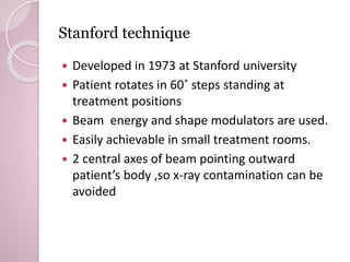
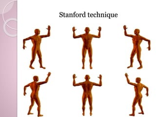
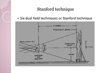
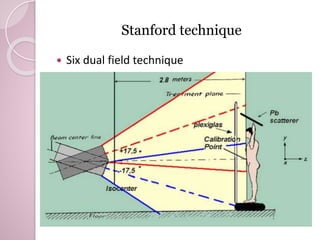
Clinical implementation of total skin electron 
beam (TSEB) therapy: A review of the relevant 
literature 
Presented by: Sehrish Inam 
Trainee Medical Physicist 
Date : May13,2014
Contents 
◦ Abstract 
◦ Introduction 
◦ Equipment requirement 
◦ Physical requirement 
◦ Single scattered electron beam therapy 
◦ Other techniques of skin therapy 
◦ Stanford technique 
◦ Dose rates 
◦ Setup problems 
◦ Dose prescription 
◦ Dosimetric setup 
◦ Dosimetric problems 
◦ Clinically acceptance objectives 
◦ Conclusion
Abstract: 
Total skin electron beam therapy has been in medical service since 
the middle of the last century in order to confront rare skin 
malignancies. Since then various techniques have been developed, 
all aiming at better clinical results in conjunction with less post-irradiation 
complications. In this article every available technique is 
presented in addition to physical parameters of technique 
establishment and common dose fractionation. This study also 
revealed the preference of the majority of institutes the last 20 
years in “six dual field technique” at a high dose rate, which is a safe 
and effective treatment.
Introduction 
•Total skin electron beam is treatment modality for 
 T-cell lymphoma 
Mycosis 
 Fungoides 
Kaposi sarcoma 
Low penetration electron beam 
Linear accelerator capable of producing large 200 cm 80 
cm uniform fields with extended SSD.
Equipment Requirement 
 Linear accelerator that can be modified in order to 
deliver a homogeneous electron field at a large 
distance from its source (2-7 m). 
 Beam degrader which ensures superficial beam 
penetration into tissue. 
 Large treatment room for large SSD. 
 ventilation that removes O3 produced by electron 
air interactions . 
 Auxiliary equipment for the proper and repeatable 
positioning. 
 Dosimetry equipments. 
 Shielding to avoid sensitivity (eyes & nails)
Physical requirements 
3steps of dosimetric checks 
1. Physical specification of field dimensions, nominal 
SSD, electron beam energies, field at treatment plane 
and dose distributions, dose rate and photon 
contamination. 
2. Dose distribution and rate for dose from electrons 
and photons. 
3. Clinical aspects that arises dose prescription, dose 
fractionation, boost fields for underdosed areas, 
shielding design
Single scattered horizontal beam 
 Requires a linear accelerator that can provide a 
homogenous electron field at an SSD of 700 cm. 
 Energy degrader for beam flattening patient is 
irradiated in standing position. 
 Requires a large treatment room
Other techniques of skin therapy 
Static large 
electron 
fields with 
patient in 
standing 
position. 
Static electron 
field, rotated 
the standing 
patient over 
360˚. 
Static 
electron field 
with patient 
translated in 
lying position
Stanford technique 
 Developed in 1973 at Stanford university 
 Patient rotates in 60˚ steps standing at 
treatment positions 
 Beam energy and shape modulators are used. 
 Easily achievable in small treatment rooms. 
 2 central axes of beam pointing outward 
patient’s body ,so x-ray contamination can be 
avoided
Stanford technique
Stanford technique 
 Six dual field techniques or Stanford technique
Stanford technique 
 Six dual field technique
Dose rates 
 High dose rate 2500-3000 cGy/min at dmax . 
 Daily treatment time reduced to 9.5min to 
15min. 
 HDR is a treatment modality in mycosis 
fungodis with good results and less time 
consuming
Setup problems 
 Room size 
 Ventilation of ozone 
 Skin sensitivity 
 Eye nail shielding
Dose prescription 
 27Gy – 40Gy(mean dose 35-36Gy) at HDR in an 
average of 9weeks,4 days per week. 
 HDR provides low toxicity ,better tolerance & 
reduces treatment time. 
 For under dosed areas boost fields of 4-26Gy 
are prescribed. 
 For vertex of scalp angled lead reflector is 
provided.
Dosimetric setup 
 Dosimeter (TLDs, ionization chambers, 
gafchromic films) 
 Solid water phantom or anthromorphic 
phantom. 
 Scanning and evaluation of gafchromic 
by Epson10000xl
Dosimetric problems 
 On extended SSD; 
Combination of partial beams in order to 
create a large field that cover patient 
dimensions. 
 Beam energy degrdading, because lowest 
energy provided in electron mode is 6MeV. 
 Thickness of degrader can vary from 3mm 
to 18mm. 
 If air volume is not sufficient use acraylic 
sheet for secondary scattering.
Clinically acceptance objectives 
 +5% of dose at dmax in a phantom on the central ray 
for atleast 80% of the nominal field area. 
 In Stanford technique dose homogeneity varies from 4% 
to 10%. 
 Prefer dosimetry by gafchromic.
Conclusion 
 Total skin electron beam irradiation is an effective 
treatment for various skin malignancies. 
 Toxicity can be reduced by HDR & appropriate shielding. 
 All techniques require linear accelerator with electron 
mode & large SSD. 
 Dosimetric technique should be carried out to ensure 
treatment quality. 
 Prescribed doses differ according to personalized 
patient needs and treatment schedules. 
 36-40 Gy dose delivered in 4 days per week for 9 
weeks at HDR.
Tseb

Tseb

  • 1.
    Clinical implementation oftotal skin electron beam (TSEB) therapy: A review of the relevant literature Presented by: Sehrish Inam Trainee Medical Physicist Date : May13,2014
  • 2.
    Contents ◦ Abstract ◦ Introduction ◦ Equipment requirement ◦ Physical requirement ◦ Single scattered electron beam therapy ◦ Other techniques of skin therapy ◦ Stanford technique ◦ Dose rates ◦ Setup problems ◦ Dose prescription ◦ Dosimetric setup ◦ Dosimetric problems ◦ Clinically acceptance objectives ◦ Conclusion
  • 3.
    Abstract: Total skinelectron beam therapy has been in medical service since the middle of the last century in order to confront rare skin malignancies. Since then various techniques have been developed, all aiming at better clinical results in conjunction with less post-irradiation complications. In this article every available technique is presented in addition to physical parameters of technique establishment and common dose fractionation. This study also revealed the preference of the majority of institutes the last 20 years in “six dual field technique” at a high dose rate, which is a safe and effective treatment.
  • 4.
    Introduction •Total skinelectron beam is treatment modality for  T-cell lymphoma Mycosis  Fungoides Kaposi sarcoma Low penetration electron beam Linear accelerator capable of producing large 200 cm 80 cm uniform fields with extended SSD.
  • 5.
    Equipment Requirement Linear accelerator that can be modified in order to deliver a homogeneous electron field at a large distance from its source (2-7 m).  Beam degrader which ensures superficial beam penetration into tissue.  Large treatment room for large SSD.  ventilation that removes O3 produced by electron air interactions .  Auxiliary equipment for the proper and repeatable positioning.  Dosimetry equipments.  Shielding to avoid sensitivity (eyes & nails)
  • 6.
    Physical requirements 3stepsof dosimetric checks 1. Physical specification of field dimensions, nominal SSD, electron beam energies, field at treatment plane and dose distributions, dose rate and photon contamination. 2. Dose distribution and rate for dose from electrons and photons. 3. Clinical aspects that arises dose prescription, dose fractionation, boost fields for underdosed areas, shielding design
  • 7.
    Single scattered horizontalbeam  Requires a linear accelerator that can provide a homogenous electron field at an SSD of 700 cm.  Energy degrader for beam flattening patient is irradiated in standing position.  Requires a large treatment room
  • 8.
    Other techniques ofskin therapy Static large electron fields with patient in standing position. Static electron field, rotated the standing patient over 360˚. Static electron field with patient translated in lying position
  • 9.
    Stanford technique Developed in 1973 at Stanford university  Patient rotates in 60˚ steps standing at treatment positions  Beam energy and shape modulators are used.  Easily achievable in small treatment rooms.  2 central axes of beam pointing outward patient’s body ,so x-ray contamination can be avoided
  • 10.
  • 11.
    Stanford technique Six dual field techniques or Stanford technique
  • 12.
    Stanford technique Six dual field technique
  • 13.
    Dose rates High dose rate 2500-3000 cGy/min at dmax .  Daily treatment time reduced to 9.5min to 15min.  HDR is a treatment modality in mycosis fungodis with good results and less time consuming
  • 14.
    Setup problems Room size  Ventilation of ozone  Skin sensitivity  Eye nail shielding
  • 15.
    Dose prescription 27Gy – 40Gy(mean dose 35-36Gy) at HDR in an average of 9weeks,4 days per week.  HDR provides low toxicity ,better tolerance & reduces treatment time.  For under dosed areas boost fields of 4-26Gy are prescribed.  For vertex of scalp angled lead reflector is provided.
  • 16.
    Dosimetric setup Dosimeter (TLDs, ionization chambers, gafchromic films)  Solid water phantom or anthromorphic phantom.  Scanning and evaluation of gafchromic by Epson10000xl
  • 17.
    Dosimetric problems On extended SSD; Combination of partial beams in order to create a large field that cover patient dimensions.  Beam energy degrdading, because lowest energy provided in electron mode is 6MeV.  Thickness of degrader can vary from 3mm to 18mm.  If air volume is not sufficient use acraylic sheet for secondary scattering.
  • 18.
    Clinically acceptance objectives  +5% of dose at dmax in a phantom on the central ray for atleast 80% of the nominal field area.  In Stanford technique dose homogeneity varies from 4% to 10%.  Prefer dosimetry by gafchromic.
  • 19.
    Conclusion  Totalskin electron beam irradiation is an effective treatment for various skin malignancies.  Toxicity can be reduced by HDR & appropriate shielding.  All techniques require linear accelerator with electron mode & large SSD.  Dosimetric technique should be carried out to ensure treatment quality.  Prescribed doses differ according to personalized patient needs and treatment schedules.  36-40 Gy dose delivered in 4 days per week for 9 weeks at HDR.

Editor's Notes

  • #9 Large electron field techniques where the patient is irradiated by large electron fields in a standing position, rotational techniques where the whole body irradiation is achieved by the rotation of the standing patient and finally translational techniques where the patient is translated in a lying position through a stationary electron field