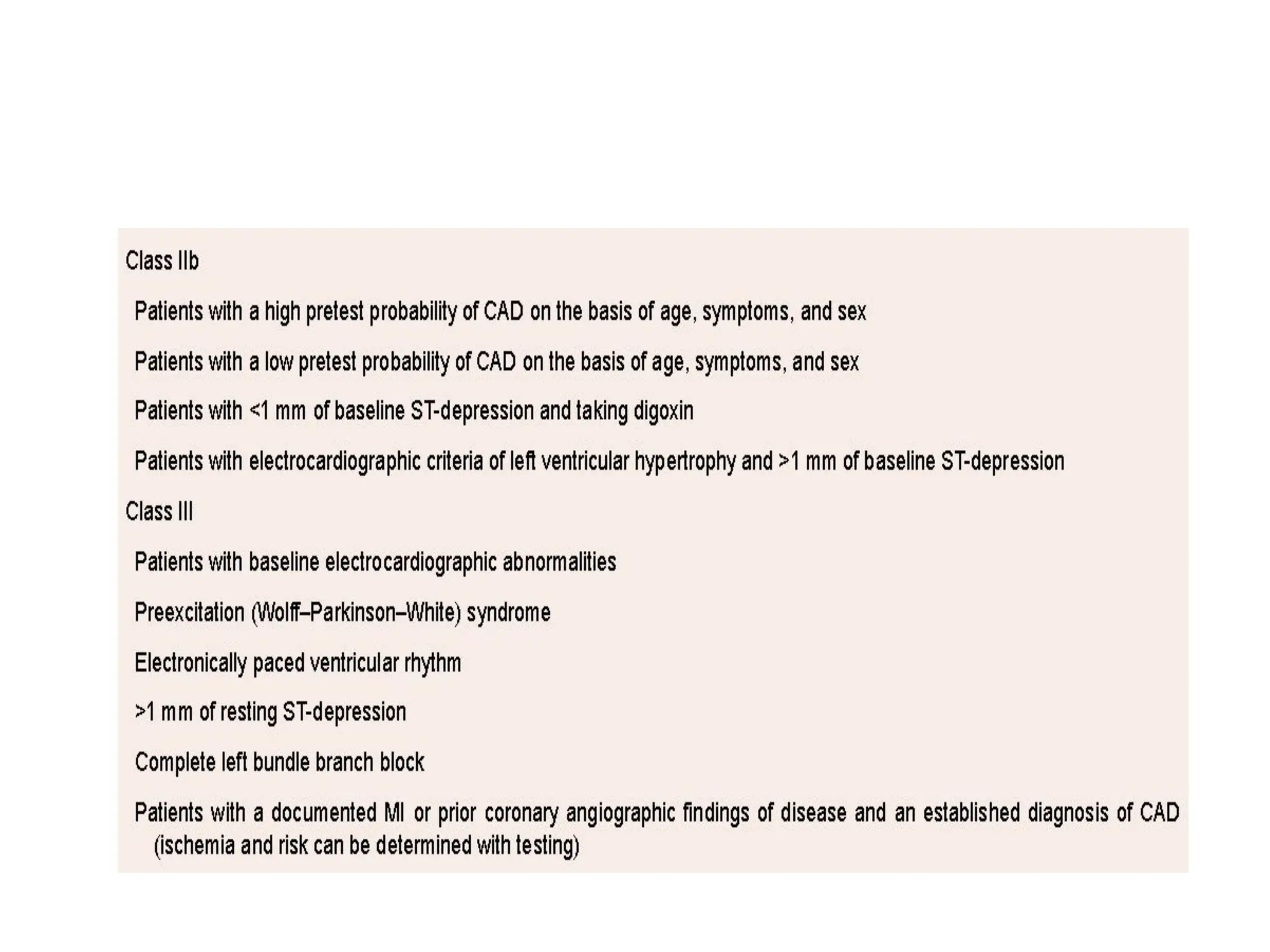
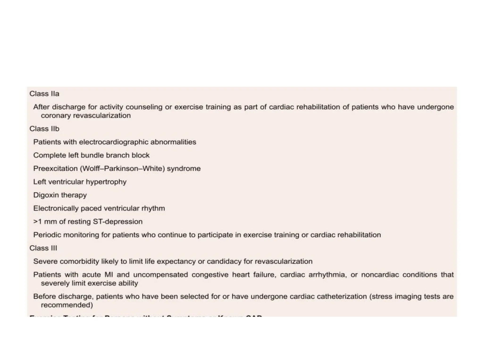
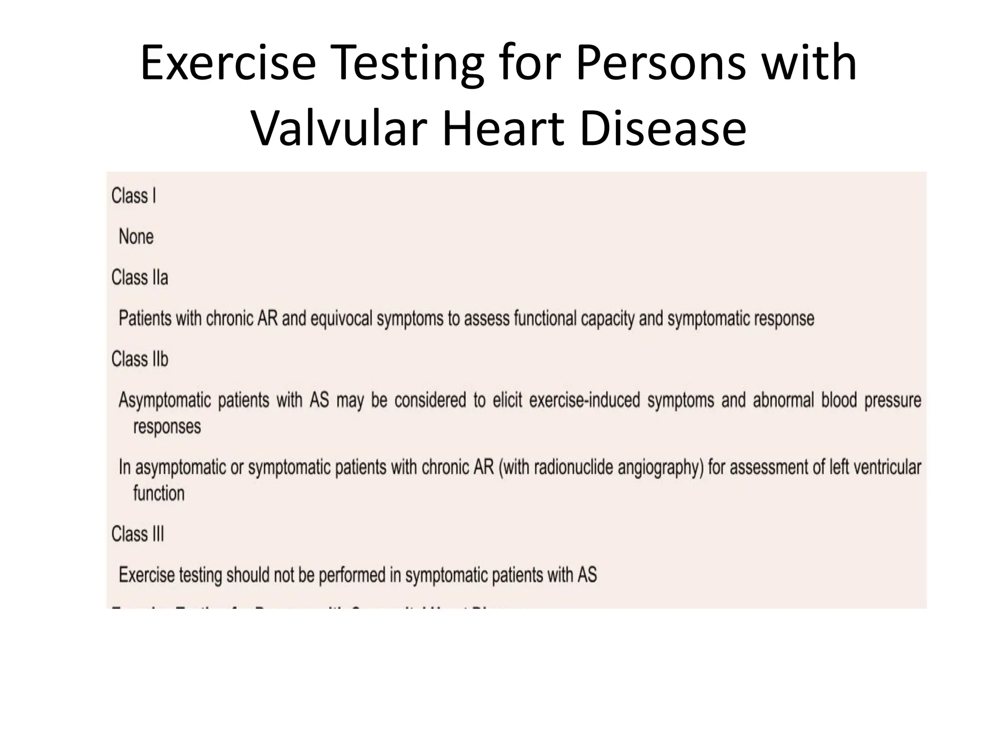
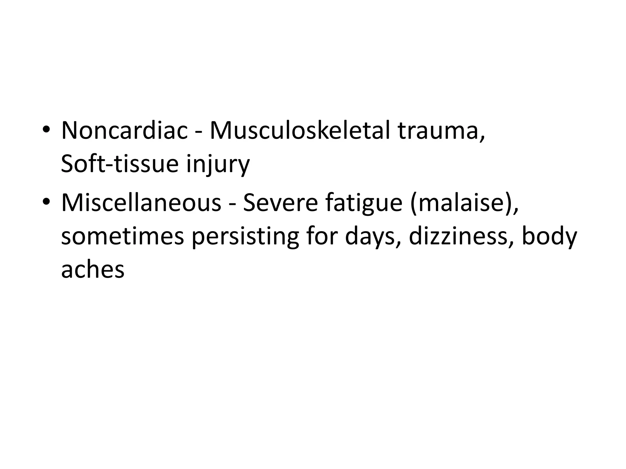
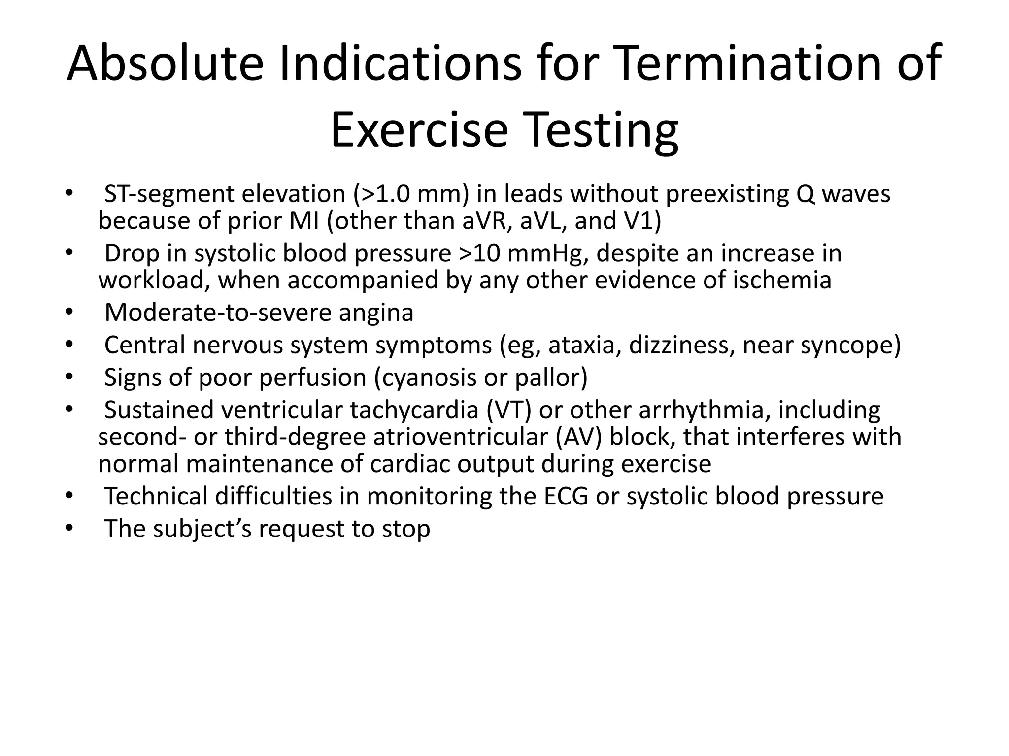
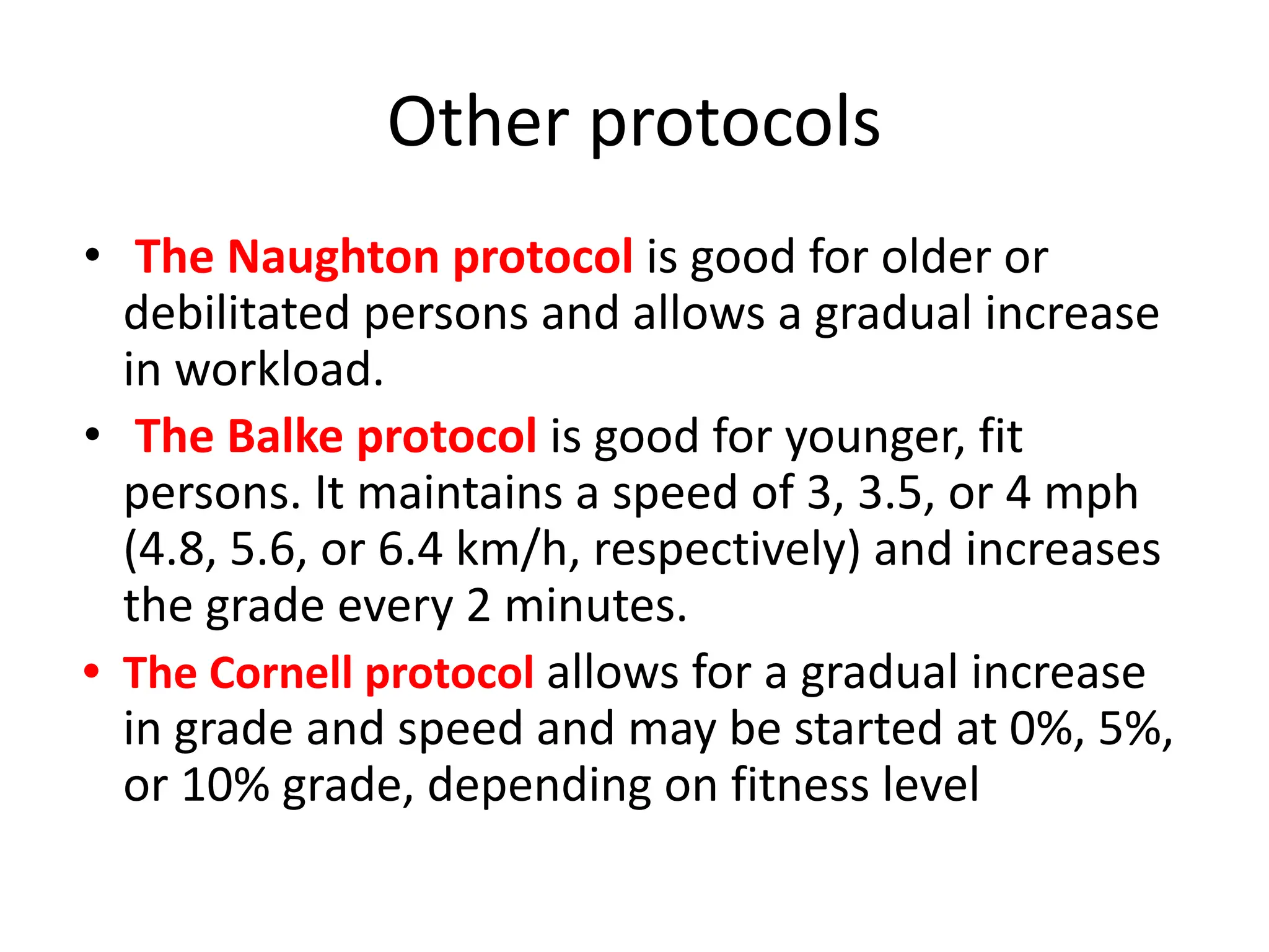
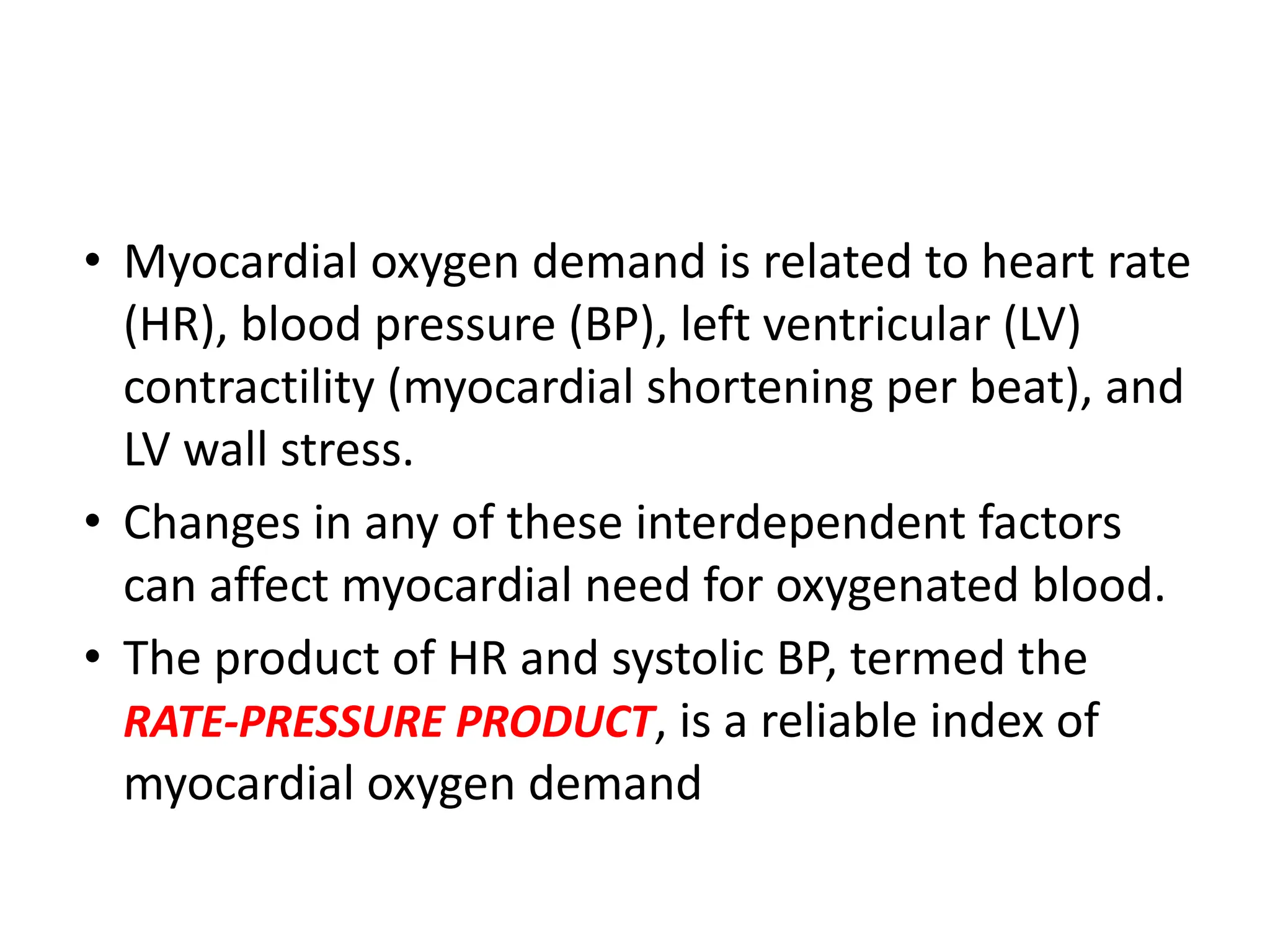
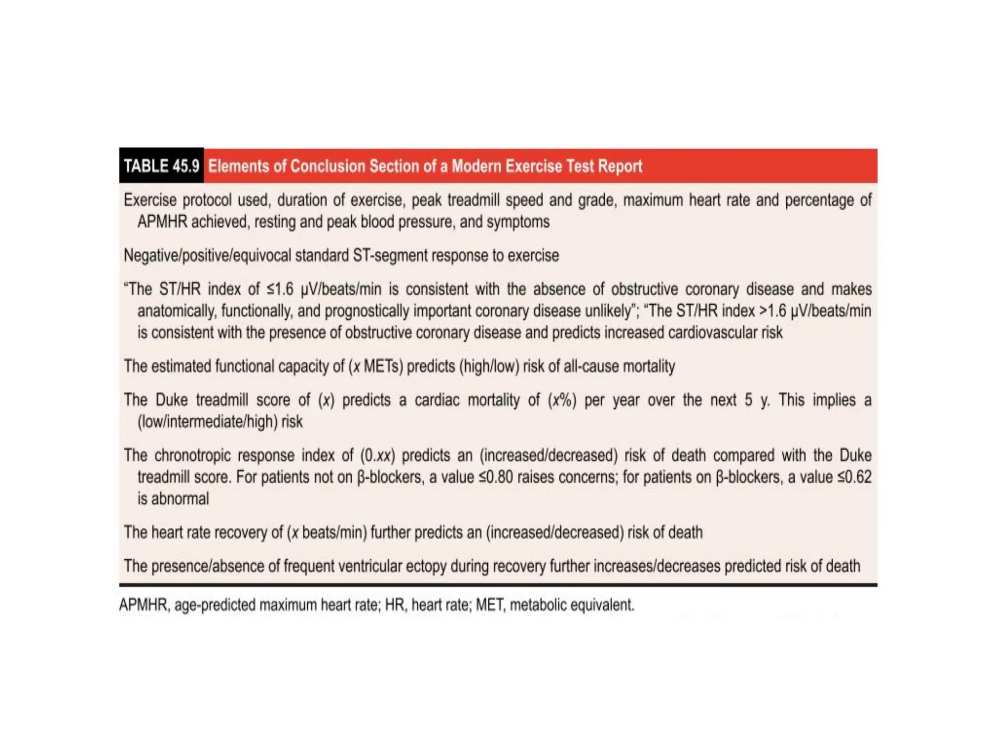
Purposes of Exercise Testing
• Detection of coronary artery disease (CAD) in patients
with chest pain (chest discomfort) syndromes or
potential symptom equivalents
• Evaluation of the anatomic and functional severity of
CAD
• Prediction of cardiovascular events and all-cause
death
• Evaluation of physical capacity and effort tolerance
• Evaluation of exercise-related symptoms
• Assessment of chronotropic competence,
arrhythmias, and response to implanted device
therapy
• Assessment of the response to medical interventions
GUIDELINES FOR EXERCISE STRESS
TESTING
Exercise Testing in the Diagnosis of
Obstructive CAD
• CLASS IA
• Adult patients (including those with complete
right bundle branch block or <1 mm of resting
ST-depression) with an intermediate pretest
probability of CAD on the basis of sex, age,
and symptoms
• Class IIa
• Patients with vasospastic angina
Risk Assessment and Prognosis Among
Patients With Symptoms or a History of
CAD
• CLASS I
• Patients undergoing initial evaluation with suspected or known
CAD (exceptions in class IIb), including those with complete right
bundle branch block or <1 mm of resting ST- depression
• Patients with suspected or known CAD previously evaluated, now
presenting with marked change in clinical status
• Low-risk unstable angina patients 8–12 h after presentation who
have been free of active ischemic or heart failure symptoms
• Intermediate-risk unstable angina patients 2–3 d after
presentation who have been free of active ischemic or heart
failure symptoms
AFTER ACUTE MI
• CLASS I
• Before discharge for prognostic assessment, activity
prescription, or evaluation of medical therapy
(submaximal at about 4– 6 d)
• Early after discharge for prognostic assessment and
cardiac rehabilitation if the predischarge exercise test
was not performed (symptom limited, about 14–21 d)
• Late after discharge for prognostic assessment, activity
prescription, evaluation of medical therapy, and
cardiac rehabilitation if the early exercise test was
submaximal (symptom limited, about 3–6 wk)
Exercise Testing for Persons without
Symptoms or Known CAD
Exercise Testing for Persons with
Valvular Heart Disease
Absolute Contraindications
• Acute myocardial infarction (MI), within 2 days
• Ongoing unstable angina
• Uncontrolled cardiac arrhythmia with hemodynamic
compromise
• Active endocarditis
• Symptomatic severe Aortic stenosis
• Decompensated heart failure
• Acute pulmonary embolism, pulmonary infarction, or deep
vein thrombosis
• Acute myocarditis or pericarditis
• Acute aortic dissection
• Physical disability that precludes safe and adequate testing
Relative Contraindications
• Known obstructive left main coronary artery stenosis
• Moderate to severe aortic stenosis with uncertain relation to
symptoms
• Tachyarrhythmias with uncontrolled ventricular rates
• Acquired advanced or complete heart block
• Hypertrophic obstructive cardiomyopathy with severe resting
gradient
• Recent stroke or transient ischemic attack
• Mental impairment with limited ability to cooperate
• Resting hypertension with systolic or diastolic blood pressures
>200/110 mmHg
• Uncorrected medical conditions, such as significant anemia,
electrolyte imbalance, and hyperthyroidism
Patient Preparation
• The subject or patient should not eat for 3 hours
before the test.
• Routine medications may be taken with small
amounts of water.
• Subjects should dress in comfortable clothing and
wear comfortable walking shoes or sneakers.
• The subject or patient should receive a detailed
explanation of the testing procedure and purpose
of the test, including the nature of the
progressive exercise, symptom and sign, end
points, and possible complications.
• A brief history and physical examination are
required to rule out contraindications to testing
and to detect important clinical signs, such as
cardiac murmur, gallop sounds, pulmonary
wheezing, or rales.
• Subjects with a history of worsening unstable
angina or decompensated heart failure should not
undergo exercise testing until their condition
stabilizes.
• Physical examination should screen for valvular or
congenital heart disease
• In patients with permanent cardiac pacemakers, it is
important to obtain information regarding the type
of pacemaker (single or dual chamber), programmed
mode, rate responsiveness, and pacing HR limits
before the test.
• In patients with Implantable Cardioverter Defibrillators
(ICDs), the peak HR during the exercise test is maintained
at least 10 beats/min below the programmed HR
threshold for antitachycardia pacing and defibrillation
PATIENT ASSESSMENT FOR
EXERCISE TESTING
HISTORY
• Medical history: including cardiovascular disease (known
existing coronary artery disease [CAD], previous myocardial
infarction, or coronary revascularization);
• Arrhythmias
• Syncope or presyncope;
• Pulmonary disease, including asthma, emphysema, and
bronchitis or recent pulmonary embolism;
• Cerebrovascular disease, including stroke;
• Peripheral artery disease;
• Current pregnancy;
• Musculoskeletal, neuromuscular, or joint disease.
• Symptoms: Angina; chest, jaw, or arm discomfort;
shortness of breath; and palpitations.
• Risk factors for atherosclerotic disease: Hypertension,
diabetes, obesity, dyslipidemia, and smoking
• If patient is without known CAD, determine the
pretest probability of CAD.
• Recent illness, hospitalization, or surgical procedure
• Medication dose and schedule
• Ability to perform physical activity
Physical Examination
• Pulse rate and regularity
• Resting blood pressure sitting and standing
• Auscultation of the lungs
• Auscultation of the heart, particularly in
patients with heart failure or valvular disease
• Examination related to orthopedic,
neurologic, or other medical conditions that
might limit exercise
PATIENT MONITORING DURING
EXERCISE TESTING
During the Exercise Period
• 12-lead ECG during last minute of each stage,
or at least every 3 minutes
• Blood pressure during last minute of each
stage, or at least every 3 minutes
• Symptom rating scales as appropriate for the
test indication and laboratory protocol
During the Recovery Period
• Monitoring for a minimum of 6 minutes after exercise in sitting or
supine position, or until near baseline heart rate, blood pressure,
ECG, and symptom measures are reached.
• A period of active cool-down may be included in the recovery
period, particularly following high levels of exercise, to minimize
the postexercise hypotensive effects of venous pooling in the
lower extremities.
• 12-lead ECG every minute
• Heart rate and blood pressure immediately after exercise, then
every 1 or 2 minutes until near-baseline measures are reached
• Symptomatic ratings every minute as long as they persist after
exercise.
• Patients should be observed until all symptoms have resolved or
returned to baseline levels.
Complications Secondary to Exercise
Testing
• CARDIAC-
• Bradyarrhythmias,Tachyarrhythmias
,Acute coronary syndromes, Heart failure,
Hypotension, syncope, and shock, Death (0.5
per 10 000 tests)
• Noncardiac - Musculoskeletal trauma,
Soft-tissue injury
• Miscellaneous - Severe fatigue (malaise),
sometimes persisting for days, dizziness, body
aches
Absolute Indications for Termination of
Exercise Testing
• ST-segment elevation (>1.0 mm) in leads without preexisting Q waves
because of prior MI (other than aVR, aVL, and V1)
• Drop in systolic blood pressure >10 mmHg, despite an increase in
workload, when accompanied by any other evidence of ischemia
• Moderate-to-severe angina
• Central nervous system symptoms (eg, ataxia, dizziness, near syncope)
• Signs of poor perfusion (cyanosis or pallor)
• Sustained ventricular tachycardia (VT) or other arrhythmia, including
second- or third-degree atrioventricular (AV) block, that interferes with
normal maintenance of cardiac output during exercise
• Technical difficulties in monitoring the ECG or systolic blood pressure
• The subject’s request to stop
Relative Indications
• Marked ST displacement (horizontal or
downsloping of >2 mm, measured 60 to 80 ms
after the J point [the end of the QRS complex])
in a patient with suspected ischemia
• Drop in systolic blood pressure >10 mmHg
(persistently below baseline) despite an
increase in workload, in the absence of other
evidence of ischemia
• Increasing chest pain
• Fatigue, shortness of breath, wheezing, leg cramps, or
claudication
• Arrhythmias other than sustained VT, including
multifocal ectopy, ventricular triplets, supraventricular
tachycardia, and bradyarrhythmias that have the
potential to become more complex or to interfere with
hemodynamic stability
• Exaggerated hypertensive response (systolic blood
pressure >250 mmHg or diastolic blood pressure >115
mmHg)
• Development of bundle-branch block that cannot
immediately be distinguished from VT
TREADMILL PROTOCOLS
BRUCE PROTOCOL
• The modified Bruce protocol employs 2 initial low level
3-minutes stages at a speed of 1.7 mph and grades 0 % and
5%,respectively, and then continues into the full Bruce protocol
• During treadmill exercise, patients should be encouraged to walk
freely and use the handrails for balance only when necessary.
• Excessive handrail gripping and support alter the BP response and
decrease the oxygen requirement (METs) per given workload,
thereby resulting in an overestimation of exercise capacity and an
inaccurate HR- and BP-to-workload relationship
Other protocols
• The Naughton protocol is good for older or
debilitated persons and allows a gradual increase
in workload.
• The Balke protocol is good for younger, fit
persons. It maintains a speed of 3, 3.5, or 4 mph
(4.8, 5.6, or 6.4 km/h, respectively) and increases
the grade every 2 minutes.
• The Cornell protocol allows for a gradual increase
in grade and speed and may be started at 0%, 5%,
or 10% grade, depending on fitness level
• 1 MET being resting energy expenditure and
defined as approximately 3.5 mL O2/kg body
weight/min
• V
̇ O2 is equal to the product of cardiac output
and oxygen extraction
• V
̇ O2max is the peak oxygen uptake achieved
during performance of the highest level of
dynamic exercise
• Myocardial oxygen demand is related to heart rate
(HR), blood pressure (BP), left ventricular (LV)
contractility (myocardial shortening per beat), and
LV wall stress.
• Changes in any of these interdependent factors
can affect myocardial need for oxygenated blood.
• The product of HR and systolic BP, termed the
RATE-PRESSURE PRODUCT, is a reliable index of
myocardial oxygen demand
DATA
• Electrocardiographic data.
• Age-predicted maximum heart rate (APMHR).
• Rating of perceived exertion (RPE)
• Blood pressure monitoring
• Symptoms
• Functional capacity
Electrocardiographic data
• ST-SEGMENT CHANGES
• Measurement of the ST-segment- it is measured 60 ms
past the J point
• ST-segment changes - measured from the isoelectric
baseline, which can be determined from the PR interval.
• If the ST-segment is elevated at rest, any depression that
occurs with exercise is still measured from the isoelectric
line;
• Early repolarization of the ST-segment at rest is normal.
• If the ST-segment is depressed at rest, any further
depression should be measured from the baseline
ST-segment
• ST-DEPRESSION - does not localize the area of
ischemia.
• ST-depression of at least 1 mm that is horizontal
or downsloping is abnormal, as is upsloping
ST-depression of at least 2.0 mm.
• Baseline ST-abnormalities are less likely to
represent exercise-induced myocardial ischemia,
and the baseline ST-depression should be
subtracted from the peak ST-depression
• NORMAL RESPONSE-
• During exercise, there is
depression of the J junction that is maximal at
peak exercise and returns to baseline during
recovery. This normal depression is upsloping
and typically <1 mm below the isoelectric line
80 ms after the J point.
Baseline Abnormalities That May Obscure
Electrocardiographic Changes during
Exercise
• Left bundle branch block
• Left ventricular hypertrophy with repolarization
abnormality
• Digitalis therapy
• Ventricular paced rhythm
• Wolff–Parkinson–White syndrome
• ST abnormality associated with supraventricular
tachycardia or atrial fibrillation
• ST-abnormalities with mitral valve prolapse and
severe anemia
Criteria that increase the probability of
ischemia
• The number of leads involved (i.e., more leads increase the
probability of ischemia),
• Workload at which the ST-depression occurs (i.e., lower workload
increases the probability)
• The angle of the slope (i.e., a downsloping angle has a higher
probability than a horizontal one)
• ST-segment adjustment relative to heart rate (ST/HR index)
• The amount of time in recovery before normalization of the
ST-segment (i.e., longer recovery increases the probability)
• The magnitude of the depression.
• Changes in the lateral leads, particularly V5, are more specific than
in any of the other leads.
• Changes in the inferior leads alone are likely to be a false-positive
result
ST-elevation
• A) ST-segment elevation with Q-waves of prior MI is a common
finding among patients who have had MI.
• It occurs among up to 50% of patients with anterior MI and 15% of
patients with previous inferior MI, and it is not caused by
ischemia.
• There may even be reciprocal ST-segment depression. Patients
with more extensive Q-waves have more pronounced
ST-elevation. These patients typically have a lower ejection
fraction than those without elevated ST-segment with a Q-wave.
• These changes do not imply ischemia (although they may imply
viability) and should be interpreted as normal.
• B) ST-segment elevation without Q-waves of prior MI represents
marked transmural myocardial ischemia. STelevation may also
indicate the location of the ischemia. This finding should be
interpreted as abnormal.
• ST-NORMALIZATION, or the lack of ST-changes
during exercise, may be a sign of ischemia.
• This phenomenon occurs when ischemic
ST-depression and ST-elevation cancel one
another.
• It should be considered in tests of patients
with no electrocardiographic changes but with
a high likelihood of CAD.
• Time to resolution of changes- the longer into
recovery time it takes for electrocardiographic
changes to resolve, the higher is the probability
that they are important.
• Rapid recovery (<1 minute) indicates less
likelihood of disease and that disease if present is
less severe.
• Bundle branch block or conduction delay-
Exercise-induced left bundle branch block is
predictive of a worse outcome
Age-predicted maximum heart rate
(APMHR)
• APMHR . HR(max) = 220 – Age
• Newer equation
• If MHR does not exceed 85% of APMHR during
testing and there are no substantial
electrocardiographic changes, the test is
usually read as nondiagnostic.
• If there are substantial electrocardiographic
changes, the test is read as abnormal,
regardless of the heart rate achieved.
CHRONOTROPIC RESPONSE INDEX
• The chronotropic response index (CRI) is a
measure of MHR in relation to chronotropic
reserve.
• A normal response is defined as a CRI of >0.8
(0.62 for patients on β-blockers)
BLOOD PRESSURE MONITORING
• It should be checked in each walking stage.
• Systolic blood pressure, Pulse pressure, HR-BP product
(Double Product), and Double-product Reserve (change in
double product from peak to rest) all increase steadily as
workload increases.
• A failure of SBP to rise with increasing workload or a drop in
SBP usually indicates the presence of CAD and is an
indication to terminate testing.
• Diastolic blood pressure decreases with exercise and may be
audible down to 0 during vigorous activity. Unlike SBP,
diastolic blood pressure is not useful in diagnosis.
SYMPTOMS
• The presence or absence of symptoms and
their change over time are included in the
final report
Exercise-Induced Symptoms
• Are the symptoms reported during the test
the same or similar to the reported historical
symptoms that prompted the exercise test?
• Exercise-induced angina is an important
clinical predictor of the presence and severity
of CAD, equal to or greater than ST-segment
depression
• Exercise-induced angina predicts an adverse
prognosis regardless of the ST-segment
response or the exercise capacity.
• Typical angina at the exercise test was a
predictor of adverse events,including death,
nonfatal MI, and revascularization
,irrespective of the absence of a positive
ST-segment response or good exercise
capacity.
Functional capacity
• Functional capacity- is a powerful marker for
prognosis.
• Strong predictor of mortality and nonfatal
cardiovascular outcomes in both men and
women with and without CAD
Postexercise recovery
• The length of the cool-down period may vary from
30 seconds to several minutes, depending on the
person.
• A general rule is to allow enough time for the
heart rate to drop to <110 beats/min.
• The exception to observing a cool-down period
may be made for exercise echocardiography, in
which it is important to image the subject when
he or she is as close as possible to MHR
Prognosis
• . The Duke nomogram is a simple chart that
factors in ST-segment deviation, amount of
angina during exercise, and exercise capacity
to give an estimate of a 5-year survival and
average annual mortality.
• The Duke treadmill score (DTS) is a numeric
form of the nomogram is an important
predictor of mortality
HEART RATE RECOVERY
• The heart rate recovery, defined as the difference in
heart rate at peak exercise and at 1 minute after
cessation of exercise.
• A heart rate recovery of 12 beats/min or less is
considered abnormal during an upright cool-down
period.
• For patients assuming an immediate supine position,
such as during exercise echocardiography, a value of
<18 beats/min is considered abnormal.
• Abnormal HRR is associated with an increase in
all-cause mortality in both asymptomatic individuals
and patients with established heart disease.
VENTRICULAR ECTOPY
• Ventricular ectopy in recovery from exercise,
including frequent ventricular ectopics
(>7/min), couplets, bigeminy, trigeminy,
ventricular tachycardia, and ventricular
fibrillation, has been shown to be predictive of
all-cause mortality.
• These findings in recovery are a better
predictor of death than ventricular ectopy
during exercise
THE EXERCISE ELECTROCARDIOGRAPHIC
TEST REPORT
EXERCISE TEST REPORT
• Exercise protocol used, duration of exercise, peak treadmill
speed and grade, maximum heart rate and percentage of
APMHR achieved, resting and peak blood pressure, and
symptoms
• Negative/positive/equivocal standard ST-segment response
to exercise
• “The ST/HR index of ≤1.6 µV/beats/min is consistent with
the absence of obstructive coronary disease and makes
anatomically, functionally, and prognostically important
coronary disease unlikely”; “The ST/HR index >1.6
µV/beats/min is consistent with the presence of obstructive
coronary disease and predicts increased cardiovascular risk
• The estimated functional capacity of (x METs) predicts
(high/low) risk of all-cause mortality The Duke treadmill
score of (x) predicts a cardiac mortality of (x%) per year over
the next 5 y. This implies a (low/intermediate/high) risk
• The chronotropic response index of (0.xx) predicts an
(increased/decreased) risk of death compared with the
Duke treadmill score. For patients not on β-blockers, a value
≤0.80 raises concerns; for patients on β-blockers, a value
≤0.62 is abnormal
• The heart rate recovery of (x beats/min) further predicts an
(increased/decreased) risk of death The presence/absence
of frequent ventricular ectopy during recovery further
increases/decreases predicted risk of deat
TMT for evaluation in chronic stable angina

TMT for evaluation in chronic stable angina

  • 2.
    Purposes of ExerciseTesting • Detection of coronary artery disease (CAD) in patients with chest pain (chest discomfort) syndromes or potential symptom equivalents • Evaluation of the anatomic and functional severity of CAD • Prediction of cardiovascular events and all-cause death • Evaluation of physical capacity and effort tolerance • Evaluation of exercise-related symptoms • Assessment of chronotropic competence, arrhythmias, and response to implanted device therapy • Assessment of the response to medical interventions
  • 3.
  • 4.
    Exercise Testing inthe Diagnosis of Obstructive CAD • CLASS IA • Adult patients (including those with complete right bundle branch block or <1 mm of resting ST-depression) with an intermediate pretest probability of CAD on the basis of sex, age, and symptoms • Class IIa • Patients with vasospastic angina
  • 6.
    Risk Assessment andPrognosis Among Patients With Symptoms or a History of CAD • CLASS I • Patients undergoing initial evaluation with suspected or known CAD (exceptions in class IIb), including those with complete right bundle branch block or <1 mm of resting ST- depression • Patients with suspected or known CAD previously evaluated, now presenting with marked change in clinical status • Low-risk unstable angina patients 8–12 h after presentation who have been free of active ischemic or heart failure symptoms • Intermediate-risk unstable angina patients 2–3 d after presentation who have been free of active ischemic or heart failure symptoms
  • 8.
    AFTER ACUTE MI •CLASS I • Before discharge for prognostic assessment, activity prescription, or evaluation of medical therapy (submaximal at about 4– 6 d) • Early after discharge for prognostic assessment and cardiac rehabilitation if the predischarge exercise test was not performed (symptom limited, about 14–21 d) • Late after discharge for prognostic assessment, activity prescription, evaluation of medical therapy, and cardiac rehabilitation if the early exercise test was submaximal (symptom limited, about 3–6 wk)
  • 10.
    Exercise Testing forPersons without Symptoms or Known CAD
  • 11.
    Exercise Testing forPersons with Valvular Heart Disease
  • 13.
    Absolute Contraindications • Acutemyocardial infarction (MI), within 2 days • Ongoing unstable angina • Uncontrolled cardiac arrhythmia with hemodynamic compromise • Active endocarditis • Symptomatic severe Aortic stenosis • Decompensated heart failure • Acute pulmonary embolism, pulmonary infarction, or deep vein thrombosis • Acute myocarditis or pericarditis • Acute aortic dissection • Physical disability that precludes safe and adequate testing
  • 14.
    Relative Contraindications • Knownobstructive left main coronary artery stenosis • Moderate to severe aortic stenosis with uncertain relation to symptoms • Tachyarrhythmias with uncontrolled ventricular rates • Acquired advanced or complete heart block • Hypertrophic obstructive cardiomyopathy with severe resting gradient • Recent stroke or transient ischemic attack • Mental impairment with limited ability to cooperate • Resting hypertension with systolic or diastolic blood pressures >200/110 mmHg • Uncorrected medical conditions, such as significant anemia, electrolyte imbalance, and hyperthyroidism
  • 15.
    Patient Preparation • Thesubject or patient should not eat for 3 hours before the test. • Routine medications may be taken with small amounts of water. • Subjects should dress in comfortable clothing and wear comfortable walking shoes or sneakers. • The subject or patient should receive a detailed explanation of the testing procedure and purpose of the test, including the nature of the progressive exercise, symptom and sign, end points, and possible complications.
  • 17.
    • A briefhistory and physical examination are required to rule out contraindications to testing and to detect important clinical signs, such as cardiac murmur, gallop sounds, pulmonary wheezing, or rales. • Subjects with a history of worsening unstable angina or decompensated heart failure should not undergo exercise testing until their condition stabilizes. • Physical examination should screen for valvular or congenital heart disease
  • 18.
    • In patientswith permanent cardiac pacemakers, it is important to obtain information regarding the type of pacemaker (single or dual chamber), programmed mode, rate responsiveness, and pacing HR limits before the test. • In patients with Implantable Cardioverter Defibrillators (ICDs), the peak HR during the exercise test is maintained at least 10 beats/min below the programmed HR threshold for antitachycardia pacing and defibrillation
  • 19.
  • 20.
    HISTORY • Medical history:including cardiovascular disease (known existing coronary artery disease [CAD], previous myocardial infarction, or coronary revascularization); • Arrhythmias • Syncope or presyncope; • Pulmonary disease, including asthma, emphysema, and bronchitis or recent pulmonary embolism; • Cerebrovascular disease, including stroke; • Peripheral artery disease; • Current pregnancy; • Musculoskeletal, neuromuscular, or joint disease.
  • 21.
    • Symptoms: Angina;chest, jaw, or arm discomfort; shortness of breath; and palpitations. • Risk factors for atherosclerotic disease: Hypertension, diabetes, obesity, dyslipidemia, and smoking • If patient is without known CAD, determine the pretest probability of CAD. • Recent illness, hospitalization, or surgical procedure • Medication dose and schedule • Ability to perform physical activity
  • 23.
    Physical Examination • Pulserate and regularity • Resting blood pressure sitting and standing • Auscultation of the lungs • Auscultation of the heart, particularly in patients with heart failure or valvular disease • Examination related to orthopedic, neurologic, or other medical conditions that might limit exercise
  • 24.
  • 25.
    During the ExercisePeriod • 12-lead ECG during last minute of each stage, or at least every 3 minutes • Blood pressure during last minute of each stage, or at least every 3 minutes • Symptom rating scales as appropriate for the test indication and laboratory protocol
  • 26.
    During the RecoveryPeriod • Monitoring for a minimum of 6 minutes after exercise in sitting or supine position, or until near baseline heart rate, blood pressure, ECG, and symptom measures are reached. • A period of active cool-down may be included in the recovery period, particularly following high levels of exercise, to minimize the postexercise hypotensive effects of venous pooling in the lower extremities. • 12-lead ECG every minute • Heart rate and blood pressure immediately after exercise, then every 1 or 2 minutes until near-baseline measures are reached • Symptomatic ratings every minute as long as they persist after exercise. • Patients should be observed until all symptoms have resolved or returned to baseline levels.
  • 27.
    Complications Secondary toExercise Testing • CARDIAC- • Bradyarrhythmias,Tachyarrhythmias ,Acute coronary syndromes, Heart failure, Hypotension, syncope, and shock, Death (0.5 per 10 000 tests)
  • 28.
    • Noncardiac -Musculoskeletal trauma, Soft-tissue injury • Miscellaneous - Severe fatigue (malaise), sometimes persisting for days, dizziness, body aches
  • 29.
    Absolute Indications forTermination of Exercise Testing • ST-segment elevation (>1.0 mm) in leads without preexisting Q waves because of prior MI (other than aVR, aVL, and V1) • Drop in systolic blood pressure >10 mmHg, despite an increase in workload, when accompanied by any other evidence of ischemia • Moderate-to-severe angina • Central nervous system symptoms (eg, ataxia, dizziness, near syncope) • Signs of poor perfusion (cyanosis or pallor) • Sustained ventricular tachycardia (VT) or other arrhythmia, including second- or third-degree atrioventricular (AV) block, that interferes with normal maintenance of cardiac output during exercise • Technical difficulties in monitoring the ECG or systolic blood pressure • The subject’s request to stop
  • 30.
    Relative Indications • MarkedST displacement (horizontal or downsloping of >2 mm, measured 60 to 80 ms after the J point [the end of the QRS complex]) in a patient with suspected ischemia • Drop in systolic blood pressure >10 mmHg (persistently below baseline) despite an increase in workload, in the absence of other evidence of ischemia • Increasing chest pain
  • 31.
    • Fatigue, shortnessof breath, wheezing, leg cramps, or claudication • Arrhythmias other than sustained VT, including multifocal ectopy, ventricular triplets, supraventricular tachycardia, and bradyarrhythmias that have the potential to become more complex or to interfere with hemodynamic stability • Exaggerated hypertensive response (systolic blood pressure >250 mmHg or diastolic blood pressure >115 mmHg) • Development of bundle-branch block that cannot immediately be distinguished from VT
  • 32.
  • 33.
  • 34.
    • The modifiedBruce protocol employs 2 initial low level 3-minutes stages at a speed of 1.7 mph and grades 0 % and 5%,respectively, and then continues into the full Bruce protocol • During treadmill exercise, patients should be encouraged to walk freely and use the handrails for balance only when necessary. • Excessive handrail gripping and support alter the BP response and decrease the oxygen requirement (METs) per given workload, thereby resulting in an overestimation of exercise capacity and an inaccurate HR- and BP-to-workload relationship
  • 35.
    Other protocols • TheNaughton protocol is good for older or debilitated persons and allows a gradual increase in workload. • The Balke protocol is good for younger, fit persons. It maintains a speed of 3, 3.5, or 4 mph (4.8, 5.6, or 6.4 km/h, respectively) and increases the grade every 2 minutes. • The Cornell protocol allows for a gradual increase in grade and speed and may be started at 0%, 5%, or 10% grade, depending on fitness level
  • 36.
    • 1 METbeing resting energy expenditure and defined as approximately 3.5 mL O2/kg body weight/min • V ̇ O2 is equal to the product of cardiac output and oxygen extraction • V ̇ O2max is the peak oxygen uptake achieved during performance of the highest level of dynamic exercise
  • 37.
    • Myocardial oxygendemand is related to heart rate (HR), blood pressure (BP), left ventricular (LV) contractility (myocardial shortening per beat), and LV wall stress. • Changes in any of these interdependent factors can affect myocardial need for oxygenated blood. • The product of HR and systolic BP, termed the RATE-PRESSURE PRODUCT, is a reliable index of myocardial oxygen demand
  • 38.
  • 39.
    • Electrocardiographic data. •Age-predicted maximum heart rate (APMHR). • Rating of perceived exertion (RPE) • Blood pressure monitoring • Symptoms • Functional capacity
  • 40.
    Electrocardiographic data • ST-SEGMENTCHANGES • Measurement of the ST-segment- it is measured 60 ms past the J point • ST-segment changes - measured from the isoelectric baseline, which can be determined from the PR interval. • If the ST-segment is elevated at rest, any depression that occurs with exercise is still measured from the isoelectric line; • Early repolarization of the ST-segment at rest is normal. • If the ST-segment is depressed at rest, any further depression should be measured from the baseline ST-segment
  • 41.
    • ST-DEPRESSION -does not localize the area of ischemia. • ST-depression of at least 1 mm that is horizontal or downsloping is abnormal, as is upsloping ST-depression of at least 2.0 mm. • Baseline ST-abnormalities are less likely to represent exercise-induced myocardial ischemia, and the baseline ST-depression should be subtracted from the peak ST-depression
  • 42.
    • NORMAL RESPONSE- •During exercise, there is depression of the J junction that is maximal at peak exercise and returns to baseline during recovery. This normal depression is upsloping and typically <1 mm below the isoelectric line 80 ms after the J point.
  • 43.
    Baseline Abnormalities ThatMay Obscure Electrocardiographic Changes during Exercise • Left bundle branch block • Left ventricular hypertrophy with repolarization abnormality • Digitalis therapy • Ventricular paced rhythm • Wolff–Parkinson–White syndrome • ST abnormality associated with supraventricular tachycardia or atrial fibrillation • ST-abnormalities with mitral valve prolapse and severe anemia
  • 44.
    Criteria that increasethe probability of ischemia • The number of leads involved (i.e., more leads increase the probability of ischemia), • Workload at which the ST-depression occurs (i.e., lower workload increases the probability) • The angle of the slope (i.e., a downsloping angle has a higher probability than a horizontal one) • ST-segment adjustment relative to heart rate (ST/HR index) • The amount of time in recovery before normalization of the ST-segment (i.e., longer recovery increases the probability) • The magnitude of the depression. • Changes in the lateral leads, particularly V5, are more specific than in any of the other leads. • Changes in the inferior leads alone are likely to be a false-positive result
  • 45.
    ST-elevation • A) ST-segmentelevation with Q-waves of prior MI is a common finding among patients who have had MI. • It occurs among up to 50% of patients with anterior MI and 15% of patients with previous inferior MI, and it is not caused by ischemia. • There may even be reciprocal ST-segment depression. Patients with more extensive Q-waves have more pronounced ST-elevation. These patients typically have a lower ejection fraction than those without elevated ST-segment with a Q-wave. • These changes do not imply ischemia (although they may imply viability) and should be interpreted as normal. • B) ST-segment elevation without Q-waves of prior MI represents marked transmural myocardial ischemia. STelevation may also indicate the location of the ischemia. This finding should be interpreted as abnormal.
  • 46.
    • ST-NORMALIZATION, orthe lack of ST-changes during exercise, may be a sign of ischemia. • This phenomenon occurs when ischemic ST-depression and ST-elevation cancel one another. • It should be considered in tests of patients with no electrocardiographic changes but with a high likelihood of CAD.
  • 47.
    • Time toresolution of changes- the longer into recovery time it takes for electrocardiographic changes to resolve, the higher is the probability that they are important. • Rapid recovery (<1 minute) indicates less likelihood of disease and that disease if present is less severe. • Bundle branch block or conduction delay- Exercise-induced left bundle branch block is predictive of a worse outcome
  • 48.
    Age-predicted maximum heartrate (APMHR) • APMHR . HR(max) = 220 – Age • Newer equation
  • 49.
    • If MHRdoes not exceed 85% of APMHR during testing and there are no substantial electrocardiographic changes, the test is usually read as nondiagnostic. • If there are substantial electrocardiographic changes, the test is read as abnormal, regardless of the heart rate achieved.
  • 50.
    CHRONOTROPIC RESPONSE INDEX •The chronotropic response index (CRI) is a measure of MHR in relation to chronotropic reserve. • A normal response is defined as a CRI of >0.8 (0.62 for patients on β-blockers)
  • 51.
    BLOOD PRESSURE MONITORING •It should be checked in each walking stage. • Systolic blood pressure, Pulse pressure, HR-BP product (Double Product), and Double-product Reserve (change in double product from peak to rest) all increase steadily as workload increases. • A failure of SBP to rise with increasing workload or a drop in SBP usually indicates the presence of CAD and is an indication to terminate testing. • Diastolic blood pressure decreases with exercise and may be audible down to 0 during vigorous activity. Unlike SBP, diastolic blood pressure is not useful in diagnosis.
  • 52.
    SYMPTOMS • The presenceor absence of symptoms and their change over time are included in the final report
  • 53.
    Exercise-Induced Symptoms • Arethe symptoms reported during the test the same or similar to the reported historical symptoms that prompted the exercise test? • Exercise-induced angina is an important clinical predictor of the presence and severity of CAD, equal to or greater than ST-segment depression
  • 54.
    • Exercise-induced anginapredicts an adverse prognosis regardless of the ST-segment response or the exercise capacity. • Typical angina at the exercise test was a predictor of adverse events,including death, nonfatal MI, and revascularization ,irrespective of the absence of a positive ST-segment response or good exercise capacity.
  • 55.
    Functional capacity • Functionalcapacity- is a powerful marker for prognosis. • Strong predictor of mortality and nonfatal cardiovascular outcomes in both men and women with and without CAD
  • 57.
    Postexercise recovery • Thelength of the cool-down period may vary from 30 seconds to several minutes, depending on the person. • A general rule is to allow enough time for the heart rate to drop to <110 beats/min. • The exception to observing a cool-down period may be made for exercise echocardiography, in which it is important to image the subject when he or she is as close as possible to MHR
  • 58.
    Prognosis • . TheDuke nomogram is a simple chart that factors in ST-segment deviation, amount of angina during exercise, and exercise capacity to give an estimate of a 5-year survival and average annual mortality. • The Duke treadmill score (DTS) is a numeric form of the nomogram is an important predictor of mortality
  • 61.
    HEART RATE RECOVERY •The heart rate recovery, defined as the difference in heart rate at peak exercise and at 1 minute after cessation of exercise. • A heart rate recovery of 12 beats/min or less is considered abnormal during an upright cool-down period. • For patients assuming an immediate supine position, such as during exercise echocardiography, a value of <18 beats/min is considered abnormal. • Abnormal HRR is associated with an increase in all-cause mortality in both asymptomatic individuals and patients with established heart disease.
  • 62.
    VENTRICULAR ECTOPY • Ventricularectopy in recovery from exercise, including frequent ventricular ectopics (>7/min), couplets, bigeminy, trigeminy, ventricular tachycardia, and ventricular fibrillation, has been shown to be predictive of all-cause mortality. • These findings in recovery are a better predictor of death than ventricular ectopy during exercise
  • 63.
  • 64.
    EXERCISE TEST REPORT •Exercise protocol used, duration of exercise, peak treadmill speed and grade, maximum heart rate and percentage of APMHR achieved, resting and peak blood pressure, and symptoms • Negative/positive/equivocal standard ST-segment response to exercise • “The ST/HR index of ≤1.6 µV/beats/min is consistent with the absence of obstructive coronary disease and makes anatomically, functionally, and prognostically important coronary disease unlikely”; “The ST/HR index >1.6 µV/beats/min is consistent with the presence of obstructive coronary disease and predicts increased cardiovascular risk
  • 65.
    • The estimatedfunctional capacity of (x METs) predicts (high/low) risk of all-cause mortality The Duke treadmill score of (x) predicts a cardiac mortality of (x%) per year over the next 5 y. This implies a (low/intermediate/high) risk • The chronotropic response index of (0.xx) predicts an (increased/decreased) risk of death compared with the Duke treadmill score. For patients not on β-blockers, a value ≤0.80 raises concerns; for patients on β-blockers, a value ≤0.62 is abnormal • The heart rate recovery of (x beats/min) further predicts an (increased/decreased) risk of death The presence/absence of frequent ventricular ectopy during recovery further increases/decreases predicted risk of deat