This document is the thesis of Guillaume Wendt submitted in October 2014 for the degree of Doctor of the University of Strasbourg. The thesis was completed in cotutelle between the University of Strasbourg, France and Saarland University, Germany under the direction of Professors Ayikoé Guy Mensah-Nyagan and Manfred J. Schmitt. The thesis assesses the neuroprotective effects of gamma-hydroxybutyrate and neurosteroids on cellular models of Alzheimer's disease.


![Table of contents
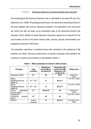
2
2.4. CELL CULTURE .........................................................................................................68
2.4.1. SH-SY5Y cells ....................................................................................................68
2.4.1.1. Routine culture........................................................................................................ 68
2.4.1.2. Freezing and unfreezing of SH-SY5Y cells............................................................. 68
2.4.2. Pichia Pastoris....................................................................................................70
2.4.2.1. Culture media.......................................................................................................... 70
2.4.2.2. Pichia pastoris culture............................................................................................. 71
2.4.3. Cell counting.......................................................................................................72
2.5. CELL VIABILITY ASSAYS.............................................................................................73
2.5.1. Trypan blue exclusion method............................................................................73
2.5.2. MTT viability assay .............................................................................................73
2.6. RT-QPCR................................................................................................................76
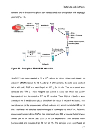
2.6.1. RNA extraction....................................................................................................76
2.6.2. RNA concentration and quality determination ....................................................78
2.6.3. Reverse transcription..........................................................................................78
2.6.4. Real Time quantitative PCR (RT-qPCR) ............................................................79
2.7. PROTEIN BASED ANALYSIS ........................................................................................81
2.7.1. Samples preparation – Protein Extraction ..........................................................81
2.7.2. Assessment of protein level with BCA assay......................................................83
2.7.3. SDS-PAGE .........................................................................................................83
2.7.4. Western analysis ................................................................................................85
2.7.4.1. “Semi dry” blotting................................................................................................... 86
2.7.4.2. Immunodetection..................................................................................................... 86
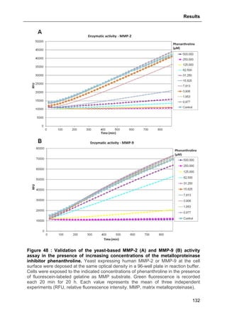
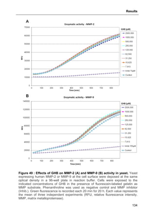
2.8. MMP-2/-9 ACTIVITY ASSAY WITH RECOMBINANT YEAST..............................................88
2.9. FLOW CYTOMETRY- AND MICROSCOPY-BASED METHODS............................................90
2.9.1. Flow cytometry (FACS) assessment of activated Caspase-3 and
TUNEL labeling ..............................................................................................................90
2.9.2. Confocal microscope analysis of apoptotic signals ............................................91
2.9.3. Calcium [Ca2+
]i imaging ......................................................................................92
2.10. STATISTICAL ANALYSIS .............................................................................................94
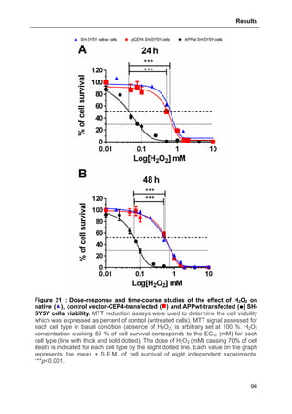
3. RESULTS.......................................................................................................................95
3.1. EFFECTS OF GHB AND/OR NEUROSTEROIDS AGAINST OXIDATIVE STRESS- AND APPWT-
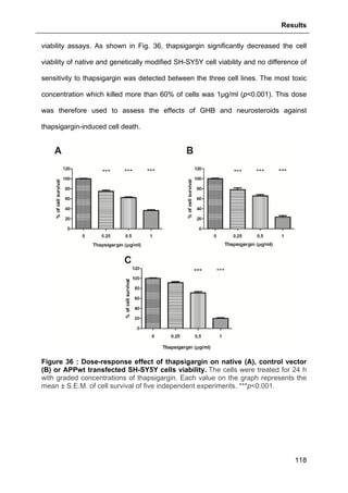
OVEREXPRESSION-INDUCED CELL DEATH ..............................................................................95
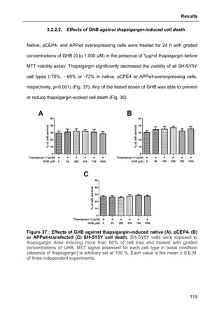
3.1.1. Effect of H2O2-induced oxidative stress on native and genetically modified SH-
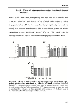
SY5Y cell viability ...........................................................................................................95
3.1.2. Trypan blue exclusion and MTT assessments of control and APPwt-
overexpressing SH-SY5Y cell viability and survival .......................................................97
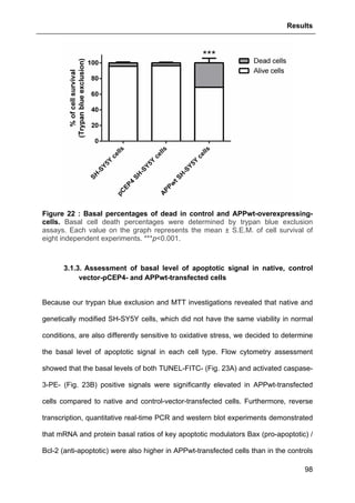
3.1.3. Assessment of basal level of apoptotic signal in native, control vector-pCEP4-
and APPwt-transfected cells...........................................................................................98
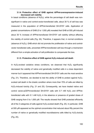
3.1.4. Protective effect of GHB against APPwt-overexpression-induced decreased cell
viability..........................................................................................................................100
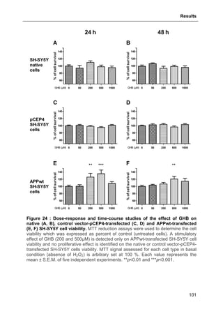
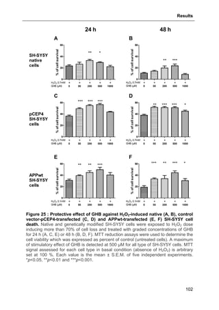
3.1.5. Protective effect of GHB against H2O2-induced cell death ...............................100
3.1.6. Protective effect of GHB against APPwt-overexpression and H2O2-evoked
apoptosis ......................................................................................................................103
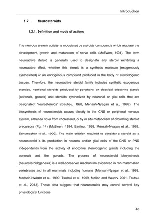
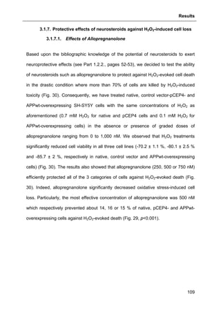
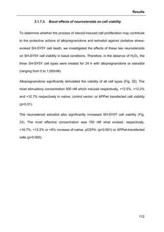
3.1.7. Protective effects of neurosteroids against H2O2-induced cell loss ..................109
3.1.7.1. Effects of Allopregnanolone .................................................................................. 109
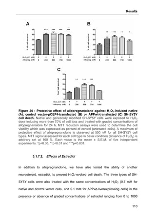
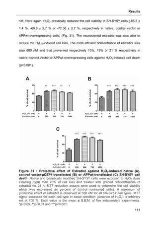
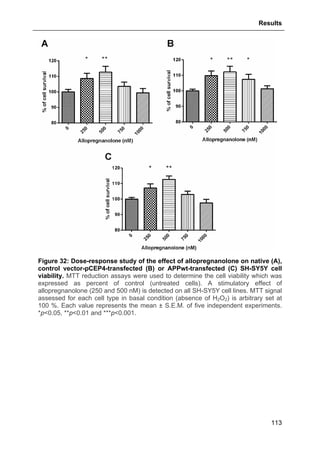
3.1.7.2. Effects of Estradiol ................................................................................................ 110
3.1.7.3. Basal effects of neurosteroids on cell viability ...................................................... 112](https://image.slidesharecdn.com/1a91c091-df65-404e-a6e2-2f0c1a7ca5fd-150303024016-conversion-gate01/85/These-Wendt-Guillaume-9-320.jpg)
![Abreviations
5
Abreviations
AD Alzheimer’s disease
[Ca2+
]i Intracellular calcium
17β-HSD 17β-Hydroxysteroid dehydrogenase
3α,5α-THP 3α, 5α-Tetrahydroxyprogesterone or allopregnanolone
3α-DIOL 3α-androstanediol
3α-HSOR 3α-Hydroxysteroid oxidoreductase
3β-HSD 3β-Hydroxysteroid dehydrogenase
5α-R 5α-Reductase
ABAD Aβ binding protein alcool dehydrogenase
ACE Angiotensin converting enzyme
acetyl-CoA Acetyl-Coenzyme A
ADAM A-disintegrin and metalloprotease
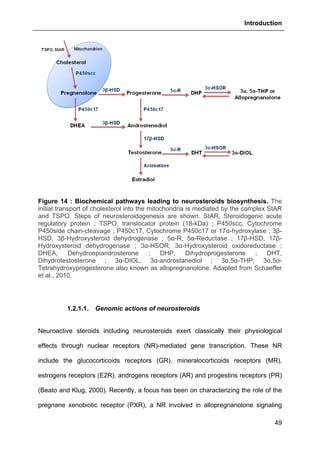
AF1 Activation function 1 domain
AF2 Activation function 2 domain
AICD APP intracellular domain
AMPAR α-amino-3-hydroxy-5-methyl-4- isoxazoleproprionic acid receptor
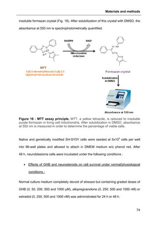
ANOVA Analysis of variance
AOX1 Alcohol oxidase 1
AOX2 Alcohol oxidase 2
AP Allopregnanolone
Apaf-1 Apoptotic protease activating factor 1
APH1 Anterior pharynx defective
APP Amyloid precursor protein
APP-CTFβ APP carboxy-terminal fragment
APPwt Wild-type APP
APS Ammonium persulfate
AR Androgens receptors
ARG Arginine
ATF4 Activating transcription factor 4
ATF6 Activating transcription factor 6
ATP Adenosine triphosphate](https://image.slidesharecdn.com/1a91c091-df65-404e-a6e2-2f0c1a7ca5fd-150303024016-conversion-gate01/85/These-Wendt-Guillaume-12-320.jpg)
![Abreviations
8
IRE1α Inositol-requiring enzyme 1α
JNK JUN amino-terminal kinase
LBDs Ligand binding regions
LTD Long term depression
LTP Long term potentiation
MAMs Mitochondria-ER associated membranes
MAPK Mitogen activated protein kinase
MAPs Microtubule-associated proteins
MCI Mild cognitive impairment
MMP-2 Matrix Metalloproteinase 2
MMP-9 Matrix Metalloproteinase 9
MOMP Mitochondrial outer membrane permeabilization
MR Mineralocorticoids receptor
mRNA Messenger ribonucleic acid
MTT 3-[4,5-dimethylthiazol-2-yl]-2,5 diphenyl tetrazolium bromide
NADH Nicotine adenine dinucleotide
NEP Neprylysin
NFTs Neurofibrillary tangles
NFκB Nuclear factor kappa-light-chain-enhancer of activated B cells
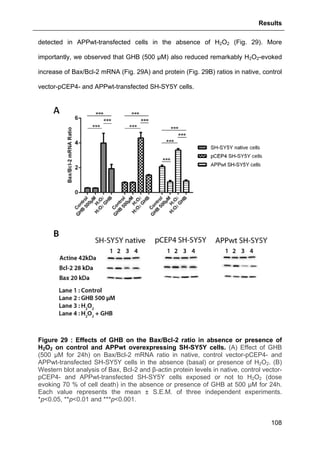
NLS Nuclear localization signal
NMDAR N-methyl-D-aspartate receptor
NR2A NMDAR subunit 2A
NR2B NMDAR subunit 2B
NR Nuclear receptors
OD Optical density
OxPhos Oxidative phosphorylation
P450c17 Cytochrome P450c17 or 17α-hydroxylase
P450scc Cytochrome P450side chain-cleavage
PBS Phosphate buffered saline
PCR Polymerase chain reaction
PDH Pyruvate dehydrogenase
PERK Protein kinase RNA-like ER kinase
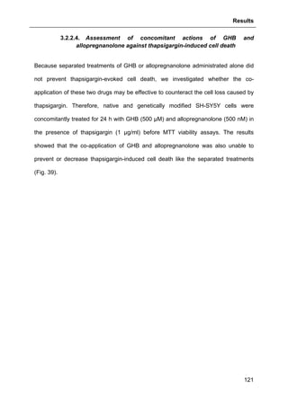
PE Phycoerythrin](https://image.slidesharecdn.com/1a91c091-df65-404e-a6e2-2f0c1a7ca5fd-150303024016-conversion-gate01/85/These-Wendt-Guillaume-15-320.jpg)

![Introduction
26
1.1.3. Biological consequences of Amyloid peptides and neurofibrillary
tangles
1.1.3.1. Synaptic failure and axonal transport impairment
Aging itself causes synaptic loss (Masliah et al., 2006), therefore, it is easy to
understand that AD is primarily a disorder of synaptic transmission (Selkoe, 2002).
As aforementioned, NFTs and synaptic loss are pivotally involed in AD
physiopathology. In parallel to Braak spreading of the disease, hippocampal
synapses begin to decline in MCI patients (Scheff et al., 2007). Some evidence
reveal a 25% decrease of presynaptic vesicle synaptophysin in mild AD (Masliah,
2001, Masliah et al., 2001). In the last Braak stages, dramatic synaptic loss is
positively correlated with dementia (Terry et al., 1991).
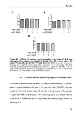
Aβ peptides are known to impair the “long term potentiation” (LTP), an experimental
indicator of memory formation (Palop and Mucke, 2010). Subsequently, signaling
molecules important to memory are also inhibited. Many studies investigated the
effects of Aβ peptides on excitatory synaptic transmission, that is tightly regulated by
the number of active N-methyl-D-aspartate receptor (NMDAR) and the α-amino-3-
hydroxy-5-methyl-4-isoxazoleproprionic acid receptor (AMPAR). NMDAR activation
has a central role in memory, by inducing LTP or long term depression (LTD),
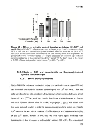
depending on the extent of the resultant intracellular calcium ([Ca2+
]i) rise in the
dendritic spines and the downstream activation of intracellular cascades (Kullmann
and Lamsa, 2007). Activation of post-synaptic NMDARs and large increases in
([Ca2+
]i) are necessary for LTP, whereas internalization of NMDARs, activation of
perisynaptic NMDARs and lower increase in [Ca2+
]i are necessary for LTD. LTP
induction promotes recruitment of AMPARs and growth of dendritic spines, whereas
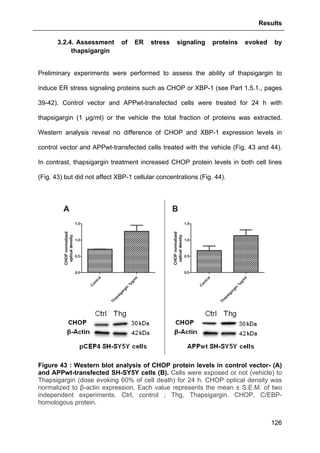
LTD induces spine shrinkage and synaptic loss (Kullmann and Lamsa, 2007).](https://image.slidesharecdn.com/1a91c091-df65-404e-a6e2-2f0c1a7ca5fd-150303024016-conversion-gate01/85/These-Wendt-Guillaume-33-320.jpg)
![Materials and methods
73
2.5. Cell viability assays
2.5.1. Trypan blue exclusion method
The trypan blue exclusion method is used to determine the number of viable cells
present in a cell suspension. It is based on the principle that live cells possess intact
cell membranes that exclude certain dyes including trypan blue, whereas dead cells
do not. In this test, a cell suspension is simply mixed with dye and then visually
examined to determine whether cells take up or exclude dye.
In our study, SH-SY5Y cells were seeded into a 24-well plate (105
cells per well) and
incubated for 48 h at 37°C under an atmosphere of 5% CO2 in DMEM supplemented
with 10% (v/v) heat-inactivated fetal calf serum. Native, control vector-pCEP4- and
APPwt-transfected SH-SY5Y cells were treated with various concentrations of H2O2
ranging from 0 to 1 mM. After 24 h, cells were detached from the plate using a 0.05%
trypsin-EDTA solution and immediately after DMEM supplemented with serum was
added. Equal volumes of the cell suspension and 0.4% (v/v) trypan blue in PBS were
mixed. Ten microliters of each mixture was transferred to a non-gridded disposable
Countess® chamber Slide and the cells were scored using a Countess® automated
cell counter (Invitrogen, Heidelberg, Germany). Each probe was counted twice. The
percent of cell survival was calculated by the Countess® software (Invitrogen,
Heidelberg, Germany) as the number of living cells divided by total cell number
(including dead and living cells).
2.5.2. MTT viability assay
The MTT (3-[4,5-dimethylthiazol-2-yl]-2,5 diphenyl tetrazolium bromide) assay is a
colorimetric assay to assess cell viability. Mitochondrial NADPH-dependent
oxidoreductase reflects the number of viable cells by reducing MTT into a purple](https://image.slidesharecdn.com/1a91c091-df65-404e-a6e2-2f0c1a7ca5fd-150303024016-conversion-gate01/85/These-Wendt-Guillaume-80-320.jpg)
![Materials and methods
84
based upon the principle that a charged molecule will migrate in an electric field
toward an electrode with opposite sign. In PAGE, proteins charged negatively by the
binding of the anionic detergent SDS separate within a matrix of polyacrylamide gel
in an electric field according to their molecular weight. Polyacrylamide is formed by
the polymerization of the monomer molecule-acrylamide crosslinked by N,N'-
methylene-bis-acrylamide (BIS). Free radicals generated by ammonium persulfate
(APS) and a catalyst acting as an oxygen scavenger (-N,N,N',N'-tetramethylethylene
diamine [TEMED]) are required to start the polymerization since acrylamide and BIS
are nonreactive by themselves or when mixed together. The distinct advantage of
acrylamide gel systems is that the initial concentrations of acrylamide and BIS control
the hardness and degree of cool, crosslinking of the gel. The hardness of a gel in
turn controls the friction that macromolecules experience as they move through the
gel in an electric field, and therefore affects the resolution of the components to be
separated. Hard gels (12-20% acrylamide) retard the migration of large molecules
more than they do with small ones. In certain cases, high concentration acrylamide
gels are so tight that they exclude large molecules from entering the gel but allow the
migration and resolution of low molecular weight components of a complex mixture.
Alternatively, in a loose gel (4-8% acrylamide), high molecular weight molecules
migrate much farther down the gel and, in some instances, can move right out of the
matrix.
In this thesis, 45 µg of proteins per sample were fractionated by 4-20% TGX SDS
pre-casted polyacrylamide gels (BioRad, Hercules, USA). Proteins were diluted in
reducing Laemmli buffer (added with β-mercaptoethanol) and heated for 5 min at
95°C. Precision Plus Protein Dual color standards (BioRad, Hercules, USA) was](https://image.slidesharecdn.com/1a91c091-df65-404e-a6e2-2f0c1a7ca5fd-150303024016-conversion-gate01/85/These-Wendt-Guillaume-91-320.jpg)

![Materials and methods
92
neuroblastoma cells were seeded onto slides at a density of 1x10⁵
cells and then
treated with GHB (500 µM) alone, H2O2 alone (0.1 mM for APPwt-transfected cells or
0.7 mM for control vector-pCEP4-transfected and native cells) or with GHB (500 µM)
and H2O2 (0.1 mM or 0.7 mM) for 1 h, 6 h, 24 h or 48 h. After treatments, the cells
were fixed in 4% paraformaldehyde for 1h, washed with PBS and permeabilized with
0.1% sodium citrate and 0.1% Triton X-100. The slides were then incubated with the
TUNEL-FITC reaction mixture for 1 h at 37°C. Negative controls were performed by
omitting TdT. Also, positive controls were made by incubating the cells with DNAse (3
UI/ml in Tris-HCl, pH 7.5 in 1 mg/ml BSA solution) during 10 min. Images were
captured using a LEICA TCS-SP confocal inverted microscope. All analyses were
carried out in comparable areas under the same optical and light conditions. Black-
and-white images were digitized and viewed on a computer with LAS software
(Leica).
2.9.3. Calcium [Ca2+
]i imaging
Intracellular calcium was measured by using FURA-2 acetoxymethyl ester (FURA-2
AM), a membrane permeable derivative of the ratiometric calcium FURA-2 which is
rapidly metabolized by cytoplasmic esterases, leading to the active dye FURA-2. The
Ca2+
unbound form of FURA-2 gets excited at 380 nm and the Ca2+
bound form of
FURA-2 at 340 nm. The emitted light is measured at 510 nm. In the presence of
augments concentrations of free calcium, the fluorescence intensity at 340 nm
increases, wheras the fluorescence intensity at 380 nm decreases. Therefore, the
340 nm/380 nm ratio increases. To assess this ratio in SH-SY5Y cells, we incubated
them with 1 µM FURA-2 AM (Invitrogen, Karlsruhe, Germany) at 37°C under 5% CO2
atmosphere for 30 min. Afterwards, the cells were washed with PBS and placed at](https://image.slidesharecdn.com/1a91c091-df65-404e-a6e2-2f0c1a7ca5fd-150303024016-conversion-gate01/85/These-Wendt-Guillaume-99-320.jpg)

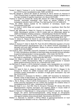
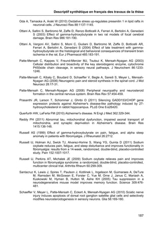
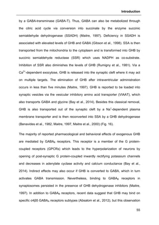
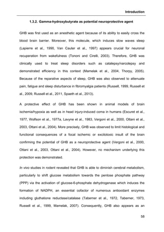
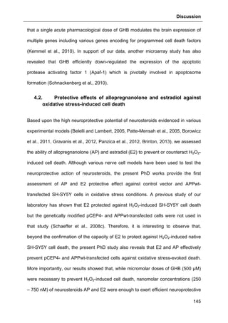
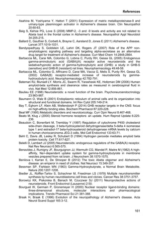
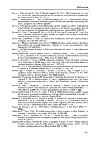
![Results
124
aimed at investigating allopregnanolone eventual action on store operated calcium
entry (SOCE) via the plasma membrane.
As shown in Fig. 41, thapsigargin induced a large cytosolic Ca2+
increase in absence
and presence of external calcium as expected. Allopregnanolone at 500 nM did not
modify calcium stores depletion or the SOCE. These data are consistent with the
absence of allopregnanolone effect on thapsigargin-evoked cell viability decrease
(see Part. 3.2.3.1., page 123).
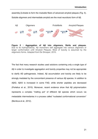
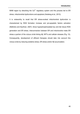
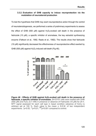
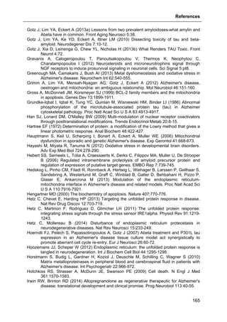
Figure 41 : Effects of allopregnanolone on thapsigargin-induced cytosolic
calcium elevations. [Ca2+
]I responses before and after calcium store depletion by
thapsirgargin (1 µg/ml, without external calcium) and after addition of thapsigargin in
the presence of extracellular calcium (store operated calcium entry, SOCE). Cells
were pre-incubated for 2 h with 500 nM allopregnanolone before the measurement.
Each value is the mean of three independent experiments (50 cells/experiment were
measured). EGTA, Ethylene glycol tetraacetic acid.](https://image.slidesharecdn.com/1a91c091-df65-404e-a6e2-2f0c1a7ca5fd-150303024016-conversion-gate01/85/These-Wendt-Guillaume-131-320.jpg)
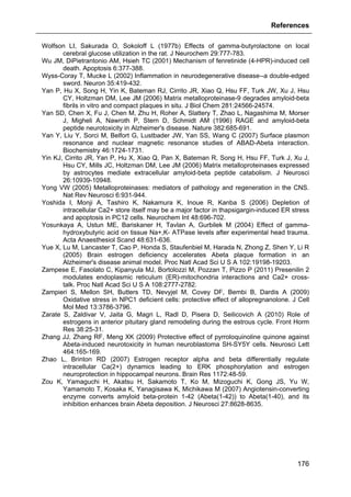
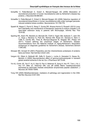
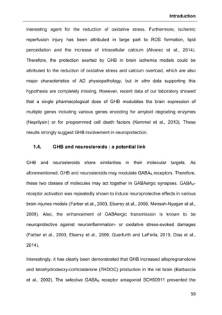
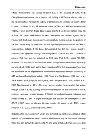
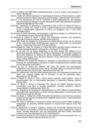
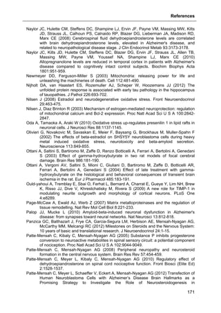
![Results
125
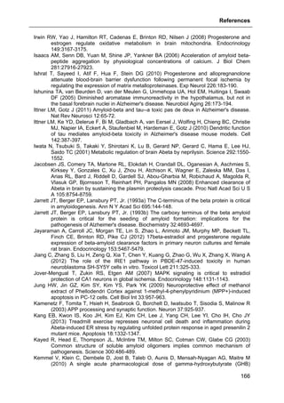
3.2.3.2. Effects of GHB
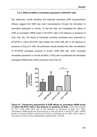
The same approach aforementioned was used to assess GHB action on
thapsigargin-induced cytosolic calcium elevations. The results showed that GHB at
500 µM (administrated as pre-treatment or maintained permanently in the incubation
medium) did not affect Ca2+
store depletion or SOCE (Fig. 42). These data are also
consistent with the absence of GHB effect on thapsigargin-evoked cell loss (see Part.
3.2.3.2., page 125).
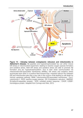
Figure 42 : Effects of GHB on thapsigargin-induced cytosolic calcium
elevations. [Ca2+
]I responses before and following calcium store depletion by
thapsirgargin (1 µg/ml, without external calcium) and after addition of thapsigargin in
the presence of extracellular calcium ([Ca2+
]I response in case of store operated
calcium entry, SOCE). Cells were either pre-incubated (for 2 h) with 500 µM GHB or
permanently exposed to GHB (500 µM) during the entire measurement. Each value
is the mean of three independent experiments (50 cells/experiment were measured).
EGTA, Ethylene glycol tetraacetic acid.](https://image.slidesharecdn.com/1a91c091-df65-404e-a6e2-2f0c1a7ca5fd-150303024016-conversion-gate01/85/These-Wendt-Guillaume-132-320.jpg)
![References
162
Brar R, Singh JP, Kaur T, Arora S, Singh AP (2014) Role of GABAergic activity of sodium
valproate against ischemia-reperfusion-induced acute kidney injury in rats. Naunyn
Schmiedebergs Arch Pharmacol 387:143-151.
Brinton RD (2013) Neurosteroids as regenerative agents in the brain: therapeutic
implications. Nat Rev Endocrinol 9:241-250.
Canevari L, Clark JB (2007) Alzheimer's disease and cholesterol: the fat connection.
Neurochem Res 32:739-750.
Cao X, Sudhof TC (2001) A transcriptionally [correction of transcriptively] active complex of
APP with Fe65 and histone acetyltransferase Tip60. Science 293:115-120.
Cardounel A, Regelson W, Kalimi M (1999) Dehydroepiandrosterone protects hippocampal
neurons against neurotoxin-induced cell death: mechanism of action. Proc Soc Exp
Biol Med 222:145-149.
Carroll JC, Rosario ER, Chang L, Stanczyk FZ, Oddo S, LaFerla FM, Pike CJ (2007)
Progesterone and estrogen regulate Alzheimer-like neuropathology in female 3xTg-
AD mice. J Neurosci 27:13357-13365.
Cente M, Filipcik P, Mandakova S, Zilka N, Krajciova G, Novak M (2009) Expression of a
truncated human tau protein induces aqueous-phase free radicals in a rat model of
tauopathy: implications for targeted antioxidative therapy. J Alzheimers Dis 17:913-
920.
Cente M, Filipcik P, Pevalova M, Novak M (2006) Expression of a truncated tau protein
induces oxidative stress in a rodent model of tauopathy. Eur J Neurosci 24:1085-
1090.
Chang HY, Nishitoh H, Yang X, Ichijo H, Baltimore D (1998) Activation of apoptosis signal-
regulating kinase 1 (ASK1) by the adapter protein Daxx. Science 281:1860-1863.
Chen S, Wang JM, Irwin RW, Yao J, Liu L, Brinton RD (2011a) Allopregnanolone promotes
regeneration and reduces beta-amyloid burden in a preclinical model of Alzheimer's
disease. PLoS One 6:e24293.
Chen SD, Yang DI, Lin TK, Shaw FZ, Liou CW, Chuang YC (2011b) Roles of Oxidative
Stress, Apoptosis, PGC-1alpha and Mitochondrial Biogenesis in Cerebral Ischemia.
Int J Mol Sci 12:7199-7215.
Cimarosti H, Zamin LL, Frozza R, Nassif M, Horn AP, Tavares A, Netto CA, Salbego C
(2005) Estradiol protects against oxygen and glucose deprivation in rat hippocampal
organotypic cultures and activates Akt and inactivates GSK-3beta. Neurochem Res
30:191-199.
Clark IM, Swingler TE, Sampieri CL, Edwards DR (2008) The regulation of matrix
metalloproteinases and their inhibitors. Int J Biochem Cell Biol 40:1362-1378.
Connelly WM, Errington AC, Crunelli V (2013) gamma-Hydroxybutyric acid (GHB) is not an
agonist of extrasynaptic GABAA receptors. PLoS One 8:e79062.
Cornejo VH, Hetz C (2013) The unfolded protein response in Alzheimer's disease. Semin
Immunopathol 35:277-292.
Costa RO, Ferreiro E, Oliveira CR, Pereira CM (2013) Inhibition of mitochondrial cytochrome
c oxidase potentiates Abeta-induced ER stress and cell death in cortical neurons. Mol
Cell Neurosci 52:1-8.
Coune P, Taleb O, Mensah-Nyagan AG, Maitre M, Kemmel V (2010) Calcium and cAMP
signaling induced by gamma-hydroxybutyrate receptor(s) stimulation in NCB-20
neurons. Neuroscience 167:49-59.
Crouch PJ, Blake R, Duce JA, Ciccotosto GD, Li QX, Barnham KJ, Curtain CC, Cherny RA,
Cappai R, Dyrks T, Masters CL, Trounce IA (2005) Copper-dependent inhibition of
human cytochrome c oxidase by a dimeric conformer of amyloid-beta1-42. J Neurosci
25:672-679.
Crunelli V, Emri Z, Leresche N (2006) Unravelling the brain targets of gamma-hydroxybutyric
acid. Curr Opin Pharmacol 6:44-52.
Cunningham LA, Wetzel M, Rosenberg GA (2005) Multiple roles for MMPs and TIMPs in
cerebral ischemia. Glia 50:329-339.](https://image.slidesharecdn.com/1a91c091-df65-404e-a6e2-2f0c1a7ca5fd-150303024016-conversion-gate01/85/These-Wendt-Guillaume-169-320.jpg)
![References
167
modifies multiple gene expression patterns in rat hippocampus and frontal cortex.
Physiol Genomics.
Kerr JF, Wyllie AH, Currie AR (1972) Apoptosis: a basic biological phenomenon with wide-
ranging implications in tissue kinetics. Br J Cancer 26:239-257.
Khlistunova I, Biernat J, Wang Y, Pickhardt M, von Bergen M, Gazova Z, Mandelkow E,
Mandelkow EM (2006) Inducible expression of Tau repeat domain in cell models of
tauopathy: aggregation is toxic to cells but can be reversed by inhibitor drugs. J Biol
Chem 281:1205-1214.
Kibaly C, Meyer L, Patte-Mensah C, Mensah-Nyagan AG (2008) Biochemical and functional
evidence for the control of pain mechanisms by dehydroepiandrosterone
endogenously synthesized in the spinal cord. FASEB J 22:93-104.
Kimoto T, Tsurugizawa T, Ohta Y, Makino J, Tamura H, Hojo Y, Takata N, Kawato S (2001)
Neurosteroid synthesis by cytochrome p450-containing systems localized in the rat
brain hippocampal neurons: N-methyl-D-aspartate and calcium-dependent synthesis.
Endocrinology 142:3578-3589.
King IA, Tabiowo A (1981) Effect of tunicamycin on epidermal glycoprotein and
glycosaminoglycan synthesis in vitro. Biochem J 198:331-338.
Kipp M, Beyer C (2009) Impact of sex steroids on neuroinflammatory processes and
experimental multiple sclerosis. Front Neuroendocrinol 30:188-200.
Kobayashi D, Zeller M, Cole T, Buttini M, McConlogue L, Sinha S, Freedman S, Morris RG,
Chen KS (2008) BACE1 gene deletion: impact on behavioral function in a model of
Alzheimer's disease. Neurobiol Aging 29:861-873.
Kole AJ, Annis RP, Deshmukh M (2013) Mature neurons: equipped for survival. Cell Death
Dis 4:e689.
Kook SY, Seok Hong H, Moon M, Mook-Jung I (2013) Disruption of blood-brain barrier in
Alzheimer disease pathogenesis. Tissue Barriers 1:e23993.
Kowald A (2001) The mitochondrial theory of aging. Biol Signals Recept 10:162-175.
Kroemer G, Reed JC (2000) Mitochondrial control of cell death. Nat Med 6:513-519.
Kullmann DM, Lamsa KP (2007) Long-term synaptic plasticity in hippocampal interneurons.
Nat Rev Neurosci 8:687-699.
Kumar S, Rezaei-Ghaleh N, Terwel D, Thal DR, Richard M, Hoch M, Mc Donald JM, Wullner
U, Glebov K, Heneka MT, Walsh DM, Zweckstetter M, Walter J (2011) Extracellular
phosphorylation of the amyloid beta-peptide promotes formation of toxic aggregates
during the pathogenesis of Alzheimer's disease. EMBO J 30:2255-2265.
Kuwana T, Newmeyer DD (2003) Bcl-2-family proteins and the role of mitochondria in
apoptosis. Curr Opin Cell Biol 15:691-699.
Laborit H, Jouany JM, Gerard J, Fabiani F (1960) [Generalities concerning the experimental
study and clinical use of gamma hydroxybutyrate of Na]. Agressologie 1:397-406.
Laird FM, Cai H, Savonenko AV, Farah MH, He K, Melnikova T, Wen H, Chiang HC, Xu G,
Koliatsos VE, Borchelt DR, Price DL, Lee HK, Wong PC (2005) BACE1, a major
determinant of selective vulnerability of the brain to amyloid-beta amyloidogenesis, is
essential for cognitive, emotional, and synaptic functions. J Neurosci 25:11693-
11709.
Lapierre O, Montplaisir J, Lamarre M, Bedard MA (1990) The effect of gamma-
hydroxybutyrate on nocturnal and diurnal sleep of normal subjects: further
considerations on REM sleep-triggering mechanisms. Sleep 13:24-30.
Lavyne MH, Hariri RJ, Tankosic T, Babiak T (1983) Effect of low dose gamma-butyrolactone
therapy on forebrain neuronal ischemia in the unrestrained, awake rat. Neurosurgery
12:430-434.
Lee BY, Ban JY, Seong YH (2005) Chronic stimulation of GABAA receptor with muscimol
reduces amyloid beta protein (25-35)-induced neurotoxicity in cultured rat cortical
cells. Neurosci Res 52:347-356.
Lee JH, Won SM, Suh J, Son SJ, Moon GJ, Park UJ, Gwag BJ (2010) Induction of the
unfolded protein response and cell death pathway in Alzheimer's disease, but not in
aged Tg2576 mice. Exp Mol Med 42:386-394.](https://image.slidesharecdn.com/1a91c091-df65-404e-a6e2-2f0c1a7ca5fd-150303024016-conversion-gate01/85/These-Wendt-Guillaume-174-320.jpg)
![References
168
Lee VM, Goedert M, Trojanowski JQ (2001) Neurodegenerative tauopathies. Annu Rev
Neurosci 24:1121-1159.
Li WW, Alexandre S, Cao X, Lee AS (1993) Transactivation of the grp78 promoter by Ca2+
depletion. A comparative analysis with A23187 and the endoplasmic reticulum
Ca(2+)-ATPase inhibitor thapsigargin. J Biol Chem 268:12003-12009.
Li YP, Bushnell AF, Lee CM, Perlmutter LS, Wong SK (1996a) Beta-amyloid induces
apoptosis in human-derived neurotypic SH-SY5Y cells. Brain Res 738:196-204.
Li YP, Bushnell AF, Lee CM, Perlmutter LS, Wong SK (1996b) Beta-amyloid induces
apoptosis in human-derived neurotypic SH-SY5Y cells. Brain Res 738:196-204.
Lim YA, Grimm A, Giese M, Mensah-Nyagan AG, Villafranca JE, Ittner LM, Eckert A, Gotz J
(2011) Inhibition of the mitochondrial enzyme ABAD restores the amyloid-beta-
mediated deregulation of estradiol. PLoS One 6:e28887.
Lim YA, Rhein V, Baysang G, Meier F, Poljak A, Raftery MJ, Guilhaus M, Ittner LM, Eckert A,
Gotz J (2010) Abeta and human amylin share a common toxicity pathway via
mitochondrial dysfunction. Proteomics 10:1621-1633.
Lin H, Bhatia R, Lal R (2001) Amyloid beta protein forms ion channels: implications for
Alzheimer's disease pathophysiology. FASEB J 15:2433-2444.
Liu B, Zhu Y, Zhou J, Wei Y, Long C, Chen M, Ling Y, Ge J, Zhuo Y (2014) Endoplasmic
reticulum stress promotes amyloid-beta peptides production in RGC-5 cells. Cell
Stress Chaperones.
Liu D, Zhang M, Yin H (2013) Signaling pathways involved in endoplasmic reticulum stress-
induced neuronal apoptosis. Int J Neurosci 123:155-162.
Lopatin AF, Riabtseva EG, Riabova VV, Lipatova T (1984) [Effect of sodium oxybutyrate on
metabolic indices in ischemic hypoxia of muscle tissue]. Farmakol Toksikol 47:53-55.
Losel R, Wehling M (2003) Nongenomic actions of steroid hormones. Nat Rev Mol Cell Biol
4:46-56.
Lovick TA, Griffiths JL, Dunn SM, Martin IL (2005) Changes in GABA(A) receptor subunit
expression in the midbrain during the oestrous cycle in Wistar rats. Neuroscience
131:397-405.
Luchetti S, di Michele F, Romeo E, Brusa L, Bernardi G, Cummings BJ, Longone P (2006)
Comparative non-radioactive RT-PCR assay: an approach to study the neurosteroids
biosynthetic pathway in humans. J Neurosci Methods 153:290-298.
Lustbader JW, Cirilli M, Lin C, Xu HW, Takuma K, Wang N, Caspersen C, Chen X, Pollak S,
Chaney M, Trinchese F, Liu S, Gunn-Moore F, Lue LF, Walker DG, Kuppusamy P,
Zewier ZL, Arancio O, Stern D, Yan SS, Wu H (2004) ABAD directly links Abeta to
mitochondrial toxicity in Alzheimer's disease. Science 304:448-452.
Machado LS, Kozak A, Ergul A, Hess DC, Borlongan CV, Fagan SC (2006) Delayed
minocycline inhibits ischemia-activated matrix metalloproteinases 2 and 9 after
experimental stroke. BMC Neurosci 7:56.
Maitre M (1997) The gamma-hydroxybutyrate signalling system in brain: organization and
functional implications. Prog Neurobiol 51:337-361.
Maitre M, Andriamampandry C, Kemmel V, Schmidt C, Hode Y, Hechler V, Gobaille S (2000)
Gamma-hydroxybutyric acid as a signaling molecule in brain. Alcohol 20:277-283.
Malhotra JD, Kaufman RJ (2007) Endoplasmic reticulum stress and oxidative stress: a
vicious cycle or a double-edged sword? Antioxid Redox Signal 9:2277-2293.
Mamelak M (2007) Alzheimer' s disease, oxidative stress and gammahydroxybutyrate.
Neurobiol Aging 28:1340-1360.
Mamelak M, Black J, Montplaisir J, Ristanovic R (2004) A pilot study on the effects of sodium
oxybate on sleep architecture and daytime alertness in narcolepsy. Sleep 27:1327-
1334.
Marchi S, Patergnani S, Pinton P (2014) The endoplasmic reticulum-mitochondria
connection: one touch, multiple functions. Biochim Biophys Acta 1837:461-469.
Martins RN, Harper CG, Stokes GB, Masters CL (1986) Increased cerebral glucose-6-
phosphate dehydrogenase activity in Alzheimer's disease may reflect oxidative
stress. J Neurochem 46:1042-1045.](https://image.slidesharecdn.com/1a91c091-df65-404e-a6e2-2f0c1a7ca5fd-150303024016-conversion-gate01/85/These-Wendt-Guillaume-175-320.jpg)
![References
173
Rumigny JF, Cash C, Mandel P, Vincendon G, Maitre M (1981) Evidence that a specific
succinic semialdehyde reductase is responsible for gamma-hydroxybutyrate
synthesis in brain tissue slices. FEBS Lett 134:96-98.
Russell AS (1999) Effect of gamma-hydroxybutyrate on pain, fatigue, and alpha sleep
anomaly in patients with fibromyalgia. J Rheumatol 26:2712.
Russell IJ, Holman AJ, Swick TJ, Alvarez-Horine S, Wang YG, Guinta D (2011) Sodium
oxybate reduces pain, fatigue, and sleep disturbance and improves functionality in
fibromyalgia: results from a 14-week, randomized, double-blind, placebo-controlled
study. Pain 152:1007-1017.
Russell IJ, Perkins AT, Michalek JE (2009) Sodium oxybate relieves pain and improves
function in fibromyalgia syndrome: a randomized, double-blind, placebo-controlled,
multicenter clinical trial. Arthritis Rheum 60:299-309.
Russell RL, Siedlak SL, Raina AK, Bautista JM, Smith MA, Perry G (1999) Increased
neuronal glucose-6-phosphate dehydrogenase and sulfhydryl levels indicate
reductive compensation to oxidative stress in Alzheimer disease. Arch Biochem
Biophys 370:236-239.
Samuel W, Masliah E, Hill LR, Butters N, Terry R (1994) Hippocampal connectivity and
Alzheimer's dementia: effects of synapse loss and tangle frequency in a two-
component model. Neurology 44:2081-2088.
Santacruz K, Lewis J, Spires T, Paulson J, Kotilinek L, Ingelsson M, Guimaraes A, DeTure
M, Ramsden M, McGowan E, Forster C, Yue M, Orne J, Janus C, Mariash A,
Kuskowski M, Hyman B, Hutton M, Ashe KH (2005) Tau suppression in a
neurodegenerative mouse model improves memory function. Science 309:476-481.
Sayeed I, Wali B, Stein DG (2007) Progesterone inhibits ischemic brain injury in a rat model
of permanent middle cerebral artery occlusion. Restor Neurol Neurosci 25:151-159.
Schaeffer V, Meyer L, Patte-Mensah C, Eckert A, Mensah-Nyagan AG (2008a) Dose-
dependent and sequence-sensitive effects of amyloid-beta peptide on
neurosteroidogenesis in human neuroblastoma cells. Neurochem Int 52:948-955.
Schaeffer V, Meyer L, Patte-Mensah C, Eckert A, Mensah-Nyagan AG (2008b) Dose-
dependent and sequence-sensitive effects of amyloid-beta peptide on
neurosteroidogenesis in human neuroblastoma cells. Neurochem Int 52:948-955.
Schaeffer V, Patte-Mensah C, Eckert A, Mensah-Nyagan AG (2006a) Modulation of
neurosteroid production in human neuroblastoma cells by Alzheimer's disease key
proteins. J Neurobiol 66:868-881.
Schaeffer V, Patte-Mensah C, Eckert A, Mensah-Nyagan AG (2006b) Modulation of
neurosteroid production in human neuroblastoma cells by Alzheimer's disease key
proteins. J Neurobiol 66:868-881.
Schaeffer V, Patte-Mensah C, Eckert A, Mensah-Nyagan AG (2008c) Selective regulation of
neurosteroid biosynthesis in human neuroblastoma cells under hydrogen peroxide-
induced oxidative stress condition. Neuroscience 151:758-770.
Scheff SW, Price DA, Schmitt FA, DeKosky ST, Mufson EJ (2007) Synaptic alterations in
CA1 in mild Alzheimer disease and mild cognitive impairment. Neurology 68:1501-
1508.
Scheuermann S, Hambsch B, Hesse L, Stumm J, Schmidt C, Beher D, Bayer TA, Beyreuther
K, Multhaup G (2001) Homodimerization of amyloid precursor protein and its
implication in the amyloidogenic pathway of Alzheimer's disease. J Biol Chem
276:33923-33929.
Schnackenberg BJ, Saini UT, Robinson BL, Ali SF, Patterson TA (2010) An acute dose of
gamma-hydroxybutyric acid alters gene expression in multiple mouse brain regions.
Neuroscience 170:523-541.
Schumacher M, Robert F, Baulieu EE (1999) [Neurosteroids: trophic effects in the nervous
system]. J Soc Biol 193:285-292.
Selkoe DJ (2002) Alzheimer's disease is a synaptic failure. Science 298:789-791.](https://image.slidesharecdn.com/1a91c091-df65-404e-a6e2-2f0c1a7ca5fd-150303024016-conversion-gate01/85/These-Wendt-Guillaume-180-320.jpg)