
Downloaded 469 times
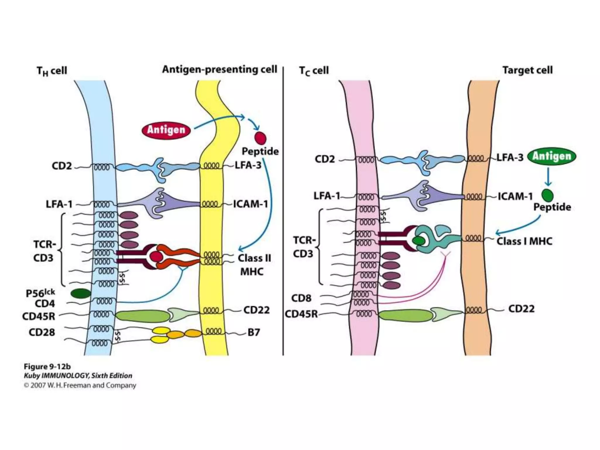
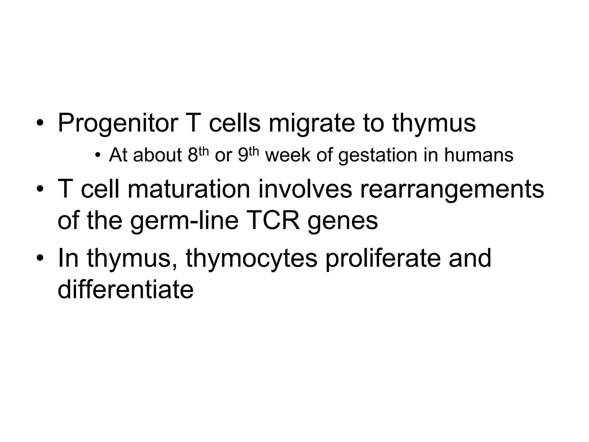
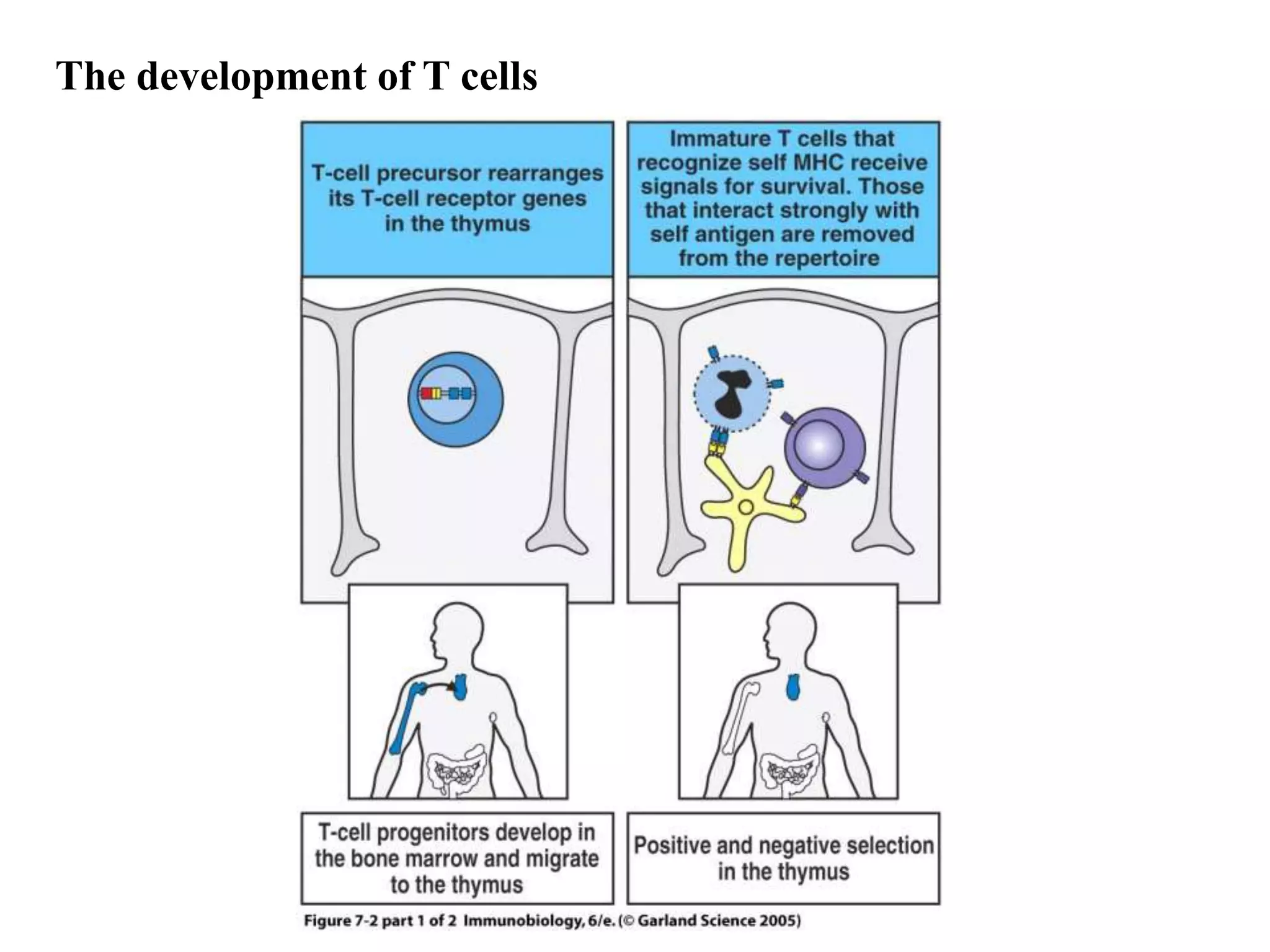
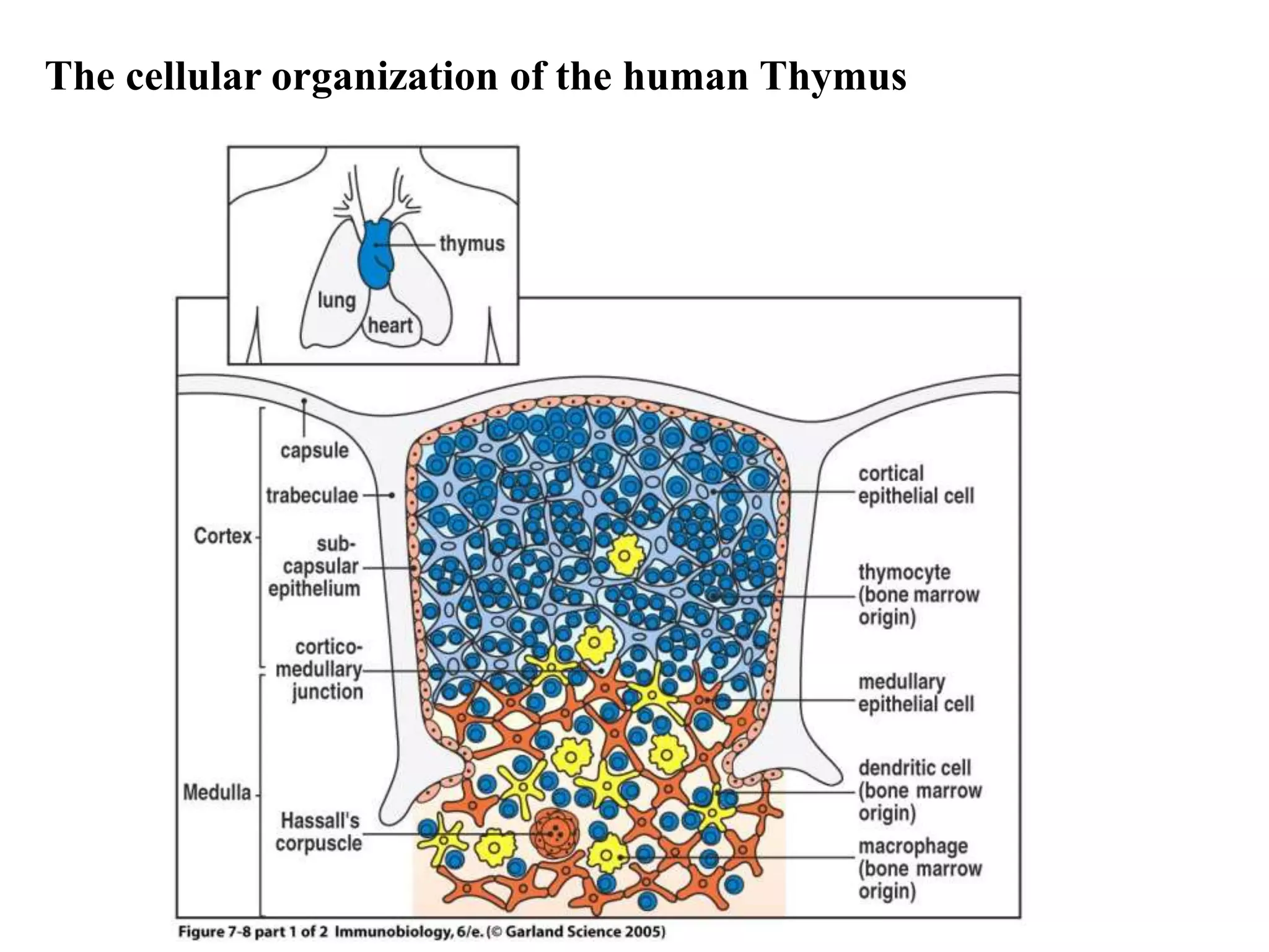
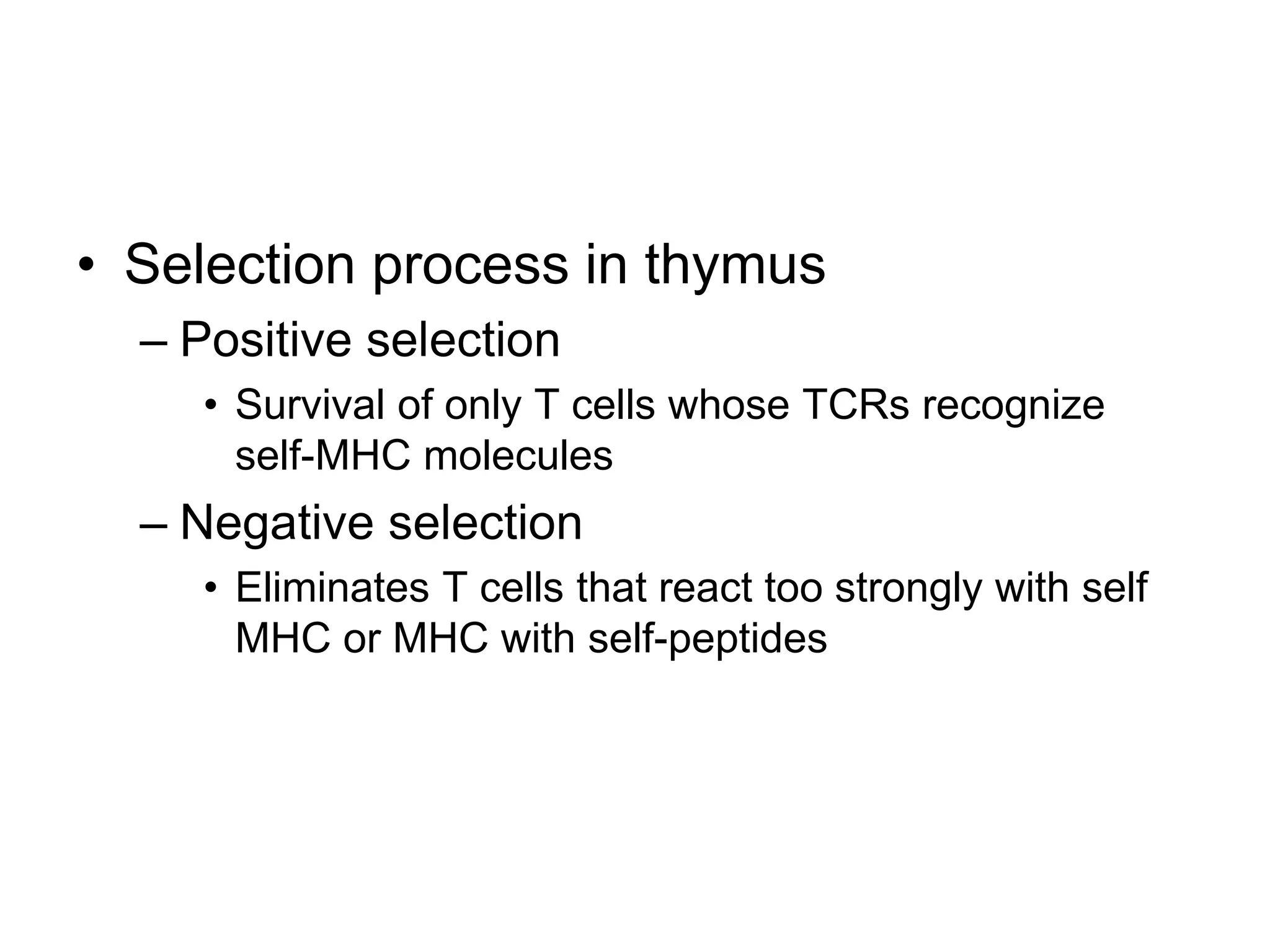
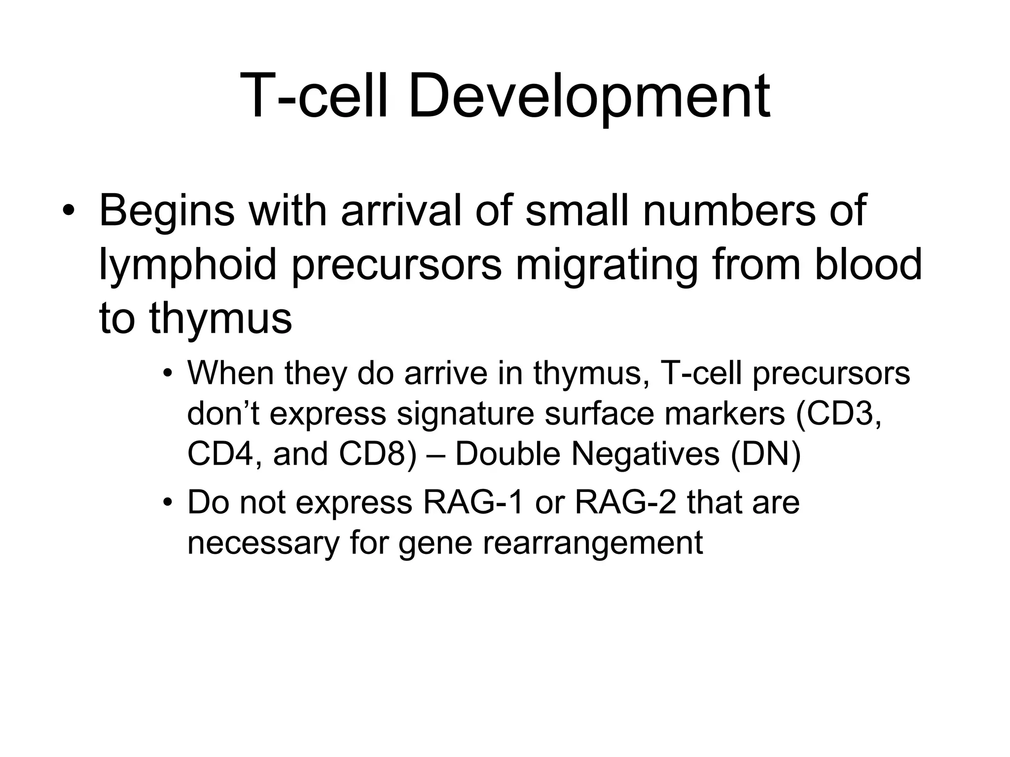
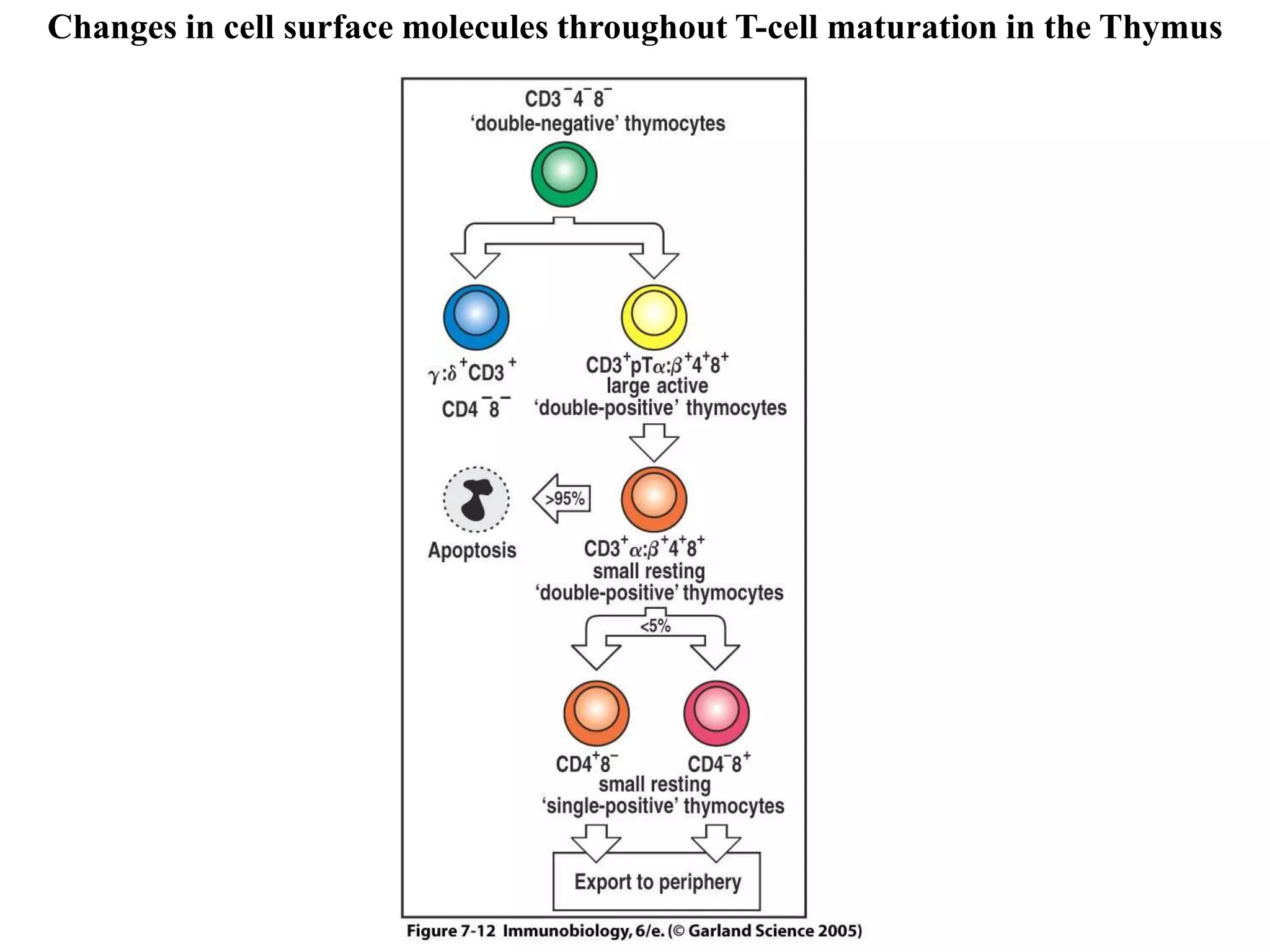
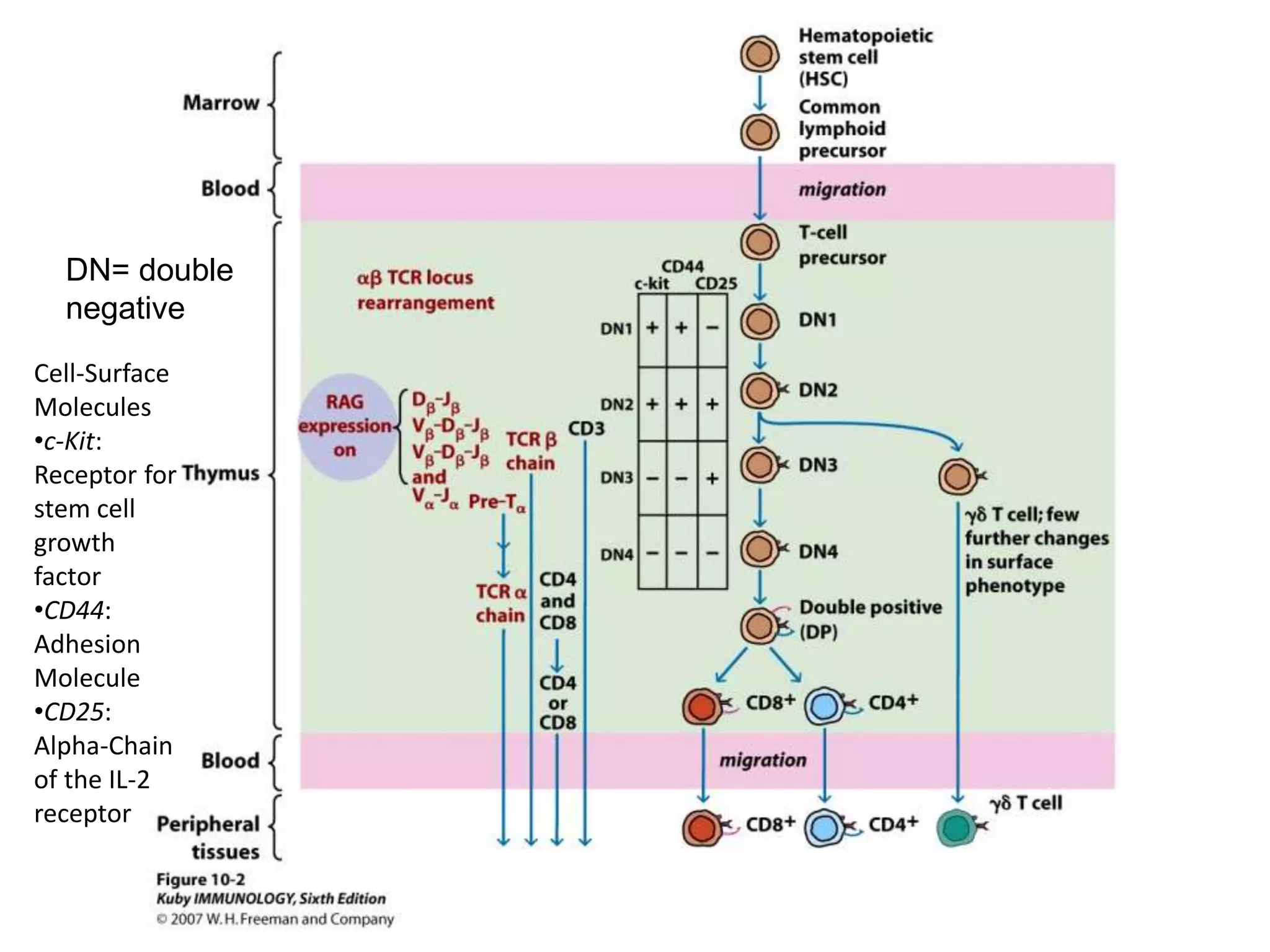
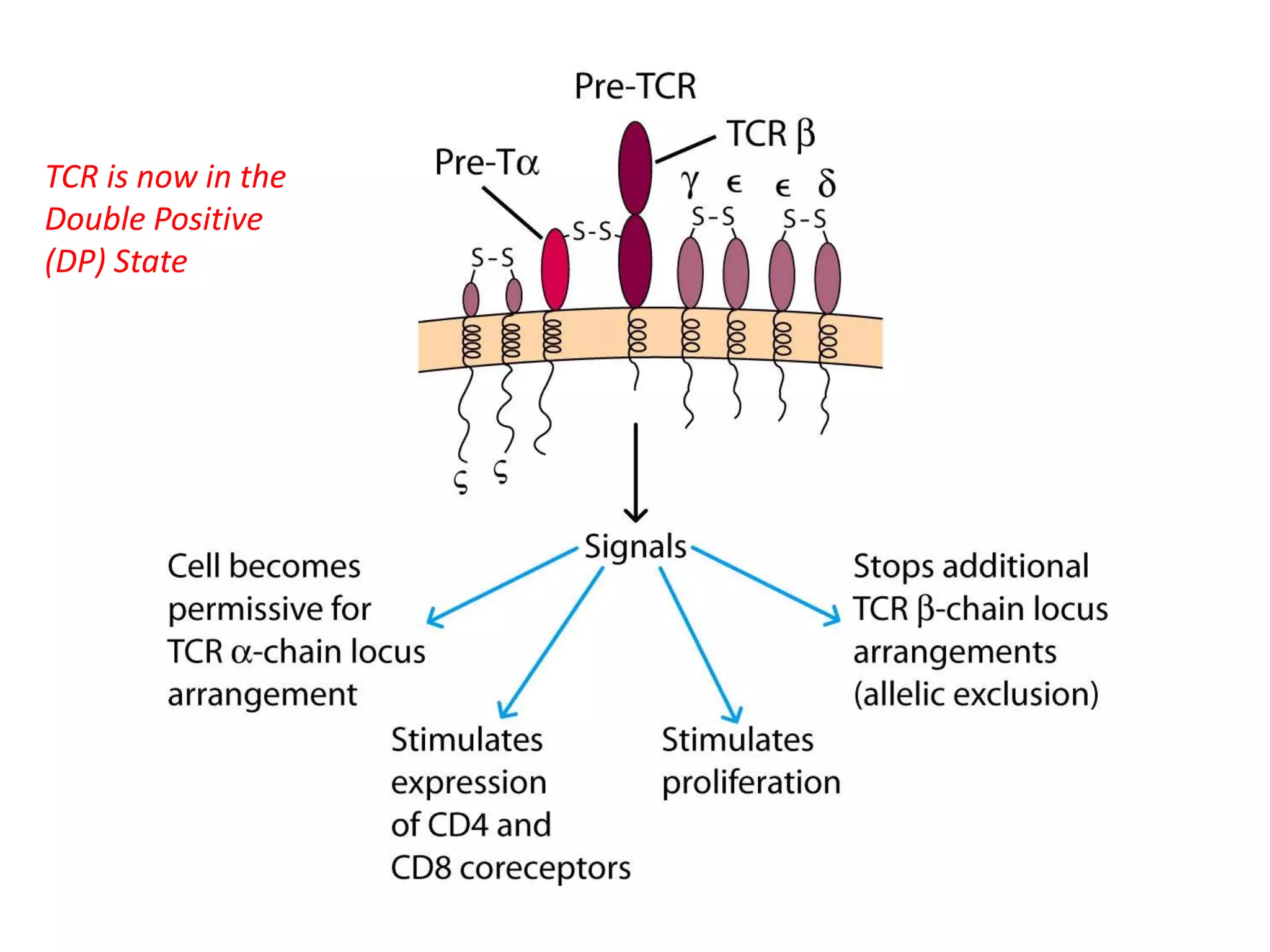
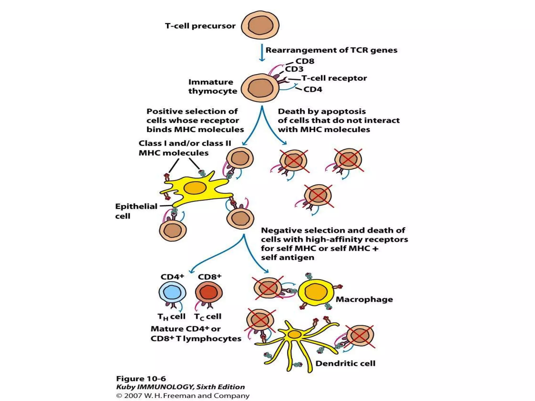
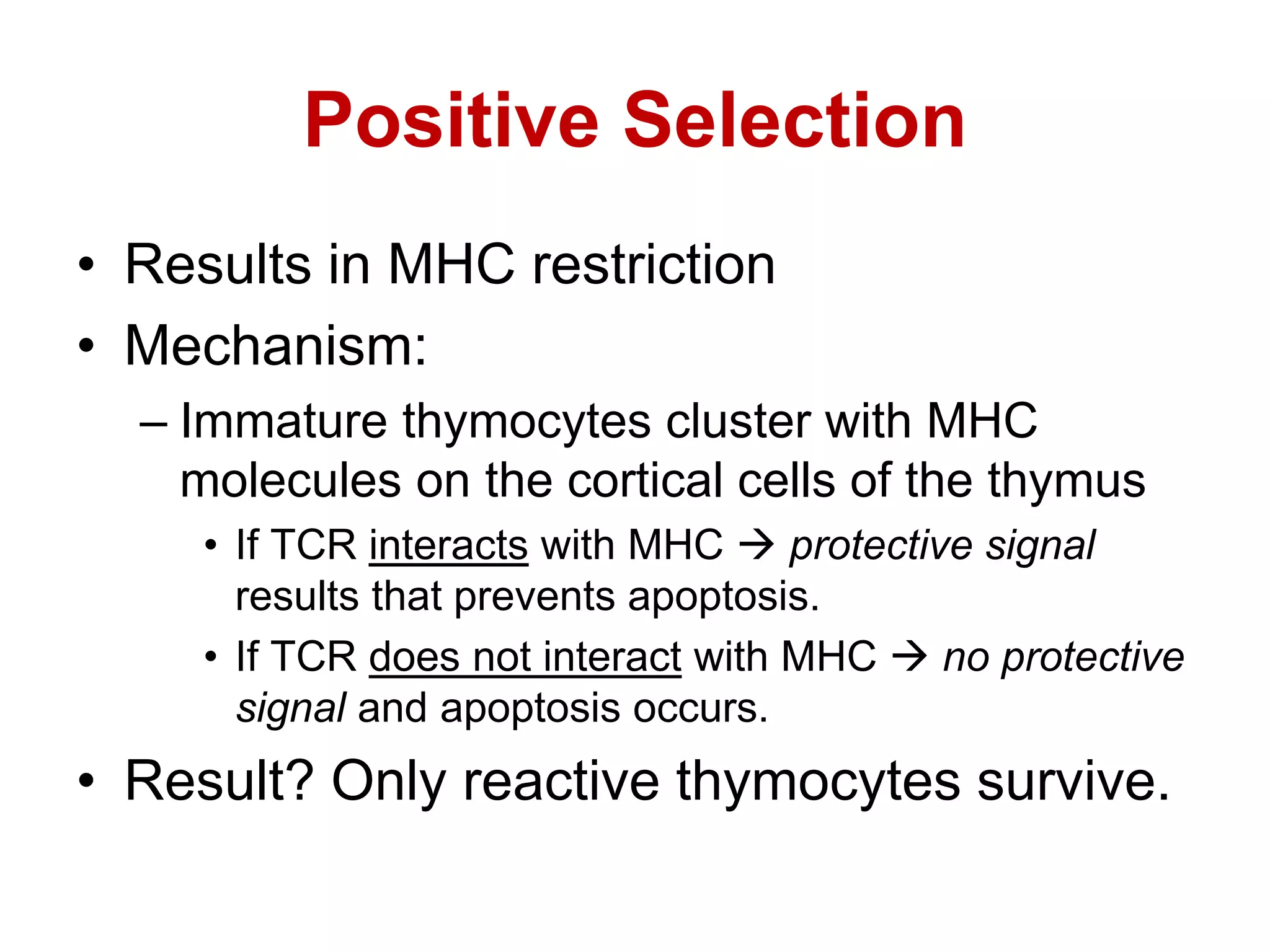
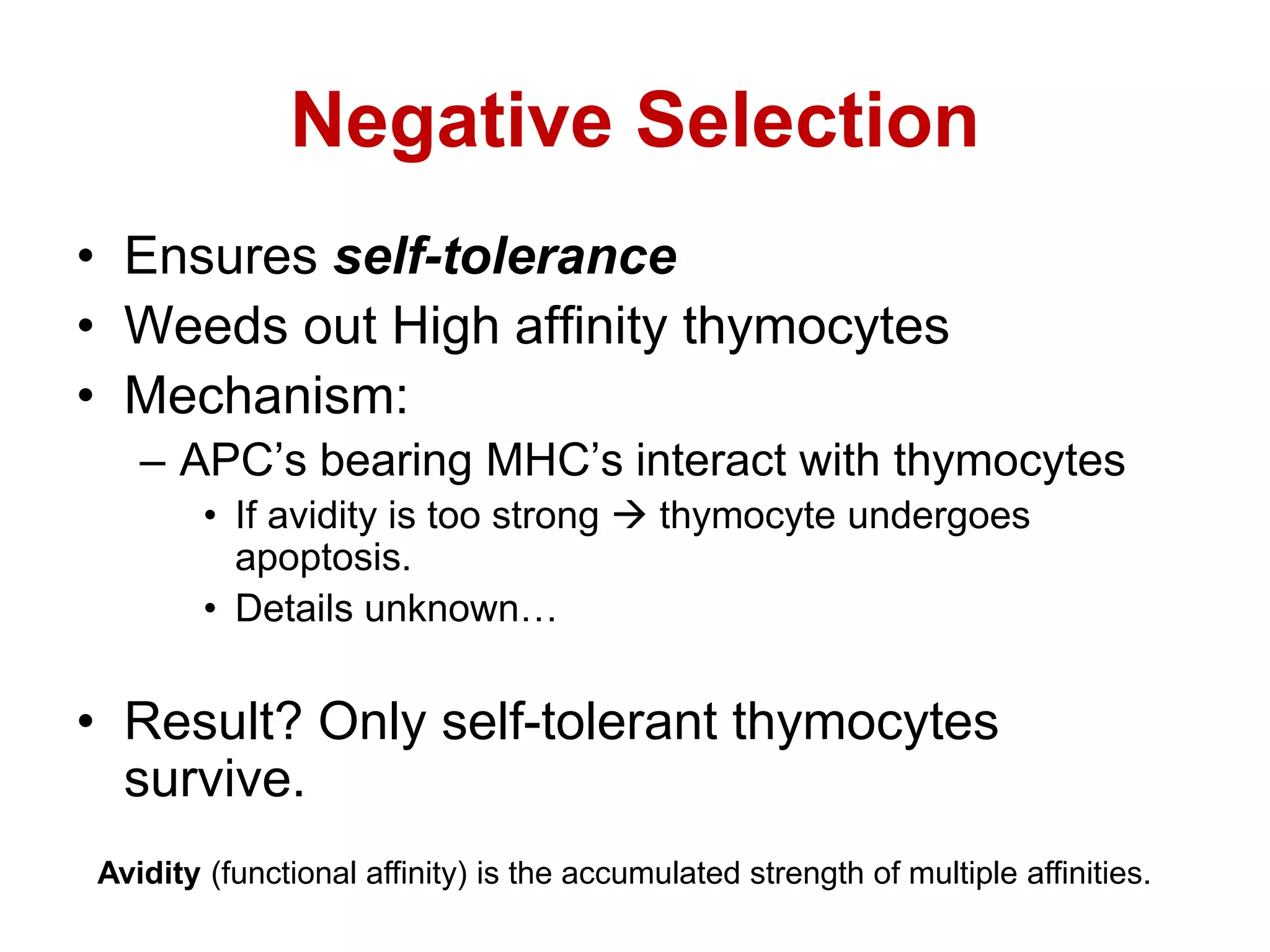
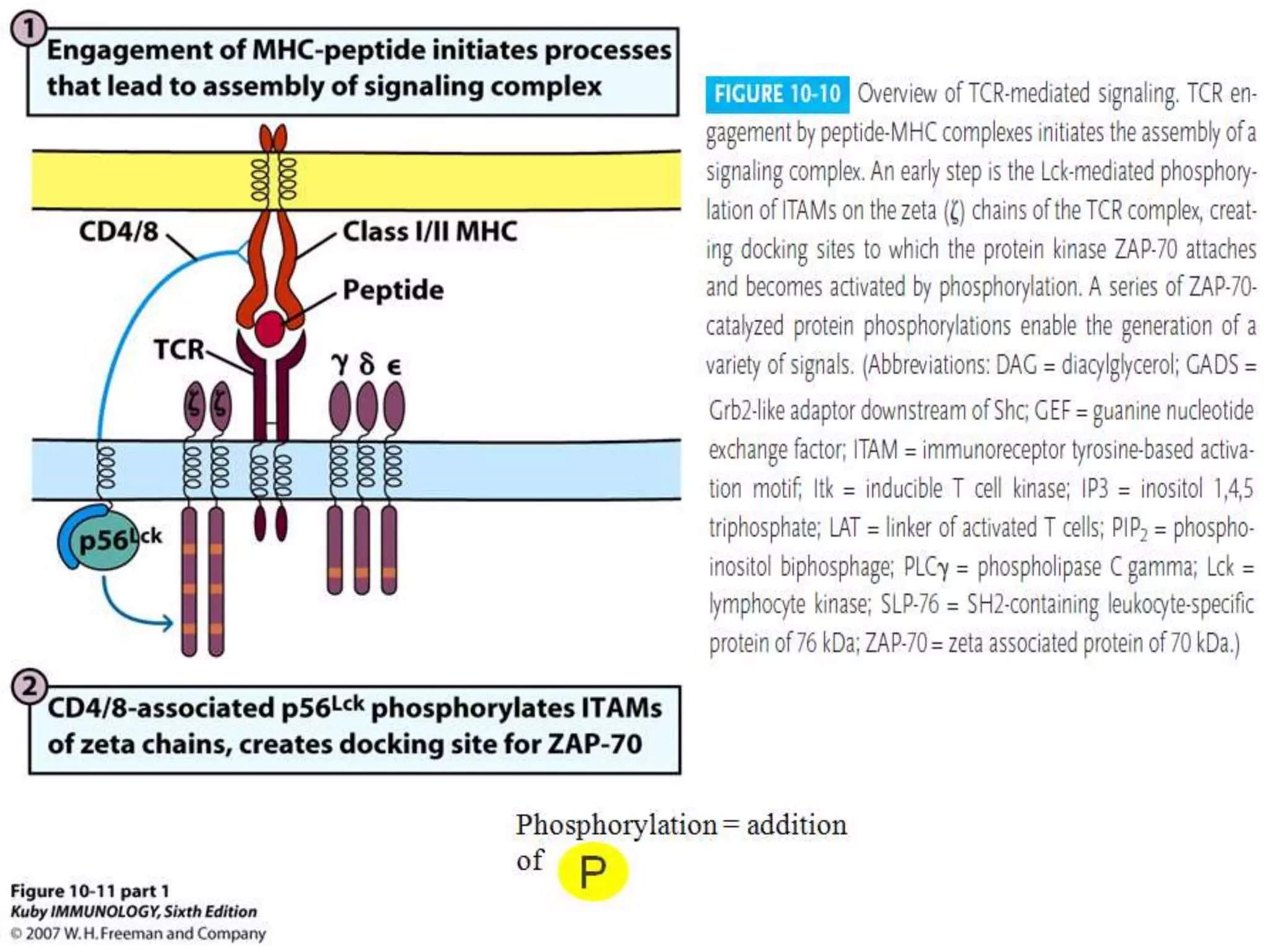
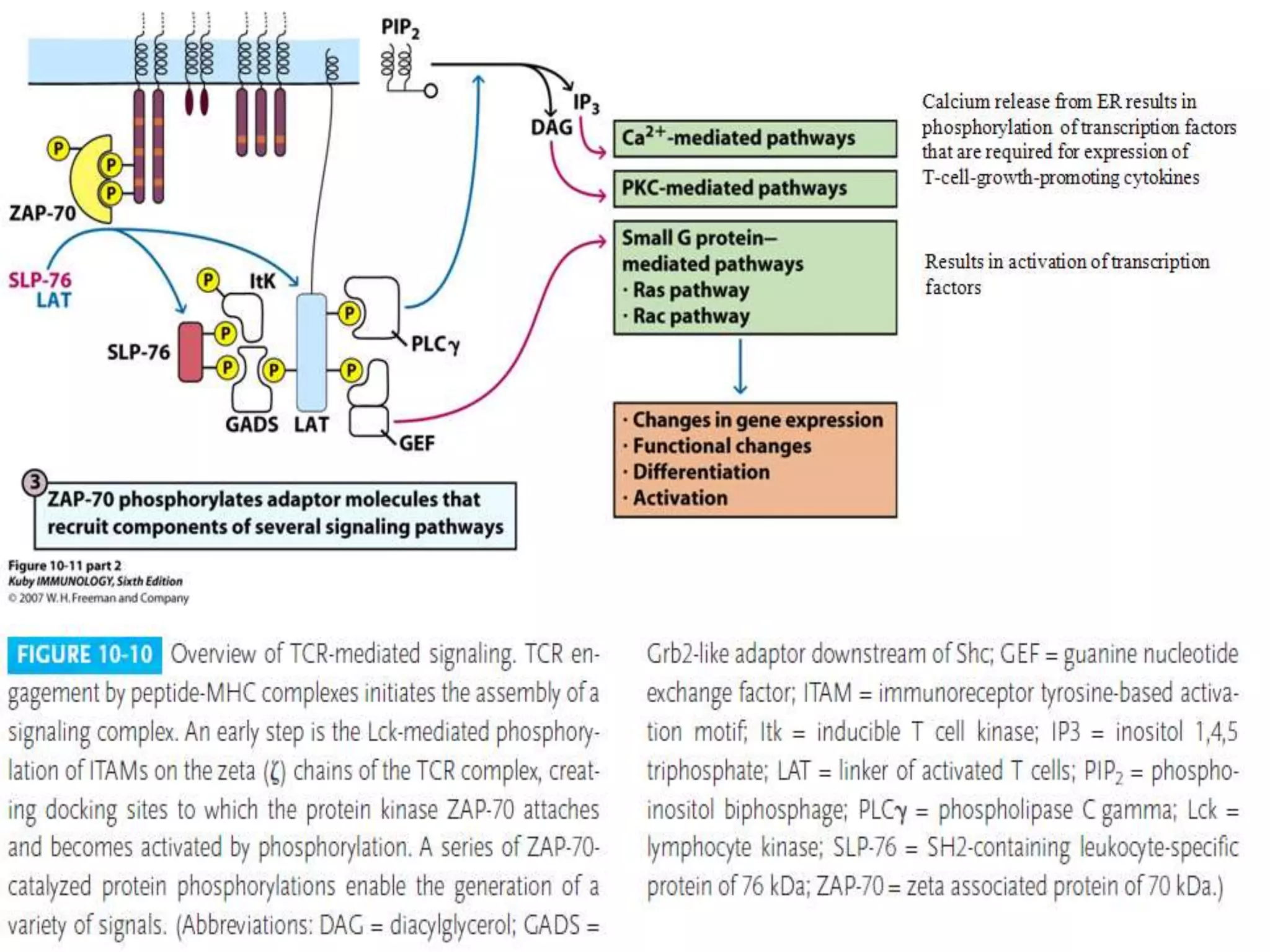

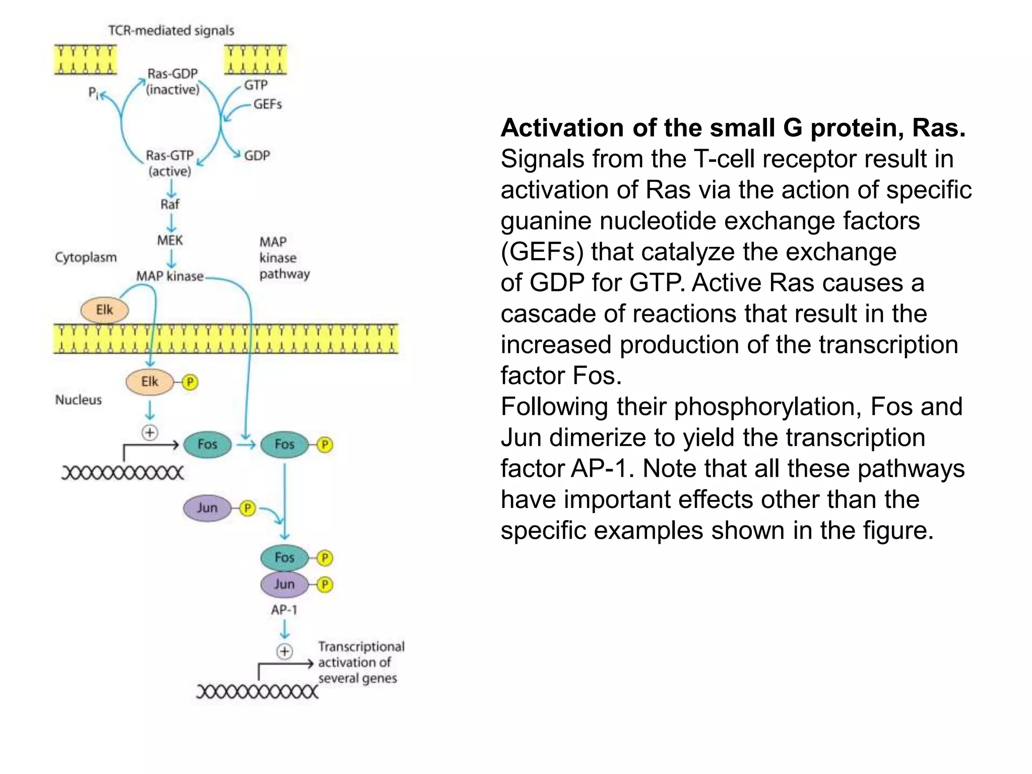

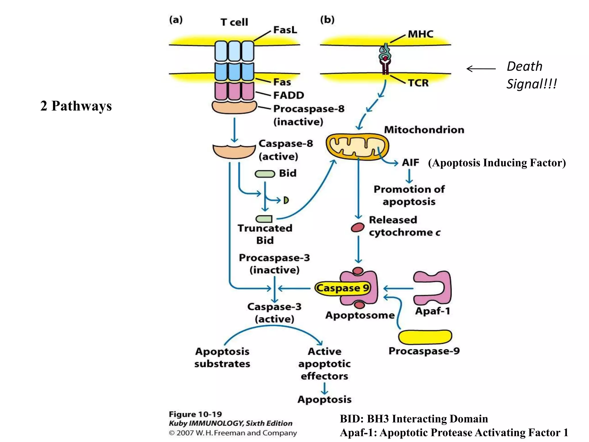

The document outlines the processes of T-cell maturation, activation, and differentiation, highlighting key stages such as early development, selection processes in the thymus, and the mechanisms of positive and negative selection. It explains the activation of T cells mediated by the TCR-CD3 complex and discusses the importance of additional signaling for full activation. The role of apoptosis in eliminating autoreactive thymocytes and maintaining T-cell populations is also emphasized.


