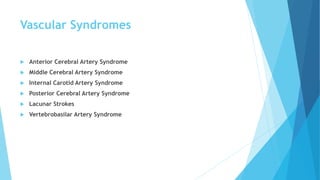
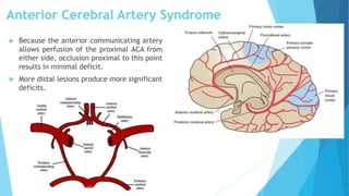
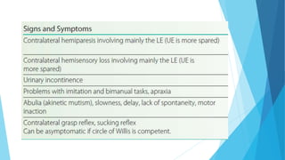
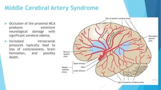
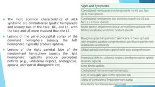
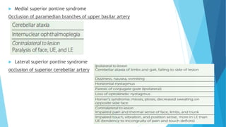
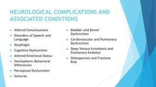
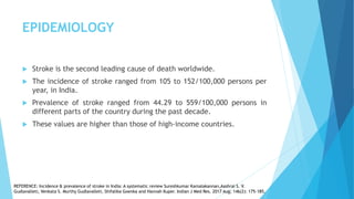
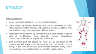
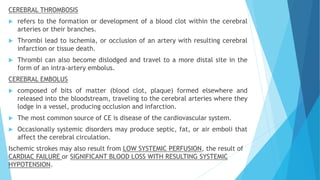
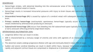
STROKE
SAYALI GUJJEWAR
MPT II
QUESTIONS
 PAPER VI
1. Clinical features of middle cerebral artery stroke. (8M)
2. Transient Ischaemic Attack. (8M)
3. Describe the blood supply of the brain and explain the investigations done for stroke. (20M)
4. Describe the clinical effects of anterior cerebral artery insufficiency. Add a note on manifestations of higher
functions. (20M)
5. List the clinical manifestations of Haemorrhagic stroke, mentioning the structures involved, resultant signs
and symptoms. Add a note on Diagnostic procedures for the same. (20M)
6. Elaborate the clinical manifestations of posterior cerebral artery infarction. Relate them to the structures
involved. (20M)
7. List the clinical manifestations of infarction of middle cerebral artery mentioning the structures involved and
resultant signs & symptoms. Add a note on diagnostic procedures for the same. (20M)
8. Discuss acute medical management of cerebro vascular accidents. (8M)
DEFINITION
 Stroke (cerebrovascular accident [CVA]) is the sudden loss of
neurological function caused by an interruption of the blood flow to
the brain.
REFERENCE: Physical rehabilitation / [edited by] Susan B. O’Sullivan, homas J. Schmitz, George D. Fulk. — 6th ed.
TYPES
 Ischemic stroke is the most common type, affecting about 80% of
individuals with stroke, and results when a clot blocks or impairs
blood flow, depriving the brain of essential oxygen and nutrients.
 Hemorrhagic stroke occurs when blood vessels rupture, causing
leakage of blood in or around the brain.
REFERENCE: Physical rehabilitation / [edited by] Susan B. O’Sullivan, homas J. Schmitz, George D. Fulk. — 6th ed.
EPIDEMIOLOGY
 Stroke is the second leading cause of death worldwide.
 The incidence of stroke ranged from 105 to 152/100,000 persons per
year, in India.
 Prevalence of stroke ranged from 44.29 to 559/100,000 persons in
different parts of the country during the past decade.
 These values are higher than those of high-income countries.
REFERENCE: Incidence & prevalence of stroke in India: A systematic review Sureshkumar Kamalakannan,Aashrai S. V.
Gudlavalleti, Venkata S. Murthy Gudlavalleti, Shifalika Goenka and Hannah Kuper. Indian J Med Res. 2017 Aug; 146(2): 175–185.
ETIOLOGY
ATHEROSCLEROSIS
 major contributory factor in cerebrovascular disease.
 characterized by plaque formation with an accumulation of lipids,
fibrin, complex carbohydrates, and calcium deposits on arterial walls
that leads to progressive narrowing of blood vessels.
 Interruption of blood flow by atherosclerotic plaques occurs at certain
sites of predilection, these generally include bifurcations,
constrictions, dilations, or angulations of arteries.
 most common sites for lesions to occur are at the origin of the
common carotid artery or at its transition into the middle cerebral
artery, at the main bifurcation of the middle cerebral artery, and at
the junction of the vertebral arteries with the basilar artery.
CEREBRAL THROMBOSIS
 refers to the formation or development of a blood clot within the cerebral
arteries or their branches.
 Thrombi lead to ischemia, or occlusion of an artery with resulting cerebral
infarction or tissue death.
 Thrombi can also become dislodged and travel to a more distal site in the
form of an intra-artery embolus.
CEREBRAL EMBOLUS
 composed of bits of matter (blood clot, plaque) formed elsewhere and
released into the bloodstream, traveling to the cerebral arteries where they
lodge in a vessel, producing occlusion and infarction.
 The most common source of CE is disease of the cardiovascular system.
 Occasionally systemic disorders may produce septic, fat, or air emboli that
affect the cerebral circulation.
Ischemic strokes may also result from LOW SYSTEMIC PERFUSION, the result of
CARDIAC FAILURE or SIGNIFICANT BLOOD LOSS WITH RESULTING SYSTEMIC
HYPOTENSION.
HEMORRHAGE
 Hemorrhagic strokes, with abnormal bleeding into the extravascular areas of the brain, are the result of
rupture of a cerebral vessel or trauma.
 Hemorrhage results in increased intracranial pressures with injury to brain tissues and restriction of distal
blood flow.
 Intracerebral hemorrhage (IH) is caused by rupture of a cerebral vessel with subsequent bleeding into the
brain.
 Primary cerebral hemorrhage (nontraumatic spontaneous hemorrhage) typically occurs in small blood
vessels weakened by atherosclerosis producing an aneurysm.
 Subarachnoid hemorrhage (SH) occurs from bleeding into the subarachnoid space typically from a saccular
or berry aneurysm affecting primarily large blood vessels.
ARTERIOVENOUS MALFORMATION (AVM)
 congenital defect that can result in stroke.
 AVM is characterized by a tortuous tangle of arteries and veins with agenesis of an interposing capillary
system.
 The abnormal vessels undergo progressive dilation with age and eventually bleed in about 50% of cases.
 Sudden and severe cerebral bleeding can result in death within hours, because intracranial pressures rise
rapidly and adjacent cortical tissues are compressed or displaced as in brainstem herniation.
RISK FACTORS
 Hypertension
 Heart Disease (HD)
 Disorders Of Heart Rhythm
 Diabetes Mellitus (DM)
 Elevated Total Blood Cholesterol
(Hypercholesterolemia)
 Elevated Low-density Lipoprotein (LDL
[“Bad”]) Cholesterol
 Low Levels Of High-density Lipoprotein
(HDL [“Good”]) Cholesterol
 Elevated Fasting Triglyceride Level
 Marked Elevations Of Hematocrit
 Family History, Age, Gender, And Race
(African American)
 Atrial fibrillation
 End-stage Renal Disease And Chronic Kidney
Disease
 Sleep Apnea
 Women With Early Menopause (Before 42
Years Of Age)
 Pregnancy, Birth, And The First 6 Weeks
Postpartum In Older Women And African
Americans
 Preeclampsia
 cigarette smoking
 Physical inactivity
 Obesity
 Diet
 Cardiac Disorders Such As Rheumatic Heart
Valvular Disease, Endocarditis, Or Cardiac
Surgery
PATHOPHYSIOLOGY
Sudden
cessation of
cerebral blood
flow
Oxygen glucose
deprivation
Neuronal death
Neuronal
activity
cessation
Infarct expands
release of excess
neurotransmitters
progressive
disturbance of energy
metabolism and
anoxic depolarization.
inability of brain cells
to produce energy,
particularly (ATP)
excess influx of
calcium ions
1. failure of the
neuronal membrane
2. stimulates the
release of nitric oxide
and cytokines
damage brain cells
ISCHEMIC CASCADE
Ischemic
strokes
cerebral
edema
elevate
intracranial
pressure
intracranial
hypertension
and neurological
deterioration
Vascular Syndromes
 Anterior Cerebral Artery Syndrome
 Middle Cerebral Artery Syndrome
 Internal Carotid Artery Syndrome
 Posterior Cerebral Artery Syndrome
 Lacunar Strokes
 Vertebrobasilar Artery Syndrome
Anterior Cerebral Artery Syndrome
 Because the anterior communicating artery
allows perfusion of the proximal ACA from
either side, occlusion proximal to this point
results in minimal deficit.
 More distal lesions produce more significant
deficits.
Middle Cerebral Artery Syndrome
 Occlusion of the proximal MCA
produces extensive
neurological damage with
significant cerebral edema.
 Increased intracranial
pressures typically lead to
loss of consciousness, brain
herniation, and possibly
death.
 The most common characteristics of MCA
syndrome are contralateral spastic hemiparesis
and sensory loss of the face, UE, and LE, with
the face and UE more involved than the LE.
 Lesions of the parieto-occipital cortex of the
dominant hemisphere (usually the left
hemisphere) typically produce aphasia.
 Lesions of the right parietal lobe of the
nondominant hemisphere (usually the right
hemisphere) typically produce perceptual
deficits (e.g., unilateral neglect, anosognosia,
apraxia, and spatial disorganization).
Posterior Cerebral Artery Syndrome
 Occlusion proximal to the posterior
communicating artery typically results in
minimal deficits owing to the collateral
blood supply from the posterior
communicating artery.
 Occlusion of thalamic branches
may produce hemianesthesia
(contralateral sensory loss) or
central post-stroke (thalamic)
pain.
 Occipital infarction produces
homonymous hemianopsia,
visual agnosia, prosopagnosia,
or, if bilateral, cortical
blindness.
 Temporal lobe ischemia results
in amnesia (memory loss).
 Contralateral hemiplegia occurs
with involvement of the
cerebral peduncle.
Internal Carotid Artery Syndrome
 Occlusion of the internal carotid artery (ICA) typically
produces massive infarction in the region of the brain
supplied by the middle cerebral artery.
 The ICA supplies both the MCA and the ACA.
 If collateral circulation to the ACA from the circle of
Willis is absent, extensive cerebral infarction in the areas
of both the ACA and MCA can occur.
 Significant edema is common with possible uncal
herniation, coma, and death (mass effect).
Lacunar Strokes
 Lacunar strokes are caused by small vessel disease deep in the
cerebral white matter (penetrating artery disease).
 They are strongly associated with hypertensive hemorrhage
and diabetic microvascular disease.
 Lacunar syndromes are consistent with specific anatomical
sites.
 Pure motor lacunar stroke is associated with involvement of
the posterior limb of the internal capsule, pons, and pyramids.
 Pure sensory lacunar stroke is associated with involvement of
the ventrolateral thalamus or thalamocortical projections.
 Dysarthria/clumsy hand syndrome (involving the base of the pons,
genu of anterior limb, or the internal capsule)
 Ataxic hemiparesis (involving the pons, genu of internal capsule,
corona radiata, or cerebellum)
 Sensory/motor stroke (involving the junction of the internal capsule
and thalamus), or
 Dystonia/involuntary movements (choreoathetosis with lacunar
infarction of the putamen or globus pallidus; hemiballismus with
involvement of thesubthalamic nucleus).
 Deficits in consciousness, language, or visual fields are not seen in
lacunar strokes because the higher cortical areas are preserved.
Vertebrobasilar Artery Syndrome
 Medial Medullary Syndrome
Occlusion of vertebral artery, medullary branch
 Lateral Medullary (Wallenburg’s) Syndrome
Occlusion of posterior inferior cerebellar artery or vertebral artery
 Complete basilar artery syndrome (locked-in syndrome)
Occlusion of Basilar artery, ventral pons
 Medial inferior pontine syndrome
Occlusion of paramedian branch of basilar artery
 Lateral inferior pontine syndrome
Occlusion of anterior inferior cerebellar artery, a branch of the basilar
artery
 Medial midpontine syndrome
Occlusion of paramedian branch of the mid-basilar artery
 Lateral midpontine syndrome
Occlusion of short circumferential artery
 Medial superior pontine syndrome
Occlusion of paramedian branches of upper basilar artery
 Lateral superior pontine syndrome
occlusion of superior cerebellar artery
NEUROLOGICAL COMPLICATIONS AND
ASSOCIATED CONDITIONS
 Altered Consciousness
 Disorders of Speech and
Language
 Dysphagia
 Cognitive Dysfunction
 Altered Emotional Status
 Hemispheric Behavioral
Differences
 Perceptual Dysfunction
 Seizures
 Bladder and Bowel
Dysfunction
 Cardiovascular and Pulmonary
Dysfunction
 Deep Venous hrombosis and
Pulmonary Embolus
 Osteoporosis and Fracture
Risk
 ALTERED CONSCIOUSNESS
 DISORDERS OF SPEECH AND LANGUAGE
o lesions involving the cortex of the dominant hemisphere (typically the left
hemisphere) demonstrate speech and language impairments.
o Aphasia is the general term used to describe an acquired communication
disorder caused by brain damage and is characterized by an impairment of
language comprehension, formulation, and use.
FLUENT APHASIA
NONFLUENT APHASIA
GLOBAL APHASIA
o Dysarthria refers to a category of motor speech disorders caused by
lesions in parts of the central or peripheral nervous system that
mediate speech production.
Stroke

Stroke

  • 1.
  • 2.
    QUESTIONS  PAPER VI 1.Clinical features of middle cerebral artery stroke. (8M) 2. Transient Ischaemic Attack. (8M) 3. Describe the blood supply of the brain and explain the investigations done for stroke. (20M) 4. Describe the clinical effects of anterior cerebral artery insufficiency. Add a note on manifestations of higher functions. (20M) 5. List the clinical manifestations of Haemorrhagic stroke, mentioning the structures involved, resultant signs and symptoms. Add a note on Diagnostic procedures for the same. (20M) 6. Elaborate the clinical manifestations of posterior cerebral artery infarction. Relate them to the structures involved. (20M) 7. List the clinical manifestations of infarction of middle cerebral artery mentioning the structures involved and resultant signs & symptoms. Add a note on diagnostic procedures for the same. (20M) 8. Discuss acute medical management of cerebro vascular accidents. (8M)
  • 3.
    DEFINITION  Stroke (cerebrovascularaccident [CVA]) is the sudden loss of neurological function caused by an interruption of the blood flow to the brain. REFERENCE: Physical rehabilitation / [edited by] Susan B. O’Sullivan, homas J. Schmitz, George D. Fulk. — 6th ed.
  • 4.
    TYPES  Ischemic strokeis the most common type, affecting about 80% of individuals with stroke, and results when a clot blocks or impairs blood flow, depriving the brain of essential oxygen and nutrients.  Hemorrhagic stroke occurs when blood vessels rupture, causing leakage of blood in or around the brain. REFERENCE: Physical rehabilitation / [edited by] Susan B. O’Sullivan, homas J. Schmitz, George D. Fulk. — 6th ed.
  • 5.
    EPIDEMIOLOGY  Stroke isthe second leading cause of death worldwide.  The incidence of stroke ranged from 105 to 152/100,000 persons per year, in India.  Prevalence of stroke ranged from 44.29 to 559/100,000 persons in different parts of the country during the past decade.  These values are higher than those of high-income countries. REFERENCE: Incidence & prevalence of stroke in India: A systematic review Sureshkumar Kamalakannan,Aashrai S. V. Gudlavalleti, Venkata S. Murthy Gudlavalleti, Shifalika Goenka and Hannah Kuper. Indian J Med Res. 2017 Aug; 146(2): 175–185.
  • 6.
    ETIOLOGY ATHEROSCLEROSIS  major contributoryfactor in cerebrovascular disease.  characterized by plaque formation with an accumulation of lipids, fibrin, complex carbohydrates, and calcium deposits on arterial walls that leads to progressive narrowing of blood vessels.  Interruption of blood flow by atherosclerotic plaques occurs at certain sites of predilection, these generally include bifurcations, constrictions, dilations, or angulations of arteries.  most common sites for lesions to occur are at the origin of the common carotid artery or at its transition into the middle cerebral artery, at the main bifurcation of the middle cerebral artery, and at the junction of the vertebral arteries with the basilar artery.
  • 7.
    CEREBRAL THROMBOSIS  refersto the formation or development of a blood clot within the cerebral arteries or their branches.  Thrombi lead to ischemia, or occlusion of an artery with resulting cerebral infarction or tissue death.  Thrombi can also become dislodged and travel to a more distal site in the form of an intra-artery embolus. CEREBRAL EMBOLUS  composed of bits of matter (blood clot, plaque) formed elsewhere and released into the bloodstream, traveling to the cerebral arteries where they lodge in a vessel, producing occlusion and infarction.  The most common source of CE is disease of the cardiovascular system.  Occasionally systemic disorders may produce septic, fat, or air emboli that affect the cerebral circulation. Ischemic strokes may also result from LOW SYSTEMIC PERFUSION, the result of CARDIAC FAILURE or SIGNIFICANT BLOOD LOSS WITH RESULTING SYSTEMIC HYPOTENSION.
  • 8.
    HEMORRHAGE  Hemorrhagic strokes,with abnormal bleeding into the extravascular areas of the brain, are the result of rupture of a cerebral vessel or trauma.  Hemorrhage results in increased intracranial pressures with injury to brain tissues and restriction of distal blood flow.  Intracerebral hemorrhage (IH) is caused by rupture of a cerebral vessel with subsequent bleeding into the brain.  Primary cerebral hemorrhage (nontraumatic spontaneous hemorrhage) typically occurs in small blood vessels weakened by atherosclerosis producing an aneurysm.  Subarachnoid hemorrhage (SH) occurs from bleeding into the subarachnoid space typically from a saccular or berry aneurysm affecting primarily large blood vessels. ARTERIOVENOUS MALFORMATION (AVM)  congenital defect that can result in stroke.  AVM is characterized by a tortuous tangle of arteries and veins with agenesis of an interposing capillary system.  The abnormal vessels undergo progressive dilation with age and eventually bleed in about 50% of cases.  Sudden and severe cerebral bleeding can result in death within hours, because intracranial pressures rise rapidly and adjacent cortical tissues are compressed or displaced as in brainstem herniation.
  • 9.
    RISK FACTORS  Hypertension Heart Disease (HD)  Disorders Of Heart Rhythm  Diabetes Mellitus (DM)  Elevated Total Blood Cholesterol (Hypercholesterolemia)  Elevated Low-density Lipoprotein (LDL [“Bad”]) Cholesterol  Low Levels Of High-density Lipoprotein (HDL [“Good”]) Cholesterol  Elevated Fasting Triglyceride Level  Marked Elevations Of Hematocrit  Family History, Age, Gender, And Race (African American)  Atrial fibrillation  End-stage Renal Disease And Chronic Kidney Disease  Sleep Apnea  Women With Early Menopause (Before 42 Years Of Age)  Pregnancy, Birth, And The First 6 Weeks Postpartum In Older Women And African Americans  Preeclampsia  cigarette smoking  Physical inactivity  Obesity  Diet  Cardiac Disorders Such As Rheumatic Heart Valvular Disease, Endocarditis, Or Cardiac Surgery
  • 10.
    PATHOPHYSIOLOGY Sudden cessation of cerebral blood flow Oxygenglucose deprivation Neuronal death Neuronal activity cessation Infarct expands
  • 11.
    release of excess neurotransmitters progressive disturbanceof energy metabolism and anoxic depolarization. inability of brain cells to produce energy, particularly (ATP) excess influx of calcium ions 1. failure of the neuronal membrane 2. stimulates the release of nitric oxide and cytokines damage brain cells ISCHEMIC CASCADE
  • 12.
  • 13.
    Vascular Syndromes  AnteriorCerebral Artery Syndrome  Middle Cerebral Artery Syndrome  Internal Carotid Artery Syndrome  Posterior Cerebral Artery Syndrome  Lacunar Strokes  Vertebrobasilar Artery Syndrome
  • 14.
    Anterior Cerebral ArterySyndrome  Because the anterior communicating artery allows perfusion of the proximal ACA from either side, occlusion proximal to this point results in minimal deficit.  More distal lesions produce more significant deficits.
  • 16.
    Middle Cerebral ArterySyndrome  Occlusion of the proximal MCA produces extensive neurological damage with significant cerebral edema.  Increased intracranial pressures typically lead to loss of consciousness, brain herniation, and possibly death.
  • 17.
     The mostcommon characteristics of MCA syndrome are contralateral spastic hemiparesis and sensory loss of the face, UE, and LE, with the face and UE more involved than the LE.  Lesions of the parieto-occipital cortex of the dominant hemisphere (usually the left hemisphere) typically produce aphasia.  Lesions of the right parietal lobe of the nondominant hemisphere (usually the right hemisphere) typically produce perceptual deficits (e.g., unilateral neglect, anosognosia, apraxia, and spatial disorganization).
  • 18.
    Posterior Cerebral ArterySyndrome  Occlusion proximal to the posterior communicating artery typically results in minimal deficits owing to the collateral blood supply from the posterior communicating artery.
  • 19.
     Occlusion ofthalamic branches may produce hemianesthesia (contralateral sensory loss) or central post-stroke (thalamic) pain.  Occipital infarction produces homonymous hemianopsia, visual agnosia, prosopagnosia, or, if bilateral, cortical blindness.  Temporal lobe ischemia results in amnesia (memory loss).  Contralateral hemiplegia occurs with involvement of the cerebral peduncle.
  • 20.
    Internal Carotid ArterySyndrome  Occlusion of the internal carotid artery (ICA) typically produces massive infarction in the region of the brain supplied by the middle cerebral artery.  The ICA supplies both the MCA and the ACA.  If collateral circulation to the ACA from the circle of Willis is absent, extensive cerebral infarction in the areas of both the ACA and MCA can occur.  Significant edema is common with possible uncal herniation, coma, and death (mass effect).
  • 21.
    Lacunar Strokes  Lacunarstrokes are caused by small vessel disease deep in the cerebral white matter (penetrating artery disease).  They are strongly associated with hypertensive hemorrhage and diabetic microvascular disease.  Lacunar syndromes are consistent with specific anatomical sites.  Pure motor lacunar stroke is associated with involvement of the posterior limb of the internal capsule, pons, and pyramids.  Pure sensory lacunar stroke is associated with involvement of the ventrolateral thalamus or thalamocortical projections.
  • 22.
     Dysarthria/clumsy handsyndrome (involving the base of the pons, genu of anterior limb, or the internal capsule)  Ataxic hemiparesis (involving the pons, genu of internal capsule, corona radiata, or cerebellum)  Sensory/motor stroke (involving the junction of the internal capsule and thalamus), or  Dystonia/involuntary movements (choreoathetosis with lacunar infarction of the putamen or globus pallidus; hemiballismus with involvement of thesubthalamic nucleus).  Deficits in consciousness, language, or visual fields are not seen in lacunar strokes because the higher cortical areas are preserved.
  • 23.
    Vertebrobasilar Artery Syndrome Medial Medullary Syndrome Occlusion of vertebral artery, medullary branch
  • 24.
     Lateral Medullary(Wallenburg’s) Syndrome Occlusion of posterior inferior cerebellar artery or vertebral artery
  • 26.
     Complete basilarartery syndrome (locked-in syndrome) Occlusion of Basilar artery, ventral pons
  • 27.
     Medial inferiorpontine syndrome Occlusion of paramedian branch of basilar artery
  • 28.
     Lateral inferiorpontine syndrome Occlusion of anterior inferior cerebellar artery, a branch of the basilar artery
  • 29.
     Medial midpontinesyndrome Occlusion of paramedian branch of the mid-basilar artery  Lateral midpontine syndrome Occlusion of short circumferential artery
  • 30.
     Medial superiorpontine syndrome Occlusion of paramedian branches of upper basilar artery  Lateral superior pontine syndrome occlusion of superior cerebellar artery
  • 31.
    NEUROLOGICAL COMPLICATIONS AND ASSOCIATEDCONDITIONS  Altered Consciousness  Disorders of Speech and Language  Dysphagia  Cognitive Dysfunction  Altered Emotional Status  Hemispheric Behavioral Differences  Perceptual Dysfunction  Seizures  Bladder and Bowel Dysfunction  Cardiovascular and Pulmonary Dysfunction  Deep Venous hrombosis and Pulmonary Embolus  Osteoporosis and Fracture Risk
  • 32.
     ALTERED CONSCIOUSNESS DISORDERS OF SPEECH AND LANGUAGE o lesions involving the cortex of the dominant hemisphere (typically the left hemisphere) demonstrate speech and language impairments. o Aphasia is the general term used to describe an acquired communication disorder caused by brain damage and is characterized by an impairment of language comprehension, formulation, and use.
  • 33.
  • 34.
  • 35.
  • 36.
    o Dysarthria refersto a category of motor speech disorders caused by lesions in parts of the central or peripheral nervous system that mediate speech production.