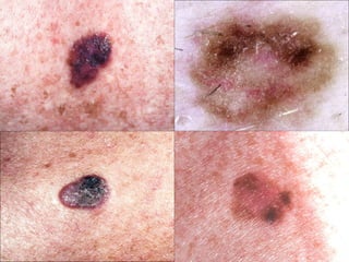
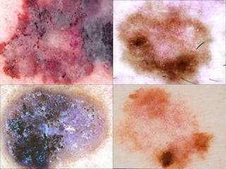
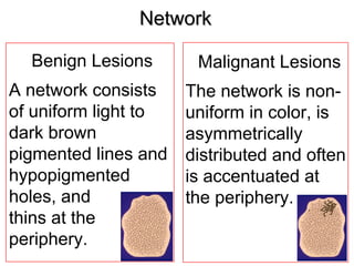
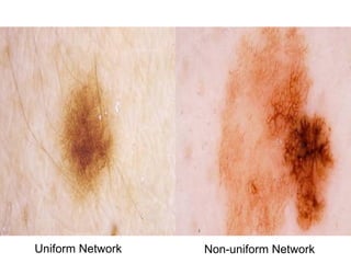
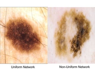
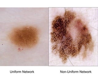
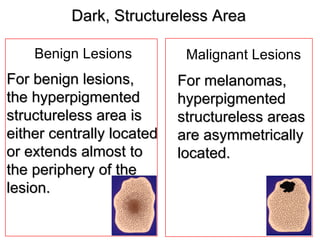
A network pattern in benign lesions consists of uniform light to dark brown pigmented lines and hypopigmented holes that thin at the periphery, while a network in malignant lesions is non-uniform in color, asymmetrically distributed, and often accentuated at the periphery. Benign lesions also have hyperpigmented structureless areas that are centrally located or extend almost to the periphery, whereas malignant lesions have asymmetrically located hyperpigmented structureless areas. Being able to recognize the appearances of different patterns is important for diagnosing melanocytic lesions.