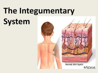
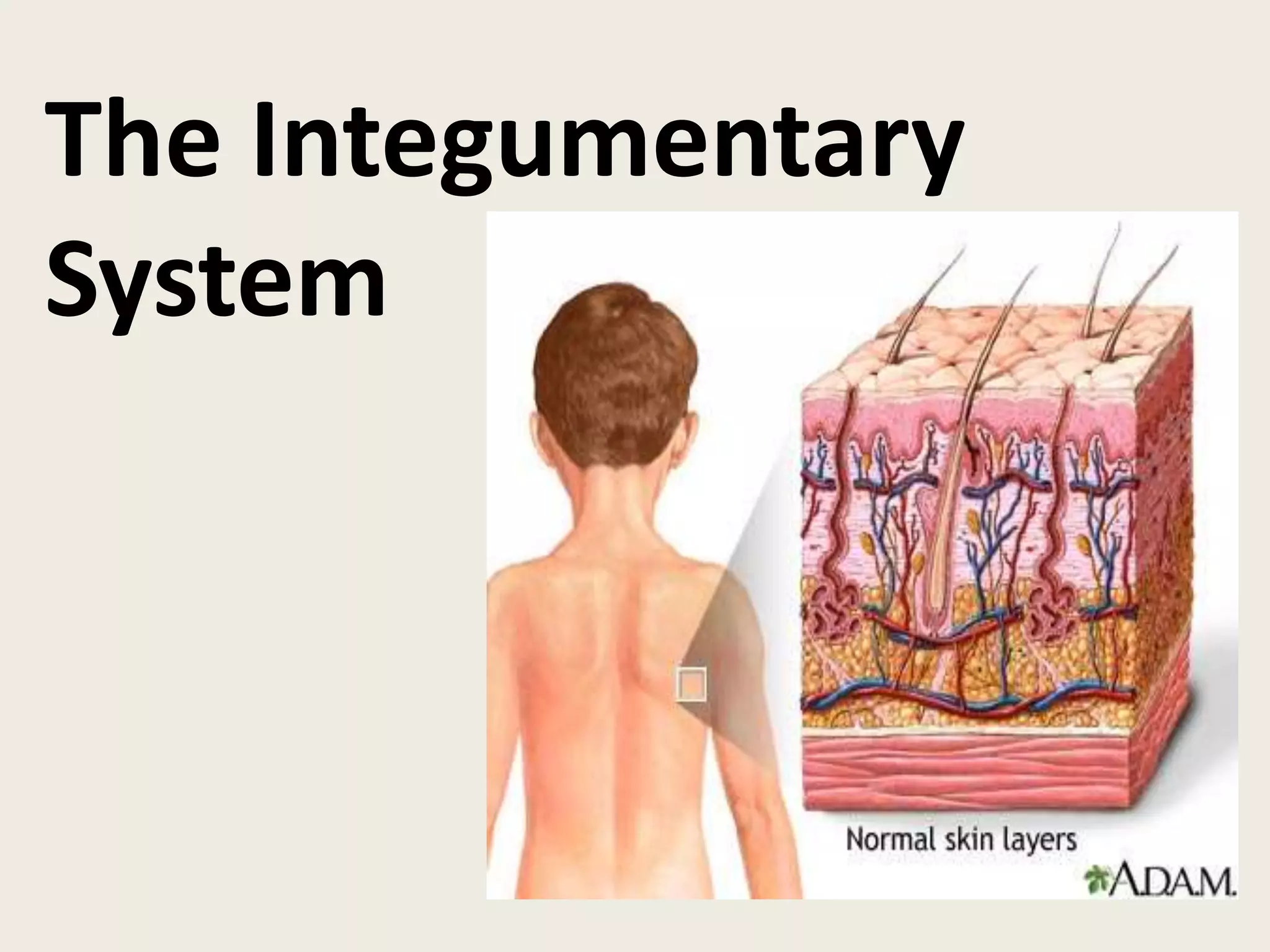
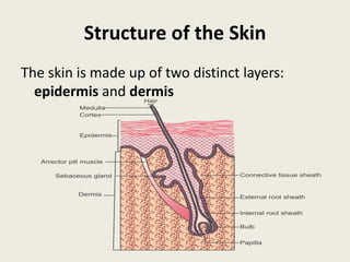
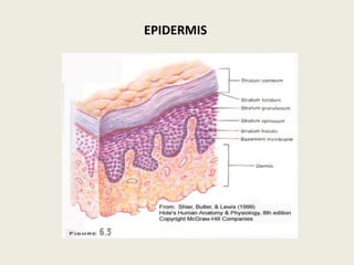
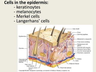
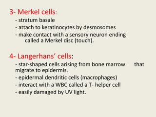
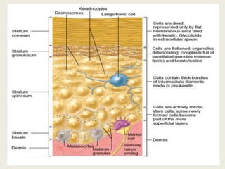
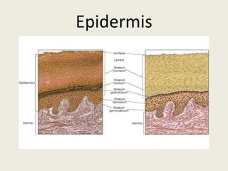
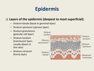
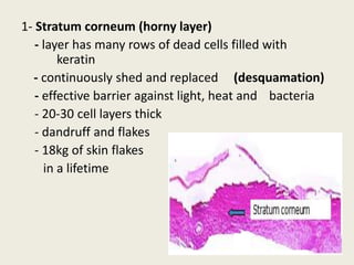
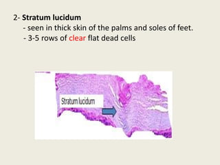
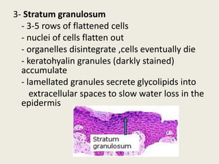
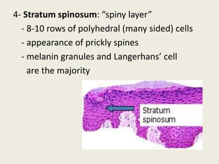
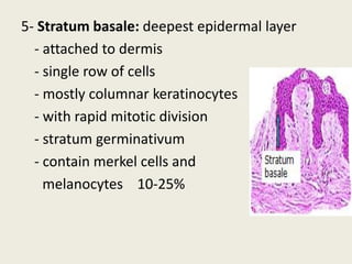
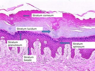
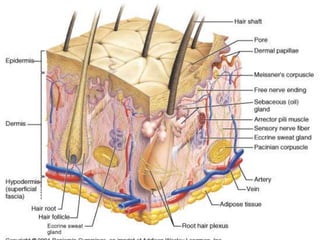
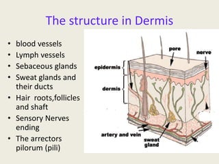
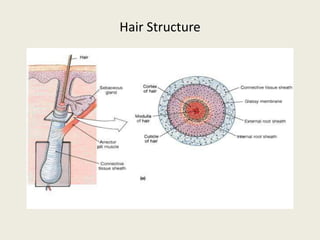
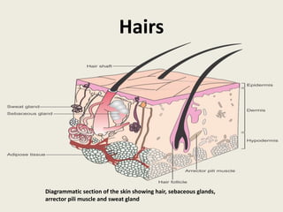
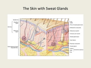
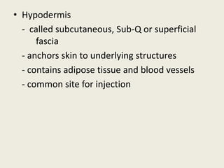
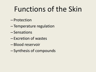
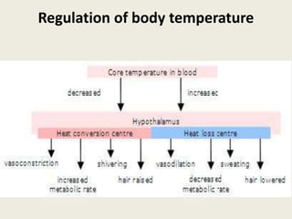
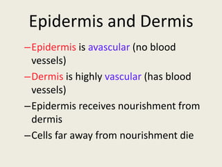
The Integumentary
System
Learning Outcomes
At the end of the lecture, the students should
be able to
1. Name the two distinct layers of the skin
2. Identify the major structures found in the
skin
3. Describe the structure and growth of hair and
nails
4. Describe the functions of the integumentary
system.
General Considerations
Definition:
– The integumentary system consists of the skin and
other structures such as glands, hairs and nails.
– Skin: the outer or external covering of the body
comprised of two parts—a superficial epidermis and a
deeper dermis
Surface area:
– Largest organ in the body with a surface area of 1.5–
2.0 m2 in an adult
– Surface area can be roughly estimated by the ‘rule of
nines’
RULE OF NINE
Structure of the Skin
The skin is made up of two distinct layers:
epidermis and dermis
Epidermis
The most superficial layer of the skin
Is avascular
Composed of keratinized stratified squamous
epithelium
Varies in thickness in different areas
Made up of 4distinct cell types and 5 layers of tissues
EPIDERMIS
Cells in the epidermis:
- keratinoytes
- melanocytes
- Merkel cells
- Langerhans’ cells
most abundant
- produce keratin (fibrous protein)
- protective; waterproofing the skin
- continuous mitosis
- form in the deepest layer called the
stratum basale
- cells push their way up to the surface
where they are dead cells filled with
keratin; will slough off. Regenerates every
25-45 days.(average 40 days)
Outer layer of dead skin cells called cuticle
1- Keratinocytes
2- Melanocytes:
- cells produce brownish/black pigment
called melanin. (8% of epidermal cells)
- stratum basale
- branching processes (dendrites)
- melanin are transported along dendrites of
the melanocytes to keratinocytes.
- melanin accumulates on the superficial
aspect of the keratinocyte shielding its
nucleus from harmful UV light.
- lack of melanin: albino
3- Merkel cells:
- stratum basale
- attach to keratinocytes by desmosomes
- make contact with a sensory neuron ending
called a Merkel disc (touch).
4- Langerhans’ cells:
- star-shaped cells arising from bone marrow that
migrate to epidermis.
- epidermal dendritic cells (macrophages)
- interact with a WBC called a T- helper cell
- easily damaged by UV light.
Epidermis
Epidermis
 Layers of the epidermis (deepest to most superficial):
– Stratum basale (basal or germinal layer)
– Stratum spinosum (spinous layer)
– Stratum granulosum
(granular cell layer)
– Stratum lucidum
(translucent layer;
usually absent in
thin skin)
– Stratum corneum
(horny layer)
1- Stratum corneum (horny layer)
- layer has many rows of dead cells filled with
keratin
- continuously shed and replaced (desquamation)
- effective barrier against light, heat and bacteria
- 20-30 cell layers thick
- dandruff and flakes
- 18kg of skin flakes
in a lifetime
2- Stratum lucidum
- seen in thick skin of the palms and soles of feet.
- 3-5 rows of clear flat dead cells
3- Stratum granulosum
- 3-5 rows of flattened cells
- nuclei of cells flatten out
- organelles disintegrate ,cells eventually die
- keratohyalin granules (darkly stained)
accumulate
- lamellated granules secrete glycolipids into
extracellular spaces to slow water loss in the
epidermis
4- Stratum spinosum: “spiny layer”
- 8-10 rows of polyhedral (many sided) cells
- appearance of prickly spines
- melanin granules and Langerhans’ cell
are the majority
5- Stratum basale: deepest epidermal layer
- attached to dermis
- single row of cells
- mostly columnar keratinocytes
- with rapid mitotic division
- stratum germinativum
- contain merkel cells and
melanocytes 10-25%
Stratum corneum
Stratum lucidum
Stratum
spinosum
Stratum
granulosum
Stratum
basale
Skin color
Colour of skin is determined by special cells
called melanocytes
 melanocytes produced pigment melanin present
within epidermis
Level of oxygenation of hemoglobin and amount
of blood circulating in the dermis – give the skin
pink color
Bile pigment in blood and carotenes in
subcutaneous fat give skin yellowish color .
Race is determined by amount of melanin
Dermis
Deep vascular layer of the skin
Made up of connective tissue containing
collagenous and elastic fibres
Blood vessels, lymphatics, nerves, hairs, nails
and glands are found in this layer
Fibroblasts, macrophages and mast cells are
the main cells
Layers of Dermis
 Papillary layer—has fingerlike projections called
dermal papillae that form the basis for
fingerprints and footprints
- contain pain receptors and touch receptors
(Meissner’s corpuscles)
Reticular layer—composed of dense connective
tissue, bundles of collagen and networks of
elastic fibres. Occupied by adipose tissue, hair
follicles, nerves, sebaceous glands and ducts of
the sweat glands
Human Anatomy, 3rd edition
Prentice Hall, © 2001
Fingerprints
The structure in Dermis
• blood vessels
• Lymph vessels
• Sebaceous glands
• Sweat glands and
their ducts
• Hair roots,follicles
and shaft
• Sensory Nerves
ending
• The arrectors
pilorum (pili)
Hairs
composed of dead columns of keratinized
cells.
distributed all over the body, except the
palms, soles, dorsal surface of distal
phalanges, umbilicus, glans penis, inner
surface of prepuce, labia minora and inner
surface of labia majora
Length, thickness and colour vary in
different parts of the body
Hair Structure
–Shaft
• Superficial portion
–Root
• Below the surface
–Cuticle
• Outermost layer of hair
–Hair develops in follicles
• Downward continuation of epidermis
Hair Structure
Hairs
Diagrammatic section of the skin showing hair, sebaceous glands,
arrector pili muscle and sweat gland
Hair follicle surrounds the root.
Bulb is the enlargement at the end of the follicle.
- Also houses the germinal layer
Papilla (nipple like) is located in the bulb and is where
the blood supply nourishes the hair.
Hair
Dark hair: mostly melanin
Blond and red hair: melanin with iron .
Gray hair: loss of pigment
White hair: air bubbles in the medullary hair shaft.
Vellus hair: fine hair
Terminal hair : coarser hair; axillary and pubic
region.
Grow in response to sex hormones
Hirsutism: excessive hairiness: increase androgens
Functions of Hair
–Protection
–Thermoregulation
–Sensory to fear and cold
–Minor role in humans
Arrector pili (pl. pilorum) is smooth muscle located in
the dermis and is attached to the side of the hair
shaft.
the muscle are stimulated by sympathetic nerve fibre
in respond to fear and cold- fright, cold and emotions
will contract muscle and pull hair in vertical position.
Causing “Goose bumps”.
Glands:
Two types of glands exist in the integument.
- Sebaceous glands (oil glands)
- Sudoriferous glands (sweat glands)
Sebaceous glands: (holocrine glands)
- connected to hair follicle
- not found on palms and soles of feet
- secretes sebum (fats, cholesterol and
proteins
- keep hair from drying out, keeps skin moist
- whiteheads, blackheads and acne
Sebaceous Glands
Sebaceous Glands
Distributed all over the dermis of the skin except
palms and soles
Abundant in scalp, face, and apertures of nose,
ears, mouth and anus
Secretes - sebum that is oily in nature:
– Keeps the hair soft and pliable
– Provides some waterproofing for skin
– Acts as a bactericidal/fungicidal agent
– Prevents drying and cracking of the skin
Sweat Glands
Two types: eccrine and apocrine
1. Eccrine sweat glands:
– Found in almost every part of the skin except for the
margins of lips, nail beds of fingers and toes
– Single tube having a highly coiled secretory part and
a straight duct
– Produce thin watery secretion
– Help in regulation of the body temperature and
excreting body salt
The Skin with Sweat Glands
Sweat Glands
Apocrine glands:
– Distributed primarily to the skin of the axilla,
eyelids,pubic region and areola of the breasts
– Cerruminous glands that produce earwax are
modified apocrine glands
– Simple branched tubular glands
– Produce a more viscous secretion with a
characteristic odour
Nails
 Hardened keratin plates
which make solid
coverings over the
dorsal surfaces of the
terminal portions of
fingers and toes
Structure of a nail:
A. Finger nail viewed from above
B. Cross-section of the finger nail and nail bed
Nails:
- Produced by cells in the epidermis
- Nail plate (body): visible portion
- Nail root: located under cuticle
- Lunula: half moon crescent shaped
white portion under cuticle
- Nail bed: located under nail plate
- Hypoxia: decreace. oxygen in blood,
nail bed will turn blue- cyanosis
Nail Structure
Nerve endings:
- Exteroceptors (stimulus outside of body)
- Pacinian (lamellated) corpuscles: deep
pressure and stretch
- Meissner’s (tactile) corpuscles: light
touch, vibration and discriminative
touch.
- hair root plexus
- free (naked) nerve endings: nociceptors
(pain) and thermoreceptors ( hot –
deep and cold- surface)
- Ruffini’s corpuscles: deep pressure
• Hypodermis
- called subcutaneous, Sub-Q or superficial
fascia
- anchors skin to underlying structures
- contains adipose tissue and blood vessels
- common site for injection
Functions of the Skin
–Protection
–Temperature regulation
–Sensations
–Excretion of wastes
–Blood reservoir
–Synthesis of compounds
1- Protection
- Physical barrier - protects underlying tissues from
injury, UV light and bacterial invasion.
- Mechanical protection- is part non specific
immunity (skin, tears and saliva).
2- Regulation of body temperature
- high temperature or strenuous exercise; sweat is
evaporated from the skin surface to cool it down.
- vasodilation (increases blood flow) and
vasoconstriction (decrease in blood flow) regulates
body temp.
Regulation of body temperature
36-37.2⁰C
3-Sensation
- nerve endings and receptor cells that detect
stimuli to temp., pain, pressure and touch.
4- Excretion
- sweat removes water and small amounts of salt,
uric acid and ammonia from the body surface
5- Blood reservoir
- dermis has an extensive network of blood vessels
carrying 8-10% of total blood flow in a resting adult.
6- Synthesis of Vitamin D (cholecalciferol)
-UV rays in sunlight stimulate the production of Vit.
D. Enzymes in the kidney and liver modify and
convert to final form; calcitriol (most active form of
Vit. D.)
Calcitriol aids in absorption of calcium from foods
and is considered a hormone.
Regulation of body temperature
conclusion
–Combination of 4 main tissues:
• Epithelial – outer layer
• Connective – underlies dermis
• Smooth Muscle – goose bumps
• Nervous – sensory receptors
Epidermis and Dermis
–Epidermis is avascular (no blood
vessels)
–Dermis is highly vascular (has blood
vessels)
–Epidermis receives nourishment from
dermis
–Cells far away from nourishment die

SKIN edit.pptx

  • 1.
  • 2.
    Learning Outcomes At theend of the lecture, the students should be able to 1. Name the two distinct layers of the skin 2. Identify the major structures found in the skin 3. Describe the structure and growth of hair and nails 4. Describe the functions of the integumentary system.
  • 3.
    General Considerations Definition: – Theintegumentary system consists of the skin and other structures such as glands, hairs and nails. – Skin: the outer or external covering of the body comprised of two parts—a superficial epidermis and a deeper dermis Surface area: – Largest organ in the body with a surface area of 1.5– 2.0 m2 in an adult – Surface area can be roughly estimated by the ‘rule of nines’
  • 4.
  • 5.
    Structure of theSkin The skin is made up of two distinct layers: epidermis and dermis
  • 6.
    Epidermis The most superficiallayer of the skin Is avascular Composed of keratinized stratified squamous epithelium Varies in thickness in different areas Made up of 4distinct cell types and 5 layers of tissues
  • 7.
  • 8.
    Cells in theepidermis: - keratinoytes - melanocytes - Merkel cells - Langerhans’ cells
  • 9.
    most abundant - producekeratin (fibrous protein) - protective; waterproofing the skin - continuous mitosis - form in the deepest layer called the stratum basale - cells push their way up to the surface where they are dead cells filled with keratin; will slough off. Regenerates every 25-45 days.(average 40 days) Outer layer of dead skin cells called cuticle 1- Keratinocytes
  • 10.
    2- Melanocytes: - cellsproduce brownish/black pigment called melanin. (8% of epidermal cells) - stratum basale - branching processes (dendrites) - melanin are transported along dendrites of the melanocytes to keratinocytes. - melanin accumulates on the superficial aspect of the keratinocyte shielding its nucleus from harmful UV light. - lack of melanin: albino
  • 12.
    3- Merkel cells: -stratum basale - attach to keratinocytes by desmosomes - make contact with a sensory neuron ending called a Merkel disc (touch). 4- Langerhans’ cells: - star-shaped cells arising from bone marrow that migrate to epidermis. - epidermal dendritic cells (macrophages) - interact with a WBC called a T- helper cell - easily damaged by UV light.
  • 14.
  • 15.
    Epidermis  Layers ofthe epidermis (deepest to most superficial): – Stratum basale (basal or germinal layer) – Stratum spinosum (spinous layer) – Stratum granulosum (granular cell layer) – Stratum lucidum (translucent layer; usually absent in thin skin) – Stratum corneum (horny layer)
  • 16.
    1- Stratum corneum(horny layer) - layer has many rows of dead cells filled with keratin - continuously shed and replaced (desquamation) - effective barrier against light, heat and bacteria - 20-30 cell layers thick - dandruff and flakes - 18kg of skin flakes in a lifetime
  • 17.
    2- Stratum lucidum -seen in thick skin of the palms and soles of feet. - 3-5 rows of clear flat dead cells
  • 18.
    3- Stratum granulosum -3-5 rows of flattened cells - nuclei of cells flatten out - organelles disintegrate ,cells eventually die - keratohyalin granules (darkly stained) accumulate - lamellated granules secrete glycolipids into extracellular spaces to slow water loss in the epidermis
  • 19.
    4- Stratum spinosum:“spiny layer” - 8-10 rows of polyhedral (many sided) cells - appearance of prickly spines - melanin granules and Langerhans’ cell are the majority
  • 20.
    5- Stratum basale:deepest epidermal layer - attached to dermis - single row of cells - mostly columnar keratinocytes - with rapid mitotic division - stratum germinativum - contain merkel cells and melanocytes 10-25%
  • 21.
  • 22.
    Skin color Colour ofskin is determined by special cells called melanocytes  melanocytes produced pigment melanin present within epidermis Level of oxygenation of hemoglobin and amount of blood circulating in the dermis – give the skin pink color Bile pigment in blood and carotenes in subcutaneous fat give skin yellowish color . Race is determined by amount of melanin
  • 23.
    Dermis Deep vascular layerof the skin Made up of connective tissue containing collagenous and elastic fibres Blood vessels, lymphatics, nerves, hairs, nails and glands are found in this layer Fibroblasts, macrophages and mast cells are the main cells
  • 25.
    Layers of Dermis Papillary layer—has fingerlike projections called dermal papillae that form the basis for fingerprints and footprints - contain pain receptors and touch receptors (Meissner’s corpuscles) Reticular layer—composed of dense connective tissue, bundles of collagen and networks of elastic fibres. Occupied by adipose tissue, hair follicles, nerves, sebaceous glands and ducts of the sweat glands
  • 26.
    Human Anatomy, 3rdedition Prentice Hall, © 2001 Fingerprints
  • 27.
    The structure inDermis • blood vessels • Lymph vessels • Sebaceous glands • Sweat glands and their ducts • Hair roots,follicles and shaft • Sensory Nerves ending • The arrectors pilorum (pili)
  • 28.
    Hairs composed of deadcolumns of keratinized cells. distributed all over the body, except the palms, soles, dorsal surface of distal phalanges, umbilicus, glans penis, inner surface of prepuce, labia minora and inner surface of labia majora Length, thickness and colour vary in different parts of the body
  • 29.
    Hair Structure –Shaft • Superficialportion –Root • Below the surface –Cuticle • Outermost layer of hair –Hair develops in follicles • Downward continuation of epidermis
  • 30.
  • 31.
    Hairs Diagrammatic section ofthe skin showing hair, sebaceous glands, arrector pili muscle and sweat gland
  • 32.
    Hair follicle surroundsthe root. Bulb is the enlargement at the end of the follicle. - Also houses the germinal layer Papilla (nipple like) is located in the bulb and is where the blood supply nourishes the hair.
  • 33.
    Hair Dark hair: mostlymelanin Blond and red hair: melanin with iron . Gray hair: loss of pigment White hair: air bubbles in the medullary hair shaft. Vellus hair: fine hair Terminal hair : coarser hair; axillary and pubic region. Grow in response to sex hormones Hirsutism: excessive hairiness: increase androgens
  • 34.
  • 35.
    Arrector pili (pl.pilorum) is smooth muscle located in the dermis and is attached to the side of the hair shaft. the muscle are stimulated by sympathetic nerve fibre in respond to fear and cold- fright, cold and emotions will contract muscle and pull hair in vertical position. Causing “Goose bumps”.
  • 37.
    Glands: Two types ofglands exist in the integument. - Sebaceous glands (oil glands) - Sudoriferous glands (sweat glands) Sebaceous glands: (holocrine glands) - connected to hair follicle - not found on palms and soles of feet - secretes sebum (fats, cholesterol and proteins - keep hair from drying out, keeps skin moist - whiteheads, blackheads and acne
  • 38.
  • 40.
    Sebaceous Glands Distributed allover the dermis of the skin except palms and soles Abundant in scalp, face, and apertures of nose, ears, mouth and anus Secretes - sebum that is oily in nature: – Keeps the hair soft and pliable – Provides some waterproofing for skin – Acts as a bactericidal/fungicidal agent – Prevents drying and cracking of the skin
  • 42.
    Sweat Glands Two types:eccrine and apocrine 1. Eccrine sweat glands: – Found in almost every part of the skin except for the margins of lips, nail beds of fingers and toes – Single tube having a highly coiled secretory part and a straight duct – Produce thin watery secretion – Help in regulation of the body temperature and excreting body salt
  • 43.
    The Skin withSweat Glands
  • 44.
    Sweat Glands Apocrine glands: –Distributed primarily to the skin of the axilla, eyelids,pubic region and areola of the breasts – Cerruminous glands that produce earwax are modified apocrine glands – Simple branched tubular glands – Produce a more viscous secretion with a characteristic odour
  • 46.
    Nails  Hardened keratinplates which make solid coverings over the dorsal surfaces of the terminal portions of fingers and toes Structure of a nail: A. Finger nail viewed from above B. Cross-section of the finger nail and nail bed
  • 47.
    Nails: - Produced bycells in the epidermis - Nail plate (body): visible portion - Nail root: located under cuticle - Lunula: half moon crescent shaped white portion under cuticle - Nail bed: located under nail plate - Hypoxia: decreace. oxygen in blood, nail bed will turn blue- cyanosis
  • 49.
  • 50.
    Nerve endings: - Exteroceptors(stimulus outside of body) - Pacinian (lamellated) corpuscles: deep pressure and stretch - Meissner’s (tactile) corpuscles: light touch, vibration and discriminative touch. - hair root plexus - free (naked) nerve endings: nociceptors (pain) and thermoreceptors ( hot – deep and cold- surface) - Ruffini’s corpuscles: deep pressure
  • 51.
    • Hypodermis - calledsubcutaneous, Sub-Q or superficial fascia - anchors skin to underlying structures - contains adipose tissue and blood vessels - common site for injection
  • 52.
    Functions of theSkin –Protection –Temperature regulation –Sensations –Excretion of wastes –Blood reservoir –Synthesis of compounds
  • 53.
    1- Protection - Physicalbarrier - protects underlying tissues from injury, UV light and bacterial invasion. - Mechanical protection- is part non specific immunity (skin, tears and saliva). 2- Regulation of body temperature - high temperature or strenuous exercise; sweat is evaporated from the skin surface to cool it down. - vasodilation (increases blood flow) and vasoconstriction (decrease in blood flow) regulates body temp.
  • 54.
    Regulation of bodytemperature
  • 55.
    Regulation of bodytemperature 36-37.2⁰C
  • 56.
    3-Sensation - nerve endingsand receptor cells that detect stimuli to temp., pain, pressure and touch.
  • 57.
    4- Excretion - sweatremoves water and small amounts of salt, uric acid and ammonia from the body surface 5- Blood reservoir - dermis has an extensive network of blood vessels carrying 8-10% of total blood flow in a resting adult. 6- Synthesis of Vitamin D (cholecalciferol) -UV rays in sunlight stimulate the production of Vit. D. Enzymes in the kidney and liver modify and convert to final form; calcitriol (most active form of Vit. D.) Calcitriol aids in absorption of calcium from foods and is considered a hormone.
  • 58.
    Regulation of bodytemperature
  • 59.
    conclusion –Combination of 4main tissues: • Epithelial – outer layer • Connective – underlies dermis • Smooth Muscle – goose bumps • Nervous – sensory receptors
  • 60.
    Epidermis and Dermis –Epidermisis avascular (no blood vessels) –Dermis is highly vascular (has blood vessels) –Epidermis receives nourishment from dermis –Cells far away from nourishment die