More Related Content

PPTX

PDF
Eritrocito, anemia y policitemia 
PPTX
Efectos sistémicos de la inflamación 
PPTX
Fisiología; Hematíes, anemia y policitemia. 
PPTX
Sistema esquelético embrionario 
PPTX

PPTX
Microscopia de Trastornos Hemodinamicos 
PDF
02- Manejo renal del Sodio What's hot

PPTX
Clase de histología del aparato genital masculino 
PPTX
Hemostasia y Coagulación de la Sangre 
PPTX
Inflamación Aguda y Crónica 
PPT

PPTX

PPT
Histología de tejido linfoide 
PPTX
Estructura de la médula ósea 
PPT

PPTX

PPTX

PPTX
Anatomía Patológica de los Vasos Sanguíneos 
PPTX
Tema 2. Citología General: ganglios linfáticos, bazo, timo, piel, tejidos bla... 
PPTX

PPTX
Electrocardiograma Normal-Fisiologia de Guyton 
PPTX

PPT
Vía Intrínseca de la Cascada de Coagulación 
PPTX
Histologia del aparato genital externo 
PPTX
Historia natural de la enfermedad de deficiencias vitaminicas 
PPT
Semiología nefrourológica bcc5_2012 
PDF
Viewers also liked

PPTX

PPT

PPT
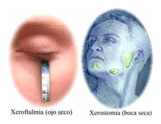
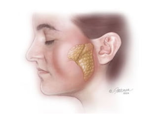
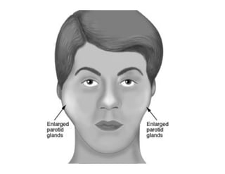
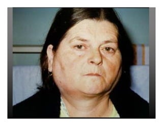
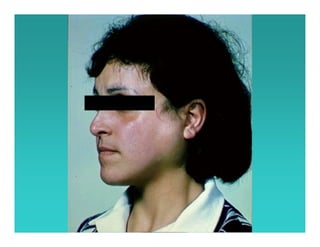
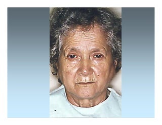
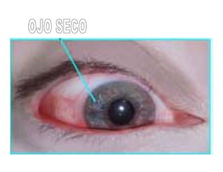
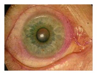
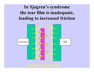
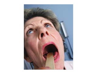
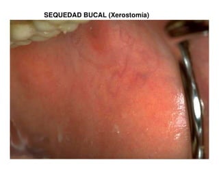
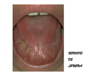
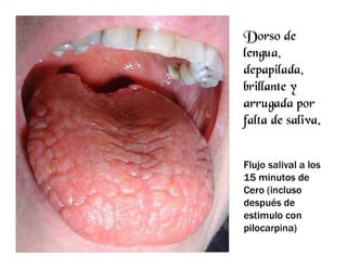
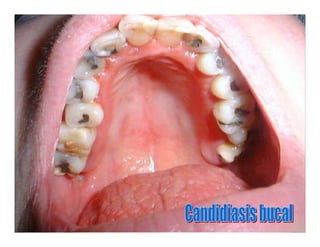
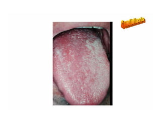
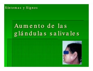
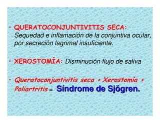
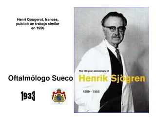
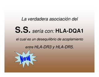
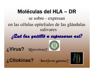
(2013-01-24) Sindrome de Sjögren ppt 
PDF

PPTX

PPTX

PPTX

PPTX

PPTX

PDF

PPT

PPTX

PPTX

PPT
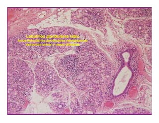
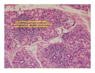
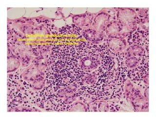
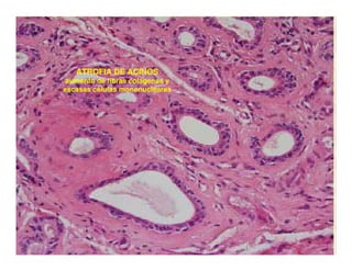
Síndrome de sjogren. seminario 10 
PPT

PPT
Fibromialgia Claudia Blázquez 
PPTX

PPTX

PPT
Powerpoint Fibromialgia C.M.C 
PPTX
More from Carlos Renato Cengarle

PPTX

PPT
Simpaticectomia Videotoracoscópica.ppt 
PPT
hemorragia subaracnoidea.ppt 
PPT
Neumona adquirida en la comunidad.ppt 
PPT
Artritis Reumatoidea Clínica.ppt 
PPT
Auditoria Basada en la Evidencia - Actualización 2008 
PPT

PPT
Reanimación Cardio Pulmonar 
PPT

PPT
Carta de los Derechos del Médico 
PPT

PPT
Diagnostico de las Poliurias 
PPT
Semiología del Derrame Pleural 
PPT

PPT
Tumores malignos del Tubo Digestivo 
PPT

PDF
Auditoría Médica, Basada en la Evidencia 
PPT
Brote Hospitalario de Infecciones Multirresistentes 
PDF

PDF