Top Rated Bangalore Call Girls Mg Road ⟟ 9332606886 ⟟ Call Me For Genuine S...
_Risk Stratification in Lung Resection (1).pdf
1. THORACIC SURGERY (G. ROCCO AND M. SCARCI, SECTION EDITORS)
Risk Stratification in Lung Resection
Michele Salati1 • Alessandro Brunelli2
Published online: 20 September 2016
The Author(s) 2016. This article is published with open access at Springerlink.com
Abstract
Purpose of Review Surgery is considered the best treat-
ment option for patients with early stage lung cancer.
Nevertheless, lung resection may cause a variable func-
tional impairment that could influence the whole cardio-
respiratory system with potential life-threatening compli-
cations. The aim of the present study is to review the most
relevant evidences about the evaluation of surgical risk
before lung resection, in order to define a practical
approach for the preoperative functional assessment in lung
cancer patients.
Recent Findings The first step in the preoperative func-
tional evaluation of a lung resection candidate is a cardiac
risk assessment. The predicted postoperative values of
forced expiratory volume in one second and carbon
monoxide lung diffusion capacity should be estimated next.
If both values are greater than 60 % of the predicted val-
ues, the patients are regarded to be at low surgical risk. If
either or both of them result in values lower than 60 %,
then a cardiopulmonary exercise test is recommended.
Patients with VO2max [20 mL/kg/min are regarded to be
at low risk, while those with VO2max 10 mL/kg/min at
high risk. Values of VO2max between 10 and 20 mL/kg/
min require further risk stratification by the VE/VCO2
slope. A VE/VCO2 35 indicates an intermediate-low risk,
while values above 35 an intermediate-high risk.
Summary The recent scientific evidence confirms that the
cardiologic evaluation, the pulmonary function test with
DLCO measurement, and the cardiopulmonary exercise
test are the cornerstones of the preoperative functional
evaluation before lung resection. We present a simplified
functional algorithm for the surgical risk stratification in
lung resection candidates.
Keywords Preoperative evaluation Cardiac risk
Co-morbidities Pulmonary function Exercise test
Operative Risk Morbidity Mortality Lung resection
Lung cancer surgery
Introduction
During the last 40 years, an increasing amount of papers
addressed the topic of perioperative risk assessment in the
field of lung surgery.
At the end of the eighties, the attention was focused on
the spirometric parameters, particularly lung volumes and
flows, as potential predictors of poor outcome when pre-
operatively impaired. Nevertheless, as it became clear in
the following decade, the spirometric evaluation was not
able to discriminate per se the surgical risk, and some other
factors, such as the lung diffusion capacity, had to be
considered for predicting the risk of morbidity and mor-
tality. These parameters were adopted as the standard of
the preoperative functional assessment before lung resec-
tion at the end of the last century. Moreover, they were
considered the first-level examination step before pro-
ceeding to more sophisticated evaluation strategies, as
This article is part of the Topical collection on Thoracic Surgery.
Alessandro Brunelli
brunellialex@gmail.com
Michele Salati
michelesalati@hotmail.com
1
Division of Thoracic Surgery, Ospedali Riuniti Ancona, Via
Conca 1, 60020 Ancona, Italy
2
Department Thoracic Surgery, St. James’s University
Hospital, Beckett Street, Leeds LS9 7TF, UK
123
Curr Surg Rep (2016) 4:37
DOI 10.1007/s40137-016-0158-x
2. reported in the most recent algorithms for the preoperative
fitness assessment. As a consequence, during the last 15
years, a growing evidence has highlighted the role of the
ergometric capacity assessment tested through the car-
diopulmonary exercise test as the ultimate evaluation tool
in order to define the surgical risk.
The present study offers an overview of the most rele-
vant papers about the risk stratification before lung resec-
tion with special attention to their clinical relapses. At the
end of each paragraph, some practical recommendations
are summarized that were condensed in order to propose a
functional evaluation algorithm useful for daily clinical
practice.
This article does not contain any studies with human or
animal subjects performed by any of the authors.
Cardiologic Evaluation
As recommended by the most recent algorithms concerning
the functional evaluation of candidates to lung resection,
the first step for estimating the surgical risk is represented
by an accurate cardiac evaluation [1••, 2].
In fact, this should be the preliminary patient assess-
ment, before proceeding with the pulmonary and ergo-
metric evaluation, since the presence of unstable cardiac
disease could per se influence an increased surgical risk. As
a consequence, an optimization of the cardiac function by
medical or surgical therapy is strongly recommended in
these patients before proceeding with the planned lung
resection.
In order to identify the category of patients with a higher
chance of postoperative cardiac adverse events due to a
pre-existent cardiac disease or some other pathologic fac-
tors, in 1999 Lee et al. developed the Revised Cardiac Risk
Index (RCRI) for stable patients undergoing non-urgent
major non-cardiac surgery [3]. This risk stratification tool
was refined in 2010 by Brunelli et al. proposing a new risk
score (ThRCRI) derived from an homogeneous population
of 1696 patients submitted exclusively to major lung
resection (1426 pulmonary lobectomies and 270 pneu-
monectomies) [4]. In order to calculate the ThRCRI of a
lung resection candidate, four different factors (each of
them having a specific weight for the final index) should be
taken into account:
1. History of coronary artery disease, 1.5 points.
2. Cerebrovascular disease, 1.5 points.
3. Serum creatinine level greater than 2 mg/dl, 1 point.
4. Pneumonectomy, 1.5 points.
Summing the points of each factor, the patient’s
aggregate ThRCRI is obtained, which ranges from a min-
imum of 0 to a maximum of 5.5. This value identifies four
different risk classes predicting an incremental risk of
cardiac morbidity:
• Class A: 0 points. Risk of cardiac complication: 1.5 %.
• Class B: 1–1.5 points. Risk of cardiac complication:
5.8 %.
• Class C: 2–2.5 points. Risk of cardiac complication:
19 %.
• Class D: [2.5 points. Risk of cardiac complication:
23 %.
In 2011, the ThRCRI was validated in two external
populations of 2621 and 1255 patients [5•, 6]. Both studies
verified that patients with a ThRCRI greater than 2.5 have a
risk of major cardiac complications, ranging from 13 to
18 %, confirming the reliability and usefulness of the
score.
Following these results, the most recent guidelines of the
ACCP about the physiologic evaluation of patients consid-
ered for resectional surgery [1••] suggested that those ones
with a ThRCRI C2 should be referred for a formal cardiol-
ogy evaluation and eventually to tests and treatments as
recommended by the American Heart Association and the
American College of Cardiology guidelines [7].
Suggestions
The cardiac evaluation is the first preliminary step of the
patient’s functional status
Calculate the ThRCRI for each lung resection candidate
In case of a ThRCRI 2, proceed with the pulmonary
functional evaluation
In case of a ThRCRI C2, optimize the cardiac function
before considering lung surgery
Forced Expiratory Volume at First Second (FEV1)
and Predicted Postoperative FEV1 (ppoFEV1)
The roles of the FEV1 and of its derived parameter ppo-
FEV1 in the functional assessment before lung resection
have considerably changed during the last decade.
Since the eighties, several papers have been published
addressing the importance of the FEV1 in defining the risk
of morbidity and mortality for lung surgery. The most
relevant ones are reported in the following list:
– 1988: Nakahara et al. Retrospective observational
study. Cohort: 157 patients submitted to anatomic lung
resection. The ppoFEV1 showed a correlation with the
postoperative respiratory complications. In the group of
patients with ppoFEV 30 %, the mortality rate was
about 60 % [8].
37 Page 2 of 9 Curr Surg Rep (2016) 4:37
123
3. – 1989: Markos et al. Retrospective observational study.
Cohort: 47 patients submitted to lobectomy (29) and
pneumonectomy (18). The ppoFEV1 was a predictor of
complications and death. No patients with a ppo-
FEV1 [40 % died, while three of six patients with a
ppoFEV1 40 % died in the perioperative period [9].
– 2005: Magdeleinat et al. Retrospective observational
study. Cohort: 106 patients submitted to lung resection
(17 sublunar resections) with a preoperative FEV1 and/
or FVC 50 %. The overall morbidity rate was 70 %
and the mortality rate 8.5 %. 21 % of patients required
prolonged mechanical ventilation (mean 11 days). The
morbidity rate raised up to 100 % for patients with a
ppoFEV1 loss [15 % [10].
– 2006: Licker et al. Retrospective observational study.
Cohort: 1239 consecutive thoracotomies. The
FEV1 60 % was an independent risk factor of respi-
ratory complications, including prolonged air leak
(OR = 2.7) and 30-day mortality (OR = 1.9) [11].
– 2008: Ferguson et al. Retrospective observational study.
Cohort: 1046 patients submitted to major lung resec-
tion. Using a classification and regression tree analysis,
FEV1 turned out to be an independent predictor of
pulmonary morbidity and cardiovascular complications.
The FEV1 was not related to mortality [12].
– 2010: Berry et al. Retrospective observational study.
Cohort: 340 patients submitted to open or video-
assisted lobectomy and with a FEV1 or a
DLCO 60 %. The overall morbidity rate was 48 %
and the mortality rate 5 %. Within the thoracotomy
patients, the level of FEV1 was inversely correlated
with the pulmonary complication rate. The FEV1 was
an independent predictor of respiratory morbidity for
the open patients but not for the ones treated with a
thoracoscopic approach [13].
Most recently, growing evidence has questioned the role
of the FEV1 in defining the risk before the surgical treat-
ment. In fact, several studies showed that the FEV1 failed
to estimate the postoperative outcome in some categories
of patients (such as the ones with an higher COPD grade).
At the same time, some Authors demonstrated the limits of
the ppoFEV1 in predicting the postoperative pulmonary
function, especially in the early postoperative period.
– 1998: Korst. Retrospective observational study. Cohort:
32 patients submitted to lobectomy. The COPD index is
inversely correlated with the residual FEV1 measured
after the operation (follow-up between 4 months and
2 years). Patients with a FEV1 60 % and a FEV1/
FVC 0.6 experienced an increase of the FEV1 after
lobectomy (mean FEV1 increase: 3.7 %) [14].
– 1999: Carretta et al. Retrospective observational study.
Cohort: 35 patients submitted to lobectomy. Patients
with an higher grade of emphysema had stable or slight
improvement of the FEV1 and FVC values after the
lobectomy (mean time of follow-up pulmonary func-
tion assessment: 4.7 months). In this group of patients,
the postoperative FEV1 increases of about 6 % in
comparison to the preoperative value [15].
– 2001: Santambrogio et al. Retrospective observational
study. Cohort: 88 patients submitted to lobectomy.
Patients encountering the spirometric criteria of COPD
(FEV1 80 %) showed a lesser reduction of the FEV1
6 months after the operation in comparison to the ones
with normal pulmonary function. The postoperative
FEV1 decrease was -3.2 % for the COPD group and
-14.9 % for the non-COPD group (p 0.001) [16].
– 2002: Brunelli et al. Retrospective observational study.
Cohort: 544 patients submitted to lobectomy (441) or
pneumonectomy (130). The postoperative complica-
tions rate (overall morbidity rate: 21.1 %, overall
mortality rate: 2.9 %) did not differ between the
patients with a preoperative FEV1 [70 % (group A:
450 pts) and the ones with a FEV1 70 % (group B:93
pts). The predictors of complications within group A
were FEV1, ppoFEV1, and COPD index. No spiromet-
ric predictors of outcome were identified for the group
B [17].
– 2003: Sekine et al. Retrospective observational study.
Cohort: 521 patients submitted to lobectomy. The
postoperative FEV1 measured 1 month after the opera-
tion showed a decrease of 13.1 % compared to the
preoperative values within the group of COPD patients
(FEV1 70 % and FEV1/FVC 0.7, 48 pts), while the
reduction for the non-COPD patients was 29.2 %
(p 0.001). The measured postoperative FEV1/ppo-
FEV1 ratio was grater than 1 for the COPD patients [18].
– 2007: Brunelli et al. Retrospective observational study.
Cohort: 200 patients submitted to lobectomy (180 pts)
and pneumonectomy (20 pts). Within the lobectomy
patients, the actual postoperative FEV1 measured at
discharge, 1 and 3 months after the operation, was
-11 %, similar, and ?6 % in comparison to the
calculated ppoFEV1. The actual postoperative FEV1
overestimated the ppoFEV1 especially for the patients
with lower expected FEV1 after the operation [19].
– 2005: Brunelli et al. Prospective observational study.
Cohort: 190 patients submitted to lobectomy (161 pts)
and pneumonectomy (29 pts). The authors presented a
regression equation in order to optimize the calculation
of the ppoFEV1 taking into account multiple correction
parameters. The estimated percentage of FEV1 reduc-
tion was obtained by the formula: [21.34–
(0.47 9 age) ? (0.49 9 percentage of functioning
parenchyma removed during operation) ? (17.91 9
COPD index)] [20].
Curr Surg Rep (2016) 4:37 Page 3 of 9 37
123
4. Taking into account the reported evidences, the FEV1
has progressively lost the role of defining independently
from other parameters the functional status and, as a con-
sequence, the risk before major lung resection.
In fact, the most recent guidelines, developed for
managing the preoperative physiologic evaluation of the
patients who were candidates to lung surgery, considered
the FEV1 as one of the factors that can lead the evalua-
tion algorithm rather than the single functional variable that
was able to select patients for surgical treatment [1••, 2–4,
5•, 6–21].
Suggestions
A formal spirometry with FEV1 measurement should be
performed for each patient who was a candidate to lung
resection.
In case of a ppoFEV1 60 %, the patient should be
considered at an higher operative risk.
In case of a ppoFEV1 60 %, the patient should be
evaluated with a second level functional test as a formal
cardiopulmonary exercise test.
Do not exclude from the operation any patient solely on
the basis of a low ppoFEV1 value.
Carbon Monoxide Lung Diffusion Capacity
(DLCO)
Evidences highlighting the DLCO as an additional and
independent lung function parameter that was able to
define the surgical risk in pulmonary resection were first
published by Ferguson et al. about 25 years ago. In the first
paper, these Authors showed the correlation between an
impaired DLCO and the development of postoperative
respiratory complications and death. In particular, analyz-
ing 237 patients submitted to major lung resection (73
pneumonectomies), they found a complication and mor-
tality rates of 40 and 20 %, respectively, in those patients
with a DLCO 60 % [22]. In 2010, Berry obtained similar
results in a retrospective study on 167 patients submitted to
open lobectomy. The logistic regression confirmed that the
DLCO was associated to pulmonary complications, which
reached the rate of about 40 % in those patients with a
DLCO 45 % [23].
Moreover, an even stronger ability to relate with the
postoperative outcome was then demonstrated for the
derived ppoDLCO, again by Ferguson in a study on 376
patients (246 lobectomies, 38 bilobectomies, 92 pneu-
monectomies). The ppoDLCO and age turned out to be the
only predictors of any type of complications and mortality
among 23 physiologic and spirometric preoperative
parameters [24]. These findings were confirmed most
recently by other studies [25, 26].
Finally, several papers documented that the DLCO main-
tains its role as risk factor before lung resection independently
fromtheCOPDstatusofthepatients.Inamulti-centricstudyon
872 patients submitted to lung resections (129 wedges/seg-
mentectomies, 611 lobectomies/bilobectomies, 132 pneu-
monectomies), Brunelli demonstrated that age and
ppoDLCO40 % were the only predictors of morbidity in the
group of patients without an airflow limitation (FEV1[80 %:
508 patients, morbidityrate for itswith ppoDLCO [40:17.5 %
vs morbidity rate for its with ppoDLCO40:37 %, p: 0.004).
Moreover, showing a low correlation coefficient between
FEV1 and DLCO for the entire population as well as for sub-
groups of analysis, the Authors recommended the DLCO
measurement before lung surgery for all the patients, irre-
spectively of the FEV1 values [27].
The central role of the ppoDLCO for the risk stratifi-
cation was corroborated by a subsequent analysis of Fer-
guson on 1008 patients submitted to anatomic major lung
resection. Dividing the population into two groups (450
COPD patients and 558 non-COPD patients, COPD was
defined as FEV1/FVC 0.7), the multivariate analysis
showed that the ppoDLCO was a significant predictor of
pulmonary complications and mortality both in patients
with and without COPD. The Authors also documented a
linear increase of pulmonary complications and mortality
with a progressive education of the ppoDLCO values
similar for the two groups of patients [28].
Suggestions
A systematic DLCO measurement should be performed
for each patient who was a candidate to lung resection
irrespectively of the FEV1 value registered.
In contrast with the FEV1, the DLCO maintains its
ability in evaluating the risk of complications indepen-
dently from the COPD status of the patients.
In case of a ppoDLCO 60 %, the patient should be
evaluated with a second level functional test as a formal
cardiopulmonary exercise test.
Do not exclude from the operation any patient solely on
the basis of a low ppoDLCO value.
Cardiopulmonary Exercise Testing (CPET)
Considering the most recent guidelines for the physiologic
evaluation before lung surgery, the formal high tech car-
diopulmonary exercise test (CPET) is considered the gold
standard for the functional assessment and the risk strati-
fication of candidates to pulmonary resection [1••].
37 Page 4 of 9 Curr Surg Rep (2016) 4:37
123
5. The first evidences addressing the role of CPET in
assessing the surgical risk were published during the
nineties. The exercise capacity expressed as percentage of
the predicted value of the maximum oxygen consumption
(VO2max %) was the first ergometric parameter found to
be associated with postoperative complication and mor-
tality. Bolliger et al. analyzed 80 patients submitted to lung
resection (14 minor resections) and evaluated by a symp-
tom-limited CPET. The VO2max % turned out to be the
best predictor of complication at the regression analysis.
Patients with a VO2max % 60 % had a high risk of
postoperative adverse events up to 89 % [29]. These data
were confirmed by a prospective trial performed from 1990
to 1997 on 125 anatomic lung resections. Among 19
demographic, spirometric, surgical, and ergonometric
parameters, the only parameters associated with postoper-
ative complications were the extent of resection and the
VO2max %. Moreover, the Authors estimated the risk of
complications at different levels of VO2max % for each
type of resection performed. In particular, in case of a
VO2max % = 60 %, they found a probability of com-
plication varying from 45 % in case of segmentectomy, to
78 % in case of pneumonectomy [30]. In 2005, Win et al.
corroborated these findings and stated that a VO2max %
threshold between 50 and 60 % should be considered the
limit, above which resections should be performed with a
low risk of complications and mortality [31].
Nevertheless, most recent papers reconsidered the
importance of the VO2max %, demonstrating that the
absolute value of the maximal oxygen consumption mea-
sured in ml/kg/min (VO2max) was the optimal ergometric
parameter in order to quantify the risk for major lung
resections. Some of the most relevant studies are reported
as follows:
– 2007: Loewen et al. Prospective multi-institutional
observational study. Cohort: 346 patients submitted to
thoracotomy wither without lung resection (73 sublobar
resections, 7 exploratory thoracotomy). The Authors
found that patients at risk for postoperative complica-
tions and high mortality rate were the ones with a
VO2max 15 ml/kg/min [32].
– 2007: Bayram et al. Prospective multi-institutional
observational study. Cohort: 55 patients submitted to
major lung resection. The Authors did not observe any
adverse events in patients with a VO2max [15 ml/kg/
min. The 28 patients with a VO2max 15 ml/kg/min
experienced a postoperative complication rate of 39 %
(2 patients died) [33].
– 2009: Brunelli et al. Retrospective observational study.
Cohort: 204 patients submitted to major lung resection
(177 lobectomies, 24 pneumonectomies). The VO2max
turned out to be the best predictor of respiratory
complications. Patients with a VO2max 12 ml/kg/min
had a mortality rate of 13 %, while no mortality was
observed in patients with a VO2max [20 ml/kg/min.
Finally,theAuthorsshowedthat,attheROCanalysis,the
best threshold for predicting both pulmonary complica-
tion and death was a VO2max 12 ml/kg/min [34].
– 2011: Licker et al. Retrospective observational study.
Cohort: 210 patients with FEV1 80 % submitted to
lung resection. The VO2max was a predictor of
cardiopulmonary complication and death at the multi-
variate analysis including preoperative clinical, surgi-
cal, and ergometric variables. Patients with a
VO2max 10 ml/kg/min had a risk of total morbidity,
cardiovascular morbidity, and cardiac morbidity of 65,
39, and 35 %, respectively, in case of major resection
[35].
Based on these evidences, the VO2max obtained at the
CPET is considered by the recent functional algorithm as
the definite and most reliable parameter stratifying the risk.
Using the VO2max value as an indicator of the global
performance status of the patients, it can be decided the
best treatment option for lung resection candidates.
Suggestions
The high tech CPET with the VO2max measurement is
the most reliable parameter for defining the surgical risk
in lung resection candidates.
Perform a formal CPET in any patient with an impaired
ppoFEV1 and ppoDLCO.
In case of a VO2max [10 ml/kg/min, the risk for a
major lung resection is acceptable varying from moder-
ate to low.
In case of a VO2max 10 ml/kg/min, the risk for a
major lung resection is high, and the patient should be
considered for minor resection or alternative non-surgi-
cal therapies.
Minute Ventilation to Carbon Dioxide Output
(VE/VCO2) Slope
Recently, several papers have been published in order to
verify if ergometric parameters other than the VO2max
have the potential for predicting the postoperative surgical
outcome, and consequently could be used as risk strati-
fication factors in patients submitted to lung resection
[36–40].
The most promising parameter is represented by the
slope of the minute ventilation to carbon dioxide output
ratio (VE/VCO2). This relationship, elsewhere reported as
ventilatory efficiency curve, describes the potential of the
Curr Surg Rep (2016) 4:37 Page 5 of 9 37
123
6. cardio-respiratory system in increasing the CO2 output
through a higher minute ventilation during the exercise. An
abnormal rise of the VE/VCO2 slope values could be
related both to pulmonary and cardiac diseases, such as
COPD, pulmonary hypertension, or heart failure [41, 42].
In 2010, Torchio et al. published a retrospective study
on 145 COPD patients submitted to major lung resection
(including 39 pneumonectomies) and evaluated them by a
formal preoperative CPET. The mortality and cardiopul-
monary morbidity rates were 3.4 and 14.5 %, respectively.
The VO2max turned out to be the best predictor of mor-
bidity after the logistic regression, while the only param-
eter associated with mortality was the VE/VCO2 slope. In
particular, a VE/VCO2 slope C34 was related to a risk of
mortality of 5.5 %. Therefore, the Authors recommended
the screening of major lung resection candidates for
potential ventilatory insufficiency to refine the risk of
mortality, irrespectively of the VO2max value reached at
the preoperative CPET.
Two years later, Brunelli et al. analyzed a cohort of 225
patients submitted to lobectomy (197) and pneumonectomy
(28) after a complete functional evaluation including a
CPET independently from the preoperative or ppo FEV1
and DLCO values. The cardiopulmonary morbidity rate
was 23 %, while a total of 25 patients (11 %) experienced a
postoperative pulmonary adverse event. This group of
patients registered a VE/VCO2 slope significantly higher in
comparison to the uncomplicated patients (34.8 vs 30.9,
p 0.001). Moreover, the Authors found that, after logistic
regression analysis the VE/VCO2 slope remained the only
predictor of respiratory complications, and those patients
with a VE/VCO2 slope C35 had a 3-fold higher probability
of experiencing respiratory complications during the post-
operative period.
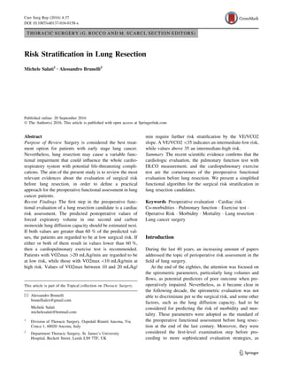
Lung resection candidate
Check cardiologic history
for calculating ThRCRI
ThRCRI 2 ThRCRI ≥ 2
Refer to cardiologist
Optimize cardiac status
Check spirometry with DLCO measurement
for calculating ppoFEV1 ppoDLCO
Either ppoFEV1 or ppoDLCO 60%
ppoFEV1 60%
and ppoDLCO 60%
Perform a CPET
for calculating VO2max VE/VCO2
VO2max 20 ml/kg/min VO2max = 10 - 20 ml/kg/min VO2max 10 ml/kg/min
Consider
compromised minor resection or
non surgical management
VE/VCO2 = 35
VE/VCO2
VE/VCO2 35
Low
Risk
Low
Risk
Low - Intermediate
Risk
Intermediate - High
Risk
High
Risk
Fig. 1 Simplified functional algorithm based on the reported evidences and authors experience (see Conclusions for explanation)
37 Page 6 of 9 Curr Surg Rep (2016) 4:37
123
7. The role of the VE/VCO2 slope as an outcome predictor
after lung resection was further confirmed by Shafiek and
coll. [43•]. In a retrospective study on 83 COPD patients,
the Authors verified that a VE/VCO2 slope [35 was the
stronger predictor of mortality and morbidity, even if tested
in association with the VO2max.
These evidences support the use of VE/VCO2 slope as a
relevant parameter for defining the preoperative risk before
lung surgery. Hopefully, further studies could strengthen
the role of this risk factor in order to include it within
functional evaluation algorithms.
Conclusions
Based on the evidences from the literature reported above
and the personal experience of the authors, we propose a
simplified functional algorithm.
As shown in Fig. 1, the flow chart starts from a cardiac
evaluation based on the estimation of the ThRCRI. If the
patient has a ThRCRI greater than 2, then a specialist
referral to a cardiologist is recommended to optimize their
cardiac status.
Following cardiac risk evaluation, a pulmonary function
test is obtained with measurement of both FEV1 and
DLCO, and split lung function is calculated based on the
planned extent of the resection.
If both ppoDLCO and ppoFEV1 are greater than 60 %
of predicted values, then no further tests are advised as they
would be regarded as low risk patients.
If either or both of these parameters are lower than
60 %, then a cardiopulmonary exercise test is
recommended.
The two parameters that should be taken into consid-
eration are the maximum oxygen consumption (VO2max)
and the efficiency slope (VE/VCO2). Patients with VO2-
max greater than 20 mL/kg/min are regarded as low risk
for surgery. Those with VO2max lower than 10 mL/kg/min
are deemed at high risk for anatomic lung resection. Pa-
tients with values of VO2max falling in between 10 and
20 mL/kg/min would be better risk stratified by the VE/
VCO2 slope. If VE/VCO2 is lower than 35, then they can
be considered at intermediate-low risk, while a value above
35 should be considered a marker of intermediate-high risk.
Funding None.
Compliance with Ethics Guidelines
Conflict of Interest Dr. Brunelli reports personal fees from Bard
Davol Inc. Dr. Salati declares no conflicts of interest relevant to this
manuscript.
Human and Animal Rights and Informed Consent This article
does not contain any studies with human or animal subjects per-
formed by any of the authors.
Open Access This article is distributed under the terms of the
Creative Commons Attribution 4.0 International License (http://
creativecommons.org/licenses/by/4.0/), which permits unrestricted
use, distribution, and reproduction in any medium, provided you give
appropriate credit to the original author(s) and the source, provide a
link to the Creative Commons license, and indicate if changes were
made.
References
Papers of particular interest, published recently, have been
highlighted as:
• Of importance
•• Of major importance
1. •• Brunelli A, Kim AW, Berger KI, Addrizzo-Harris DJ. Physi-
ologic evaluation of the patient with lung cancer being considered
for resectional surgery. Chest 2013;143:166S–190S. The Authors
performed a rigorous review of the medical literature proposing
a list of evidence based recommendations and an exhaustive
algorithm for the preoperative physiologic assessment in patients
candidates to lung resection for NSCLC. Once performed the
cardiac and spirometric evaluations with the estimation of the
FEV1 and DLCO predictive postoperative values, the final risk
stratification should be obtained considering the VO2max mea-
sured at the cardiopulmonary exercise test.
2. Brunelli A, Charloux A, Bolliger CT, Rocco G, Sculier JP, Varela
G, Licker M, Ferguson MK, Faivre-Finn C, Huber RM, Clini EM,
Win T, De Ruysscher D, European Respiratory Society and
European Society of Thoracic Surgeons joint task force on fitness
for radical therapy. ERS/ESTS clinical guidelines on fitness for
radical therapy in lung cancer patients (surgery and chemo-ra-
diotherapy). Eur Respir J. 2009;34:17–41.
3. Lee TH, Marcantonio ER, Mangione CM, Thomas EJ, Polanczyk
CA, Cook EF, Sugarbaker DJ, Donaldson MC, Poss R, Ho KK,
Ludwig LE, Pedan A, Goldman L. Derivation and prospective
validation of a simple index for prediction of cardiac risk of
major noncardiac surgery. Circulation. 1999;100:1043–9.
4. Brunelli A, Varela G, Salati M, Jimenez MF, Pompili C, Novoa
N, Sabbatini A. Recalibration of the revised cardiac risk index in
lung resection candidates. Ann Thorac Surg. 2010;90:199–203.
5. • Brunelli A, Cassivi SD, Fibla J, Halgren LA, Wigle DA, Allen
MS, Nichols FC, Shen KR, Deschamps C. External validation of
the recalibrated thoracic revised cardiac risk index for predicting
the risk of major cardiac complications after lung resection. Ann
Thorac Surg. 2011;92:445–448. The Authors validated the use of
a multiparametric cardiac risk index (ThRCRI) developed to
assess the risk of adverse cardiac events after lung resec-
tion. They found, in a cohort of 2,621 patients, a progressive
increase of observed complication rate in line with the class of
risk assigned to the patients (class A: cardiac complication rate
0.9%, B: 4.2%, C: 8%, D: 18%). This study confirm the reliability
of the ThRCRI.
6. Ferguson MK, Celauro AD, Vigneswaran WT. Validation of a
modified scoring system for cardiovascular risk associated with
major lung resection. Eur J Cardiothorac Surg. 2012;41:598–602.
7. Fleisher LA, Beckman JA, Brown KA, Calkins H, Chaikof EL,
Fleischmann KE, Freeman WK, Froehlich JB, Kasper EK,
Curr Surg Rep (2016) 4:37 Page 7 of 9 37
123
8. Kersten JR, Riegel B, Robb JF, Smith SC Jr, Jacobs AK, Adams
CD, Anderson JL, Antman EM, Buller CE, Creager MA, Ettinger
SM, Faxon DP, Fuster V, Halperin JL, Hiratzka LF, Hunt SA,
Lytle BW, Nishimura R, Ornato JP, Page RL, Riegel B, Tark-
ington LG, Yancy CW. American College of Cardiology/Amer-
ican Heart Association Task Force on Practice Guidelines
(Writing Committee to Revise the 2002 Guidelines on Periop-
erative Cardiovascular Evaluation for Noncardiac Surgery);
American Society of Echocardiography; American Society of
Nuclear Cardiology; Heart Rhythm Society; Society of Cardio-
vascular Anesthesiologists; Society for Cardiovascular Angiog-
raphy and Interventions; Society for Vascular Medicine and
Biology; Society for Vascular Surgery. ACC/AHA 2007 guide-
lines on perioperative cardiovascular evaluation and care for
noncardiac surgery: a report of the American College of Cardi-
ology/American Heart Association Task Force on Practice
Guidelines (Writing Committee to Revise the 2002 Guidelines on
Perioperative Cardiovascular Evaluation for Noncardiac Sur-
gery): developed in collaboration with the American Society of
Echocardiography, American Society of Nuclear Cardiology,
Heart Rhythm Society, Society of Cardiovascular Anesthesiolo-
gists, Society for Cardiovascular Angiography and Interventions,
Society for Vascular Medicine and Biology, and Society for
Vascular Surgery. Circulation 2007;116:e418–99
8. Nakahara K, Ohno K, Hashimoto J, Miyoshi S, Maeda H, Mat-
sumura A, Mizuta T, Akashi A, Nakagawa K, Kawashima Y.
Prediction of postoperative respiratory failure in patients under-
going lung resection for lung cancer. Ann Thorac Surg.
1988;46:549–52.
9. Markos J, Mullan BP, Hillman DR, Musk AW, Antico VF,
Lovegrove FT, Carter MJ, Finucane KE. Preoperative assessment
as a predictor of mortality and morbidity after lung resection. Am
Rev Respir Dis. 1989;139:902–10.
10. Magdeleinat P, Seguin A, Alifano M, Boubia S, Regnard JF.
Early and long-term results of lung resection for non-small-cell
lung cancer in patients with severe ventilatory impairment. Eur J
Cardiothorac Surg. 2005;27:1099–105.
11. Licker MJ, Widikker I, Robert J, Frey JG, Spiliopoulos A,
Ellenberger C, Schweizer A, Tschopp JM. Operative mortality
and respiratory complications after lung resection for cancer:
impact of chronic obstructive pulmonary disease and time trends.
Ann Thorac Surg. 2006;81:1830–7.
12. Ferguson MK, Siddique J, Karrison T. Modeling major lung
resection outcomes using classification trees and multiple impu-
tation techniques. Eur J Cardiothorac Surg. 2008;34:1085–9.
13. Berry MF, Hanna J, Tong BC, Burfeind WR Jr, Harpole DH,
D’Amico TA, Onaitis MW. Risk factors for morbidity after
lobectomy for lung cancer in elderly patients. Ann Thorac Surg.
2009;88:1093–9.
14. Korst RJ, Ginsberg RJ, Ailawadi M, Bains MS, Downey RJ Jr,
Rusch VW, Stover D. Lobectomy improves ventilatory function
in selected patients with severe COPD. Ann Thorac Surg.
1998;66:898–902.
15. Carretta A, Zannini P, Puglisi A, Chiesa G, Vanzulli A, Bianchi
A, Fumagalli A, Bianco S. Improvement of pulmonary function
after lobectomy for non-small cell lung cancer in emphysematous
patients. Eur J Cardiothorac Surg. 1999;15:602–7.
16. Santambrogio L, Nosotti M, Baisi A, Ronzoni G, Bellaviti N,
Rosso L. Pulmonary lobectomy for lung cancer: a prospective
study to compare patients with forced expiratory volume in 1 s
more or less than 80 % of predicted. Eur J Cardiothorac Surg.
2001;20:684–7.
17. Brunelli A, Al Refai M, Monteverde M, Sabbatini A, Xiume F,
Fianchini A. Predictors of early morbidity after major lung
resection in patients with and without airflow limitation. Ann
Thorac Surg. 2002;74:999–1003.
18. Sekine Y, Iwata T, Chiyo M, Yasufuku K, Motohashi S, Yoshida
S, Suzuki M, Iizasa T, Saitoh Y, Fujisawa T. Minimal alteration
of pulmonary function after lobectomy in lung cancer patients
with chronic obstructive pulmonary disease. Ann Thorac Surg.
2003;76:356–62.
19. Brunelli A, Refai M, Salati M, Xiumé F, Sabbatini A. Predicted
versus observed FEV1 and DLCO after major lung resection: a
prospective evaluation at different post- operative periods. Ann
Thorac Surg. 2007;83:1134–9.
20. Brunelli A, Sabbatini A, Xiume F, Al Refai M, Borri A, Salati M,
Marasco RD, Fianchini A. A model to predict the decline of the
forced expiratory volume in one second and the carbon monoxide
lung diffusion capacity early after major lung resection. Interact
CardioVasc Thorac Surg. 2005;4:61–5.
21. Lim E, Baldwin D, Beckles M, Duffy J, Entwisle J, Faivre-Finn
C, Kerr K, Macfie A, McGuigan J, Padley S, Popat S, Screaton N,
Snee M, Waller D, Warburton C, Win T, British Thoracic
Society, Society for Cardiothoracic Surgery in Great Britain and
Ireland. Guidelines on the radical management of patients with
lung cancer. Thorax. 2010;65:iii1–27.
22. Ferguson MK, Little L, Rizzo L, Popovich KJ, Glonek GF, Leff
A, Manjoney D, Little AG. Diffusing capacity predicts morbidity
and mortality after pulmonary resection. J Thorac Cardiovasc
Surg. 1988;96:894–900.
23. Berry MF, Villamizar-Ortiz NR, Tong BC, Burfeind WR Jr,
Harpole DH, D’Amico TA, Onaitis MW. Pulmonary function
tests do not predict pulmonary complications after thoracoscopic
lobectomy. Ann Thorac Surg. 2010;89:1044–51.
24. Ferguson MK, Reeder LB, Mick R. Optimizing selection of
patients for major lung resection. J Thorac Cardiovasc Surg.
1995;109:275–81.
25. Santini M, Fiorello A, Vicidomini G, Di Crescenzo VG, Laperuta
P. Role of diffusing capacity in predicting complications after
lung resection for cancer. J Thorac Cardiovasc Surg.
2007;55:391–4.
26. Ferguson MK, Gaissert HA, Grab JD, Sheng S. Pulmonary
complications after lung resection in the absence of chronic
obstructive pulmonary disease: the predictive role of diffusing
capacity. J Thorac Cardiovasc Surg. 2009;138:1297–302.
27. Brunelli A, Refai MA, Salati M, Sabbatini A, Morgan-Hughes
NJ, Rocco G. Carbon monoxide lung diffusion capacity improves
risk stratification in patients without airflow limitation: evidence
for systematic measurement before lung resection. Eur J Car-
diothorac Surg. 2006;29:567–70.
28. Ferguson MK, Vigneswaran WT. Diffusing capacity predicts
morbidity after lung resection in patients without obstructive lung
disease. Ann Thorac Surg. 2008;85:1158–64.
29. Bolliger CT, Jordan P, Solèr M, Stulz P, Grädel E, Skarvan K,
Elsasser S, Gonon M, Wyser C, Tamm M. Exercise capacity as a
predictor of postoperative complications in lung resection can-
didates. Am J Respir Crit Care Med. 1995;151:1472–80.
30. Brutsche MH, Spiliopoulos A, Bolliger CT, Licker M, Frey JG,
Tschopp JM. Exercise capacity and extent of resection as pre-
dictors of surgical risk in lung cancer. Eur Respir J.
2000;15:828–32.
31. Win T, Jackson A, Sharples L, Groves AM, Wells FC, Ritchie
AJ, Laroche CM. Cardiopulmonary exercise tests and lung cancer
surgical outcome. Chest. 2005;127:1159–65.
32. Loewen GM, Watson D, Kohman L, Herndon JE 2nd, Shennib H,
Kernstine K, Olak J, Mador MJ, Harpole D, Sugarbaker D, Green
M. Preoperative exercise Vo2 measurement for lung resection
candidates: results of Cancer and Leukemia Group B Protocol
9238. J Thorac Oncol. 2007;2:619–25.
33. Bayram AS, Candan T, Gebitekin C. Preoperative maximal
exercise oxygen consumption test predicts postoperative
37 Page 8 of 9 Curr Surg Rep (2016) 4:37
123
9. pulmonary morbidity following major lung resection. Respirol-
ogy. 2007;12:505–10.
34. Brunelli A, Belardinelli R, Refai M, Salati M, Socci L, Pompili
C, Sabbatini A. Peak oxygen consumption during cardiopul-
monary exercise test improves risk stratification in candidates to
Major lung resection. Chest. 2009;135:1260–7.
35. Licker M, Schnyder JM, Frey JG, Diaper J, Cartier V, Inan C,
Robert J, Bridevaux PO, Tschopp JM. Impact of aerobic exercise
capacity and procedure-related factors in lung cancer surgery. Eur
Respir J. 2011;37:1189–98.
36. Campione A, Terzi A, Bobbio M, Rosso GL, Scardovi AB, Feola
M. Oxygen pulse as a predictor of cardiopulmonary events in
lung resection. Asian Cardiovasc Thorac Ann. 2010;18:147–52.
37. Kasikcioglu E, Toker A, Tanju S, Arzuman P, Kayserilioglu A,
Dilege S, Kalayci G. Oxygen uptake kinetics during cardiopul-
monary exercise testing and postoperative complications in
patients with lung cancer. Lung Cancer. 2009;66:85–8.
38. Wang JS, Abboud RT, Evans KG, Finley RJ, Graham BL. Role of
CO diffusing capacity during exercise in the preoperative eval-
uation for lung resection. Am J Respir Crit Care Med.
2000;162:1435–44.
39. Torchio R, Guglielmo M, Giardino R, Ardissone F, Ciacco C,
Gulotta C, Veljkovic A, Bugiani M. Exercise ventilatory ineffi-
ciency and mortality in patients with chronic obstructive pul-
monary disease undergoing surgery for non small-cell lung
cancer. Eur J Cardiothorac Surg. 2010;38:14–9.
40. Brunelli A, Belardinelli R, Pompili C, Xiumé F, Refai M, Salati
M, Sabbatini A. Minute ventilation-to-carbon dioxide output
(VE/VCO2) slope is the strongest predictor of respiratory com-
plications and death after pulmonary resection. Ann Thorac Surg.
2012;93:1802–6.
41. Arena R, Myers J, Hsu L, Peberdy MA, Pinkstaff S, Bensimhon
D, Chase P, Vicenzi M, Guazzi M. The minute ventilation/carbon
dioxide production slope is prognostically superior to the oxygen
uptake efficiency slope. J Card Fail. 2007;13:462–9.
42. Reindl I, Wernecke KD, Opitz C, Wensel R, Konig D, Dengler T,
Schimke I, Kleber FX. Impaired ventilatory efficiency in chronic
heart failure: possible role of pulmonary vasoconstriction. Am
Heart J. 1998;136:778–85.
43. • Shafiek H, Valera JL, Togores B, Torrecilla JA, Sauleda J,
Cosio BG. Risk of postoperative complications in chronic
obstructive lung disease patients considered fit for lung surgery:
beyond oxygen consumption. Eur J Cardiothorac Surg 2016;
doi:10/1093/ejcts/ezw104. The Authors analyzed a selected
cohort of patients submitted to lung resection after a complete
preoperative functional evaluation including the cardiopul-
monary exercise test. They found that the minute ventilation to
carbon dioxide output (VE/VCO2) slope was the variable most
strongly associated to the postoperative complication and the
mortality. This questioned the use of the maximal oxygen con-
sumption as the optimal parameter to define the risk in candidates
to major lung resection.
Curr Surg Rep (2016) 4:37 Page 9 of 9 37
123
![reported in the most recent algorithms for the preoperative
fitness assessment. As a consequence, during the last 15
years, a growing evidence has highlighted the role of the
ergometric capacity assessment tested through the car-
diopulmonary exercise test as the ultimate evaluation tool
in order to define the surgical risk.
The present study offers an overview of the most rele-
vant papers about the risk stratification before lung resec-
tion with special attention to their clinical relapses. At the
end of each paragraph, some practical recommendations
are summarized that were condensed in order to propose a
functional evaluation algorithm useful for daily clinical
practice.
This article does not contain any studies with human or
animal subjects performed by any of the authors.
Cardiologic Evaluation
As recommended by the most recent algorithms concerning
the functional evaluation of candidates to lung resection,
the first step for estimating the surgical risk is represented
by an accurate cardiac evaluation [1••, 2].
In fact, this should be the preliminary patient assess-
ment, before proceeding with the pulmonary and ergo-
metric evaluation, since the presence of unstable cardiac
disease could per se influence an increased surgical risk. As
a consequence, an optimization of the cardiac function by
medical or surgical therapy is strongly recommended in
these patients before proceeding with the planned lung
resection.
In order to identify the category of patients with a higher
chance of postoperative cardiac adverse events due to a
pre-existent cardiac disease or some other pathologic fac-
tors, in 1999 Lee et al. developed the Revised Cardiac Risk
Index (RCRI) for stable patients undergoing non-urgent
major non-cardiac surgery [3]. This risk stratification tool
was refined in 2010 by Brunelli et al. proposing a new risk
score (ThRCRI) derived from an homogeneous population
of 1696 patients submitted exclusively to major lung
resection (1426 pulmonary lobectomies and 270 pneu-
monectomies) [4]. In order to calculate the ThRCRI of a
lung resection candidate, four different factors (each of
them having a specific weight for the final index) should be
taken into account:
1. History of coronary artery disease, 1.5 points.
2. Cerebrovascular disease, 1.5 points.
3. Serum creatinine level greater than 2 mg/dl, 1 point.
4. Pneumonectomy, 1.5 points.
Summing the points of each factor, the patient’s
aggregate ThRCRI is obtained, which ranges from a min-
imum of 0 to a maximum of 5.5. This value identifies four
different risk classes predicting an incremental risk of
cardiac morbidity:
• Class A: 0 points. Risk of cardiac complication: 1.5 %.
• Class B: 1–1.5 points. Risk of cardiac complication:
5.8 %.
• Class C: 2–2.5 points. Risk of cardiac complication:
19 %.
• Class D: [2.5 points. Risk of cardiac complication:
23 %.
In 2011, the ThRCRI was validated in two external
populations of 2621 and 1255 patients [5•, 6]. Both studies
verified that patients with a ThRCRI greater than 2.5 have a
risk of major cardiac complications, ranging from 13 to
18 %, confirming the reliability and usefulness of the
score.
Following these results, the most recent guidelines of the
ACCP about the physiologic evaluation of patients consid-
ered for resectional surgery [1••] suggested that those ones
with a ThRCRI C2 should be referred for a formal cardiol-
ogy evaluation and eventually to tests and treatments as
recommended by the American Heart Association and the
American College of Cardiology guidelines [7].
Suggestions
The cardiac evaluation is the first preliminary step of the
patient’s functional status
Calculate the ThRCRI for each lung resection candidate
In case of a ThRCRI 2, proceed with the pulmonary
functional evaluation
In case of a ThRCRI C2, optimize the cardiac function
before considering lung surgery
Forced Expiratory Volume at First Second (FEV1)
and Predicted Postoperative FEV1 (ppoFEV1)
The roles of the FEV1 and of its derived parameter ppo-
FEV1 in the functional assessment before lung resection
have considerably changed during the last decade.
Since the eighties, several papers have been published
addressing the importance of the FEV1 in defining the risk
of morbidity and mortality for lung surgery. The most
relevant ones are reported in the following list:
– 1988: Nakahara et al. Retrospective observational
study. Cohort: 157 patients submitted to anatomic lung
resection. The ppoFEV1 showed a correlation with the
postoperative respiratory complications. In the group of
patients with ppoFEV 30 %, the mortality rate was
about 60 % [8].
37 Page 2 of 9 Curr Surg Rep (2016) 4:37
123](data:image/gif;base64,R0lGODlhAQABAIAAAAAAAP///yH5BAEAAAAALAAAAAABAAEAAAIBRAA7)