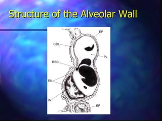
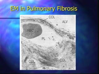
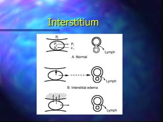
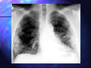
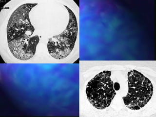
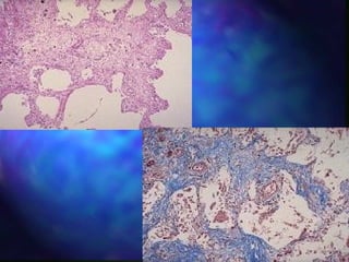
RESTRICTIVE LUNG DISEASE


     ssharma.com
       By Sat Sharma
     Assistant Professor
    University of Manitoba
Background
The lung volumes are reduced either because of:

1. Alteration in lung parenchyma.

2. Diseases of the pleura, chest wall or neuromuscular
   apparatus.

   Physiologically restrictive lung diseases are
   defined by reduced total lung capacity, vital
   capacity and functional residual capacity, but
   with preserved air flow.
Restrictive lung diseases may be divided into the
 following groups:

s   Intrinsic lung diseases (diseases of the lung
    parenchyma)


s   Extrinsic disorders (extra-parenchymal
    diseases)
Intrinsic Lung Diseases
These diseases cause either:
s Inflammation and/or scarring of lung tissue
  (interstitial lung disease)
                            or
s Fill the air spaces with exudate and debris
  (pneumonitis).

s   These diseases are classified further according
    to the etiological factor.
Extrinsic Disorders
The chest wall, pleura and respiratory muscles
 are the components of respiratory pump.

Disorders of these structures will cause lung
  restriction and impair ventilatory function.

These are grouped as:
s Non-muscular diseases of the chest wall.
s Neuromuscular disorders.
Pathophysiology
Intrinsic lung diseases:
s Diffuse parenchymal disorders cause reduction in all
  lung volumes.
s This is produced by excessive elastic recoil of the
  lungs.
s Expiratory flows are reduced in proportion to lung
  volumes.
s Arterial hypoxemia is caused by ventilation/perfusion
  mismatch.
s Impaired diffusion of oxygen will cause exercise-
  induced desaturation.
s Hyperventilation at rest secondary to reflex
  stimulation.
Extrinsic Disorders
s Diseases of the pleura, thoracic cage, decrease
  compliance of respiratory system.
s There is reduction in lung volumes.
s Secondarily, atelectasis occurs leading to V/Q
  mismatch § hypoxemia.
s The thoracic cage and neuromuscular
  structures are a part of respiratory system.
s Any disease of these structures will cause
  restrictive disease and ventilatory dysfunction.
Diseases of the Lung
    Parenchyma
Structure of the Alveolar Wall
EM in Pulmonary Fibrosis
Interstitium
Diffuse Interstitial Pulmonary
                  Fibrosis
s   Synonyms: idiopathic pulmonary fibrosis, interstitial
    pneumonia, cryptogenic fibrosing alveolitis.


                          Pathology
s   Thickening of interstitium.
s   Initially, infiltration with lymphocytes and plasma cells.
s   Later fibroblasts lay down thick collagen bundles.
s   These changes occur irregularly within the lung.
s   Eventually alveolar architecture is destroyed –
    honeycomb lung
Etiology
Unknown, may be immunological reaction.
                  Clinical Features
s Uncommon disease, affects adults in late
  middle age.
s Progressive exertional dyspnea, later at rest.
s Non-productive cough.
s Physical examination shows finger clubbing,
  fine inspiratory crackles throughout both lungs.
s Patient may develop respiratory failure
  terminally.
s The disease progresses insidiously, median
  survival 4-6 years.
Pulmonary Function
s Spirometry reveals a
  restrictive pattern. FVC is
  reduced, but FEV1/FVC
  supernormal.
s All lung volumes – TLC,
  FRC, RV – are reduced.
s Pressure volume curve of
  the lung is displaced
  downward and flattened.
Gas Exchange
s Arterial PaO2 and PaCO2 are reduced, pH
  normal.
s On exercise PaO2 decreases dramatically.
s Physiologic dead space and physiologic shunt
  and VQ mismatch are increased.
s Diffuse impairment contributes to hypoxemia
  on exercise.
s There is marked reduction in diffusing capacity
  due to thickening of blood gas barrier and VQ
  mismatch.
Diagnosis
s   Diagnosis is often suggested by history, chest
    radiograph and high resolution CT scan of the
    lungs.

s   If old chest x-rays show classical disease,
    absence of other disease processes on history
    and no occupational or environmental
    exposure – clinical diagnosis can be made.

s   In other cases a surgical lung biopsy is
    obtained.
Treatment
s   Each patient is individually assessed.
s   Patients are treated if they have symptoms or
    progressive dysfunction on pulmonary function tests.
s   Corticosteroids (Prednisone 1 mg/kg) is standard
    therapy.
s   Prednisone dose is lowered over 6-8 weeks and
    continued at 15 mg for 1-2 years.
s   Addition of Imuran may benefit survival.
s   Cyclophosphamide occasionally used.
s   Antifibrotics such as colchicine may be used.
s   Ancillary therapies such as oxygen, rehabilitation,
    psychosocial aspects are helpful.
Sarcoidosis
s A disease characterized by the presence of
  granulomatous tissue.
s This is a systemic disease which involves eyes,
  brain, heart, lungs, bones and kidneys, skin,
  liver and spleen.
s On pathology a non-caseating granuloma
  composed of histiocytes, giant cells and
  lymphocytes.
s In advanced lung disease fibrotic changes are
  seen.
Etiology
s   Unknown, likely immunological basis.


                 Clinical Features
Four stages are identified:
s Stage 0: No obvious intrathoracic involvement
s Stage 1: Bilateral hilar lymphadenopathy, often
  accompanied by arthritis, uveitis and erythema
  nodosum.
s Stage 2: Pulmonary parenchyma is also involved,
  changes in mid and upper zones.
s Stage 3: Pulmonary infiltrates and fibrosis without
  adenopathy.
Non-caseating granulomas
      in Sarcoidosis
Stage I
(bilateral hilar adenopathy)
Stage II
Reticular nodules and BHL
HRCT – subpleural nodules
Pulmonary Function
s No impairment occurs in stages 0 and 1.
s In stages 2 and 3 restrictive changes are seen.

             Treatment and Prognosis
s 85% of these patients improve spontaneously,
  but 15% may develop progressive fibrosis and
  respiratory failure.
s Treatment is other observation, but in
  symptomatic patients or deteriorating PFT’s –
  treatment recommended.
s Prednisone 0.5- 1 mg/kg initially, then tapered
  and continued for 6 months to 1 year.
Hypersensitivity Pneumonitis
s Also known as extrinsic allergic alveolitis.
s Hypersensitivity reaction in the lung occurs in
  response to inhaled organic dust.
s Example is farmer’s lung.
s The exposure may be occupational or
  environmental.
s The disease occurs from type III and type IV
  hypersensitivity reactions.
s Farmer’s lung is due to thermophilic
  actinomyces in moldy hay.
s Bird fancier’s lung is caused by avian antigen.
Pathology

s   There is infiltration of alveolar walls with
    lymphocytes, plasma cells and histiocytes.
s   There are loosely formed granulomas.
s   Fibrotic changes occur in advanced disease.
Clinical Features
s   The disease may occur in acute or chronic forms.

                        Acute HP
s   Dyspnea, fever, malaise and cough appear 4-6 hours
    after exposure.
s   These symptoms continue for 24-48 hours.
s   Physical examination shows fine crackles throughout
    the lungs.
s   These patients present with progressive dyspnea over
    a period of years.
s   Chest radiograph may be normal, but may show
    reticular nodular infiltration.
HRCT in Acute HP
Chronic HP

s These patients present with progressive
  dyspnea.
s Physical examination shows bilateral inspiratory
  crackles.
s Chest x-ray shows reticular nodular infiltration
  and fibrosis predominantly in upper lobes.
s Pulmonary function tests – restrictive pattern.
s Gas exchange shows hypoxemia which worsens
  on exercise.
Interstitial Disease Caused by
    Drugs, Poisons and Radiation

s Various drugs cause acute pulmonary reaction
  – proceeding to interstitial fibrosis.
s These drugs are busulfan, nitrofurantoin,
  amiodarone, bleomycin.
s High oxygen concentration – interstitial
  fibrosis.
s Radiation exposure – acute pneumonitis –
  fibrosis.
Collagen Vascular Diseases
s Several collagen vascular diseases particularly
  systemic sclerosis and lupus and rheumatoid
  arthritis may lead to systemic sclerosis.
s Dyspnea is often severe.
s A definite diagnosis requires surgical lung
  biopsy.
s Treatment is corticosteroids plus cytotoxic
  therapy.
Pleural Diseases
s Pneumothorax could be either primary or
  secondary.
s Pleural effusion can be acute or chronic.
s Pleural effusion is divided into exudate and
  transudate.
s Pleural thickening – longstanding pleural
  effusion results in fibrotic pleura which splints
  the lung and prevents its expansion.
s If the disease is bilateral – may cause
  restrictive lung diease.
s Treatment may be decortication.
Diseases of the Chest Wall
s   Deformity of thoracic cage such as kyphoscoliosis and
    ankylosing spondylitis.
s   Scoliosis – lateral curvature of spine, kyphosis –
    posterior curvature.
s   Cause is unknown, polio and previous tuberculosis.
s   Patients develop exertional dyspnea, rapid shallow
    breathing.
s   Hypoxemia, hypercapnia and cor-pulmonale supervene.
s   Pulmonary function tests show RVP with normal
    diffusion.
s   Cause of death is respiratory failure or intracurrent
    pulmonary infection.
s   Treatment is non-invasive or invasive chronic
    ventilation.
Neuromuscular Disorders
s   Diseases affecting muscles of respiration or their nerve
    supply.
s   Poliomyelitis, Guillain-Barre syndrome, ALS,
    myasthenia gravis, muscular dystrophies.
s   All these lead to dyspnea and respiratory failure.
s   PFT’s show reduced FVC, TLC and FEV1.
s   The progress of disease can be monitored by FVC and
    blood gases.
s   Maximal inspiratory and expiratory pressures are
    reduced.
s   Treatment is either treating the underlying cause or
    assisted ventilation.
RESTRICTIVE LUNG DISEASE


     ssharma.com
       By Sat Sharma
     Assistant Professor
    University of Manitoba

Restrictive lung disease

  • 1.
    RESTRICTIVE LUNG DISEASE ssharma.com By Sat Sharma Assistant Professor University of Manitoba
  • 2.
    Background The lung volumesare reduced either because of: 1. Alteration in lung parenchyma. 2. Diseases of the pleura, chest wall or neuromuscular apparatus. Physiologically restrictive lung diseases are defined by reduced total lung capacity, vital capacity and functional residual capacity, but with preserved air flow.
  • 3.
    Restrictive lung diseasesmay be divided into the following groups: s Intrinsic lung diseases (diseases of the lung parenchyma) s Extrinsic disorders (extra-parenchymal diseases)
  • 4.
    Intrinsic Lung Diseases Thesediseases cause either: s Inflammation and/or scarring of lung tissue (interstitial lung disease) or s Fill the air spaces with exudate and debris (pneumonitis). s These diseases are classified further according to the etiological factor.
  • 5.
    Extrinsic Disorders The chestwall, pleura and respiratory muscles are the components of respiratory pump. Disorders of these structures will cause lung restriction and impair ventilatory function. These are grouped as: s Non-muscular diseases of the chest wall. s Neuromuscular disorders.
  • 6.
    Pathophysiology Intrinsic lung diseases: sDiffuse parenchymal disorders cause reduction in all lung volumes. s This is produced by excessive elastic recoil of the lungs. s Expiratory flows are reduced in proportion to lung volumes. s Arterial hypoxemia is caused by ventilation/perfusion mismatch. s Impaired diffusion of oxygen will cause exercise- induced desaturation. s Hyperventilation at rest secondary to reflex stimulation.
  • 7.
    Extrinsic Disorders s Diseasesof the pleura, thoracic cage, decrease compliance of respiratory system. s There is reduction in lung volumes. s Secondarily, atelectasis occurs leading to V/Q mismatch § hypoxemia. s The thoracic cage and neuromuscular structures are a part of respiratory system. s Any disease of these structures will cause restrictive disease and ventilatory dysfunction.
  • 8.
    Diseases of theLung Parenchyma
  • 9.
    Structure of theAlveolar Wall
  • 10.
  • 11.
  • 12.
    Diffuse Interstitial Pulmonary Fibrosis s Synonyms: idiopathic pulmonary fibrosis, interstitial pneumonia, cryptogenic fibrosing alveolitis. Pathology s Thickening of interstitium. s Initially, infiltration with lymphocytes and plasma cells. s Later fibroblasts lay down thick collagen bundles. s These changes occur irregularly within the lung. s Eventually alveolar architecture is destroyed – honeycomb lung
  • 13.
    Etiology Unknown, may beimmunological reaction. Clinical Features s Uncommon disease, affects adults in late middle age. s Progressive exertional dyspnea, later at rest. s Non-productive cough. s Physical examination shows finger clubbing, fine inspiratory crackles throughout both lungs. s Patient may develop respiratory failure terminally. s The disease progresses insidiously, median survival 4-6 years.
  • 18.
    Pulmonary Function s Spirometryreveals a restrictive pattern. FVC is reduced, but FEV1/FVC supernormal. s All lung volumes – TLC, FRC, RV – are reduced. s Pressure volume curve of the lung is displaced downward and flattened.
  • 19.
    Gas Exchange s ArterialPaO2 and PaCO2 are reduced, pH normal. s On exercise PaO2 decreases dramatically. s Physiologic dead space and physiologic shunt and VQ mismatch are increased. s Diffuse impairment contributes to hypoxemia on exercise. s There is marked reduction in diffusing capacity due to thickening of blood gas barrier and VQ mismatch.
  • 20.
    Diagnosis s Diagnosis is often suggested by history, chest radiograph and high resolution CT scan of the lungs. s If old chest x-rays show classical disease, absence of other disease processes on history and no occupational or environmental exposure – clinical diagnosis can be made. s In other cases a surgical lung biopsy is obtained.
  • 21.
    Treatment s Each patient is individually assessed. s Patients are treated if they have symptoms or progressive dysfunction on pulmonary function tests. s Corticosteroids (Prednisone 1 mg/kg) is standard therapy. s Prednisone dose is lowered over 6-8 weeks and continued at 15 mg for 1-2 years. s Addition of Imuran may benefit survival. s Cyclophosphamide occasionally used. s Antifibrotics such as colchicine may be used. s Ancillary therapies such as oxygen, rehabilitation, psychosocial aspects are helpful.
  • 22.
    Sarcoidosis s A diseasecharacterized by the presence of granulomatous tissue. s This is a systemic disease which involves eyes, brain, heart, lungs, bones and kidneys, skin, liver and spleen. s On pathology a non-caseating granuloma composed of histiocytes, giant cells and lymphocytes. s In advanced lung disease fibrotic changes are seen.
  • 23.
    Etiology s Unknown, likely immunological basis. Clinical Features Four stages are identified: s Stage 0: No obvious intrathoracic involvement s Stage 1: Bilateral hilar lymphadenopathy, often accompanied by arthritis, uveitis and erythema nodosum. s Stage 2: Pulmonary parenchyma is also involved, changes in mid and upper zones. s Stage 3: Pulmonary infiltrates and fibrosis without adenopathy.
  • 24.
  • 25.
  • 26.
  • 27.
  • 28.
    Pulmonary Function s Noimpairment occurs in stages 0 and 1. s In stages 2 and 3 restrictive changes are seen. Treatment and Prognosis s 85% of these patients improve spontaneously, but 15% may develop progressive fibrosis and respiratory failure. s Treatment is other observation, but in symptomatic patients or deteriorating PFT’s – treatment recommended. s Prednisone 0.5- 1 mg/kg initially, then tapered and continued for 6 months to 1 year.
  • 29.
    Hypersensitivity Pneumonitis s Alsoknown as extrinsic allergic alveolitis. s Hypersensitivity reaction in the lung occurs in response to inhaled organic dust. s Example is farmer’s lung. s The exposure may be occupational or environmental. s The disease occurs from type III and type IV hypersensitivity reactions. s Farmer’s lung is due to thermophilic actinomyces in moldy hay. s Bird fancier’s lung is caused by avian antigen.
  • 30.
    Pathology s There is infiltration of alveolar walls with lymphocytes, plasma cells and histiocytes. s There are loosely formed granulomas. s Fibrotic changes occur in advanced disease.
  • 32.
    Clinical Features s The disease may occur in acute or chronic forms. Acute HP s Dyspnea, fever, malaise and cough appear 4-6 hours after exposure. s These symptoms continue for 24-48 hours. s Physical examination shows fine crackles throughout the lungs. s These patients present with progressive dyspnea over a period of years. s Chest radiograph may be normal, but may show reticular nodular infiltration.
  • 33.
  • 34.
    Chronic HP s Thesepatients present with progressive dyspnea. s Physical examination shows bilateral inspiratory crackles. s Chest x-ray shows reticular nodular infiltration and fibrosis predominantly in upper lobes. s Pulmonary function tests – restrictive pattern. s Gas exchange shows hypoxemia which worsens on exercise.
  • 35.
    Interstitial Disease Causedby Drugs, Poisons and Radiation s Various drugs cause acute pulmonary reaction – proceeding to interstitial fibrosis. s These drugs are busulfan, nitrofurantoin, amiodarone, bleomycin. s High oxygen concentration – interstitial fibrosis. s Radiation exposure – acute pneumonitis – fibrosis.
  • 36.
    Collagen Vascular Diseases sSeveral collagen vascular diseases particularly systemic sclerosis and lupus and rheumatoid arthritis may lead to systemic sclerosis. s Dyspnea is often severe. s A definite diagnosis requires surgical lung biopsy. s Treatment is corticosteroids plus cytotoxic therapy.
  • 37.
    Pleural Diseases s Pneumothoraxcould be either primary or secondary. s Pleural effusion can be acute or chronic. s Pleural effusion is divided into exudate and transudate. s Pleural thickening – longstanding pleural effusion results in fibrotic pleura which splints the lung and prevents its expansion. s If the disease is bilateral – may cause restrictive lung diease. s Treatment may be decortication.
  • 38.
    Diseases of theChest Wall s Deformity of thoracic cage such as kyphoscoliosis and ankylosing spondylitis. s Scoliosis – lateral curvature of spine, kyphosis – posterior curvature. s Cause is unknown, polio and previous tuberculosis. s Patients develop exertional dyspnea, rapid shallow breathing. s Hypoxemia, hypercapnia and cor-pulmonale supervene. s Pulmonary function tests show RVP with normal diffusion. s Cause of death is respiratory failure or intracurrent pulmonary infection. s Treatment is non-invasive or invasive chronic ventilation.
  • 40.
    Neuromuscular Disorders s Diseases affecting muscles of respiration or their nerve supply. s Poliomyelitis, Guillain-Barre syndrome, ALS, myasthenia gravis, muscular dystrophies. s All these lead to dyspnea and respiratory failure. s PFT’s show reduced FVC, TLC and FEV1. s The progress of disease can be monitored by FVC and blood gases. s Maximal inspiratory and expiratory pressures are reduced. s Treatment is either treating the underlying cause or assisted ventilation.
  • 41.
    RESTRICTIVE LUNG DISEASE ssharma.com By Sat Sharma Assistant Professor University of Manitoba