Radiography Images.pdf
•
0 likes•6 views
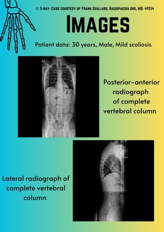
This radiology report documents x-rays of a 30-year-old male patient's complete vertebral column from both a posterior-anterior and lateral view. The x-rays reveal a mild scoliosis in the patient's spine.
Report
Share
Report
Share
Download to read offline
Recommended
Educational resource transcript .pdf
This educational resource provides an overview of how to identify structures within the human thorax on a radiograph. It discusses how dense tissues like bones appear brighter on radiographs since they absorb more x-rays, while less dense tissues like lungs appear darker. The document then labels key bones, organs and other structures visible on a chest x-ray, including the ribs, heart, lungs, trachea and intercostal muscles.
Educational resource transcript .docx
This educational resource provides an overview of how to identify structures within the human thorax on a radiograph. It discusses how dense tissues like bones appear brighter on radiographs since they absorb more x-rays, while less dense tissues like lungs appear darker. The resource then labels key bones, organs and other structures visible on a chest x-ray, including the ribs, heart, lungs, trachea and intercostal muscles.
Computed Tomography Reflection.pdf
1) The student witnessed a micro CT machine scan a temporal bone, which is used for paleontology research rather than medical purposes like they initially assumed.
2) Micro CT scans are much more expensive than typical hospital CT scans, costing £44-80 per hour to operate. However, they allow delicate fossils to be scanned without damaging them.
3) The student was impressed by the high quality 3D image produced from the thousands of angle projections captured, showing how detailed information can be extracted from specimens using CT technology.
Computed Tomography Factsheet.pdf
Computed tomography (CT) was first developed in the 1970s by Godfrey Hounsfield and Allan Cormack. The first CT scan was performed on a human patient in 1971. Today, CT scans provide detailed internal images of the body and take only a few minutes to perform, with patients typically receiving results within a couple weeks. CT scans use X-rays and computer processing to generate 3D images of the body by combining many X-ray images taken at different angles. Contrast agents may be used to make soft tissues more visible on scans.
Whole Portfolio.pdf
This document provides information on the history and development of anatomical illustration, photography, radiography, and computed tomography. It discusses key figures like Galen, Vesalius, Gray, and others and how their work advanced the fields. It also reflects on the author's experience in practical sessions for illustration, photography, and radiography where they were able to apply their learnings and experiment with different techniques. The author gained a better understanding of the arts and sciences behind each modality and how they capture anatomy in different dimensional representations.
Radiography Factsheet.pdf
Wilhelm Roentgen accidentally discovered x-rays in 1895 while experimenting with cathode ray tubes, noticing a fluorescent screen lighting up from invisible rays he called x-rays. His first x-ray image was of his wife's hand showing her wedding ring. Radiography uses x-rays, a form of electromagnetic radiation, to visualize internal body structures by passing through tissues of varying densities. Electrons are emitted from a cathode in an x-ray tube and accelerated toward an anode, where 1% of their kinetic energy is converted to x-rays emitted in a beam for imaging. While quick and non-invasive, x-rays produce ionizing radiation which in excessive exposure can damage tissues and increase cancer risk.
Microscopy Factsheet.pdf
Microscopy involves using a microscope to view objects too small to see with the naked eye, such as cells, organelles, viruses, and proteins. Microscopes work by using either light or electron beams to illuminate samples, with electron microscopes providing higher magnification and resolution than optical microscopes. Key aspects of microscopy include magnification, resolution, and the different types of microscopes such as transmission electron microscopes and scanning electron microscopes.
Photography Factsheet.pdf
The first photograph was taken in 1826 by Joseph Nicéphore Niépce, capturing "A view from the window Le Gras" using a camera obscura and a bitumen-coated plate that took hours of exposure to develop the image. Niépce dissolved leftover bitumen to make the image permanent, though the process was difficult and time-consuming. Advancing technology has since reduced the time needed to take a photograph from hours to less than a second.
Photography Images.pdf
This document contains a list of 7 image numbers. It also notes that image 7 has had its background removed and that all images are the property of the University of Bristol School of Anatomy.
Magnetic Resonance Imaging Factsheet.pdf
Raymond Damadian invented the MRI machine and performed the first MRI scan on a live patient in 1977. Through his research on sodium and potassium molecules in living cells, he experimented with nuclear magnetic resonance where atomic nuclei absorb and emit electromagnetic radiation in a magnetic field. He theorized that tumor and normal tissue could be distinguished by their prolonged relaxation times measured by NMR.
Ultrasound Factsheet.pdf
Italian physicist Lazaro Spallanzani theorized in 1794 that sound waves could be used to detect objects, inspired by how bats navigate with echolocation. French scientists Jacques and Pierre Curie later experimented with producing ultrasonic waves using quartz crystals and electrical currents. In 1956, Ian Donald first used ultrasound for medical diagnosis, measuring a fetus's head and establishing the field of sonography. Ultrasound produces live images of internal movement like the heartbeat in real time through high frequency sound wave emissions that are reflected back at different speeds through tissues.
Ultrasound Reflection.pdf
The student was excited to participate in an ultrasound practical having seen one before. They observed others scanning and provided feedback, making them confident to scan their volunteer classmate. Applying gel improved the image quality, showing ultrasound's live nature. Scanning the neck was interesting as the student could identify structures from previous anatomy lessons like arteries and veins. They also saw vocal cords vibrating in real time and the internal jugular vein enlarging during a breathing maneuver. Scanning the arm was less informative since upper limb anatomy had not been covered yet. Ultrasound requires specific training to interpret images clearly and know what to look for with the probe. The student wants more ultrasound experience to gain confidence in structure recognition.
Magnetic Resonance Imaging Reflection.pdf
The document discusses the author's experience with an MRI practical. They found watching lectures and applying the concepts hands-on in a role-playing activity helped them better understand magnetic fields over radiation. The practical highlighted factors that can affect MRI eligibility, such as certain tattoo inks containing metal. The author enjoyed identifying abnormalities on exposed cadavers by comparing MRI images to physical anatomy. They learned MRI and CT scans each have strengths for spatial and contrast resolution that make them useful for different diagnostic purposes.
Photography Reflection.pdf
Prior to taking photographs in a practical session, the author was confident in their photography skills from using smartphone cameras but found the scientific concepts behind DSLR cameras daunting. During the practical, the author focused on experimenting with different camera settings and found their photos improved as they gained experience. The author realized photography involves more skill than they initially thought and wishes they had more time to experiment further. They discuss ethical guidelines that were followed around photographing human anatomy.
Radiography Reflection.pdf
The document discusses the author's experience and learning about radiography. Before taking radiography classes, the author incorrectly thought only bones were visible on x-rays. Through personal experiences getting x-rays for broken bones and appendicitis, as well as learning about x-ray attenuation and viewing more radiographs, the author gained an understanding that soft tissues can also be seen. While soft tissues are less clear on x-rays than CT or MRI scans, radiographs remain very useful for diagnosing broken bones as a quick, non-invasive method.
Illustration Images.pdf
The document lists a series of drawing exercises including 15 minute drawings with the dominant hand, 1 minute drawings with the dominant hand, 1 minute one line drawings with the dominant hand, blind drawings with the non-dominant hand, basic shape drawings, a 15 minute drawing with the non-dominant hand, and a charcoal drawing with the dominant hand. The images from these exercises are the property of the University of Bristol School of Anatomy.
Illustration Factsheet.pdf
Galen, known as the 'Father of anatomy', made discoveries in the 2nd century by dissecting barbary apes since human dissection was forbidden. In the 1500s, Vesalius found inaccuracies in Galen's work due to his reliance on animal not human anatomy. He published the seven volume book "De humani corporis fabrica" with detailed human anatomy illustrations. In 1858, Gray and Carter published the first edition of "Gray's Anatomy" containing hand drawn illustrations which has since been updated through 40 editions and remains an important anatomical reference text.
The Evolution and Impact of Hip Hop a cultural and artistic
RGMDEZFE by RGM DEZ streaming everywhere Spotify, Apple Music, iTunes ,Tidal and etc
➒➌➎➏➑➐➋➑➐➐ Satta Matka Dpboss Matka Guessing Indian Matka
➒➌➎➏➑➐➋➑➐➐ Satta Matka Dpboss Matka Guessing Indian Matka➒➌➎➏➑➐➋➑➐➐Dpboss Matka Guessing Satta Matka Kalyan Chart Indian Matka
➒➌➎➏➑➐➋➑➐➐ Satta Matka Dpboss Matka Guessing Indian Matka KALYAN MATKA | MATKA RESULT | KALYAN MATKA TIPS | SATTA MATKA | MATKA.COM | MATKA PANA JODI TODAY | BATTA SATKA | MATKA PATTI JODI NUMBER | MATKA RESULTS | MATKA CHART | MATKA JODI | SATTA COM | FULL RATE GAME | MATKA GAME | MATKA WAPKA | ALL MATKA RESULT LIVE ONLINE | MATKA RESULT | KALYAN MATKA RESULT | DPBOSS MATKA 143 | MAIN MATKA➒➌➍➑➊➑➏➍➋➒ Satta Matka Satta result marka result Satta Matka Satta result mar...
➒➌➍➑➊➑➏➍➋➒ Satta Matka Satta result marka result Satta Matka Satta result mar...➒➌➍➑➊➑➏➍➋➒ Satta Matka Satta result marka result
➒➌➍➑➊➑➏➍➋➒ Satta Matka Satta result marka result Satta Matka Satta result marka result Dp Boss sattamatka341 satta143 Satta Matka Sattamatka New Mumbai Ratan Satta Matka Fast Matka Milan Market Kalyan Matka Results Satta Game Matka Game Satta Matka Kalyan Satta Matka Mumbai Main Online Matka Results Satta Matka Tips Milan Chart Satta Matka Boss New Star Day Satta King Live Satta Matka Results Satta Matka Company Indian Matka Satta Matka Kalyan Night Matka
Kalyan Result Final ank Satta 143 Kalyan final Kalyan panel chart Kalyan Result guessing Time bazar Kalyan guessing Kalyan satta sattamatka
Satta Matka Sattamatka Satta matka Satta result Matka result Satta result matkaresult Satta matka result Matka 420 Matka420 matka guessing matka guessing satta matta matka satta matta matka Kalyan chart Kalyan chart Satta matta Matka 143 SattamattaMatka143 Satta live Satta live Kalyan open Kalyan open Kalyan final Kalyan final Kalyan chart Kalyan chart Kalyan Panel Chart Kalyan Panel Chart Dp Boss india matka india matka
Dpboss Matka Guessing Satta Matta Matka Kalyan Chart Indian Matka
Dpboss Matka Guessing Satta Matta Matka Kalyan Chart Indian Matka➒➌➎➏➑➐➋➑➐➐Dpboss Matka Guessing Satta Matka Kalyan Chart Indian Matka
9356872877Sattamatka.satta.matka.satta matka.kalyan weekly chart.kalyan chart.kalyan jodi chart.kalyan penal chart.kalyan today.kalyan open.fix satta.fix fix fix Satta matka nambar. Dpboss Matka Guessing Satta Matta Matka Kalyan Chart Indian MatkaMatka Guessing Satta Matta Matka Kalyan Chart Indian Matka Dpboss
Matka Guessing Satta Matta Matka Kalyan Chart Indian Matka Dpboss➒➌➎➏➑➐➋➑➐➐Dpboss Matka Guessing Satta Matka Kalyan Chart Indian Matka
9356872877Sattamatka.satta.matka.satta matka.kalyan weekly chart.kalyan chart.kalyan jodi chart.kalyan penal chart.kalyan today.kalyan open.fix satta.fix fix fix Satta matka nambar. Dpboss Matka Guessing Satta Matta Matka Kalyan Chart Indian Matka➒➌➎➏➑➐➋➑➐➐ Satta Matka Dpboss Matka Guessing Indian Matka
➒➌➎➏➑➐➋➑➐➐ Satta Matka Dpboss Matka Guessing Indian Matka➒➌➎➏➑➐➋➑➐➐Dpboss Matka Guessing Satta Matka Kalyan Chart Indian Matka
➒➌➎➏➑➐➋➑➐➐ Satta Matka Dpboss Matka Guessing Indian Matka
KALYAN MATKA | MATKA RESULT | KALYAN MATKA TIPS | SATTA MATKA | MATKA.COM | MATKA PANA JODI TODAY | BATTA SATKA | MATKA PATTI JODI NUMBER | MATKA RESULTS | MATKA CHART | MATKA JODI | SATTA COM | FULL RATE GAME | MATKA GAME | MATKA WAPKA | ALL MATKA RESULT LIVE ONLINE | MATKA RESULT | KALYAN MATKA RESULT | DPBOSS MATKA 143 | MAIN MATKAARNAUVALERY RECORD STORE SCAVENGER HUNT.
This is my presentation for the Record Store Scavenger Hunt.
➒➌➎➏➑➐➋➑➐➐ Satta Matka Dpboss Matka Guessing
➒➌➎➏➑➐➋➑➐➐ Satta Matka Dpboss Matka Guessing➒➌➎➏➑➐➋➑➐➐Dpboss Matka Guessing Satta Matka Kalyan Chart Indian Matka
SATTA MATKA SATTA FAST RESULT KALYAN TOP MATKA RESULT KALYAN SATTA MATKA FAST RESULT MILAN RATAN RAJDHANI MAIN BAZAR MATKA FAST TIPS RESULT MATKA CHART JODI CHART PANEL CHART FREE FIX GAME SATTAMATKA ! MATKA MOBI SATTA 143 spboss.in TOP NO1 RESULT FULL RATE MATKA ONLINE GAME PLAY BY APP SPBOSS一比一原版迪肯大学毕业证(DU毕业证书)学历如何办理
多少钱办理【微信号:176555708】【多少钱办理(DU毕业证书)】【微信号:176555708】《成绩单、外壳、offer、真实留信官方学历认证(永久存档/真实可查)》采用学校原版纸张、特殊工艺完全按照原版一比一制作(包括:隐形水印,阴影底纹,钢印LOGO烫金烫银,LOGO烫金烫银复合重叠,文字图案浮雕,激光镭射,紫外荧光,温感,复印防伪)行业标杆!精益求精,诚心合作,真诚制作!多年品质 ,按需精细制作,24小时接单,全套进口原装设备,十五年致力于帮助留学生解决难题,业务范围有加拿大、英国、澳洲、韩国、美国、新加坡,新西兰等学历材料,包您满意。
【我们承诺采用的是学校原版纸张(纸质、底色、纹路)我们拥有全套进口原装设备,特殊工艺都是采用不同机器制作,仿真度基本可以达到100%,所有工艺效果都可提前给客户展示,不满意可以根据客户要求进行调整,直到满意为止!】
【业务选择办理准则】
一、工作未确定,回国需先给父母、亲戚朋友看下文凭的情况,办理一份就读学校的毕业证【微信号:176555708】文凭即可
二、回国进私企、外企、自己做生意的情况,这些单位是不查询毕业证真伪的,而且国内没有渠道去查询国外文凭的真假,也不需要提供真实教育部认证。鉴于此,办理一份毕业证【微信号:176555708】即可
三、进国企,银行,事业单位,考公务员等等,这些单位是必需要提供真实教育部认证的,办理教育部认证所需资料众多且烦琐,所有材料您都必须提供原件,我们凭借丰富的经验,快捷的绿色通道帮您快速整合材料,让您少走弯路。
留信网认证的作用:
1:该专业认证可证明留学生真实身份
2:同时对留学生所学专业登记给予评定
3:国家专业人才认证中心颁发入库证书
4:这个认证书并且可以归档倒地方
5:凡事获得留信网入网的信息将会逐步更新到个人身份内,将在公安局网内查询个人身份证信息后,同步读取人才网入库信息
6:个人职称评审加20分
7:个人信誉贷款加10分
8:在国家人才网主办的国家网络招聘大会中纳入资料,供国家高端企业选择人才
留信网服务项目:
1、留学生专业人才库服务(留信分析)
2、国(境)学习人员提供就业推荐信服务
3、留学人员区块链存储服务
【关于价格问题(保证一手价格)】
我们所定的价格是非常合理的,而且我们现在做得单子大多数都是代理和回头客户介绍的所以一般现在有新的单子 我给客户的都是第一手的代理价格,因为我想坦诚对待大家 不想跟大家在价格方面浪费时间
对于老客户或者被老客户介绍过来的朋友,我们都会适当给一些优惠。
选择实体注册公司办理,更放心,更安全!我们的承诺:客户在留信官方认证查询网站查询到认证通过结果后付款,不成功不收费!
Mr. Brainwash ❤️ Beautiful Girl _ FRANK FLUEGEL GALERIE.pdf
Mr. Brainwash Beautiful Girl / Mixed Media / signed / Unique
Year: 2023
Format: 96,5 x 127 cm / 37.8 x 50 inch
Material: Fine Art Paper with hand-torn edges.
Method: Mixed Media, Stencil, Spray Paint.
Edition: Unique
Other: handsigned by Mr. Brainwash front and verso.
Beautiful Girl by Mr. Brainwash is a mixed media artwork on paper done in 2023. It is unique and of course signed by Mr. Brainwash. The picture is a tribute to his own most successful work of art, the Balloon Girl. In this new creation, however, the theme of the little girl is slightly modified.
In Mr. Brainwash’s mixed media artwork titled “Beautiful Girl,” we are presented with a captivating depiction of a little girl adorned in a summer dress, with two playful pigtails framing her face. The artwork exudes a sense of innocence and whimsy, as the girl is shown in a dreamy state, lifting one end of her skirt and looking down as if she were about to dance. Through the use of mixed media, Mr. Brainwash skillfully combines different artistic elements to create a visually striking composition. The vibrant colors and bold brushstrokes bring the artwork to life, evoking a sense of joy and happiness. The attention to detail in the girl’s expression and body language adds depth and character to the piece, allowing viewers to connect with the young protagonist on a personal and emotional level. “Beautiful Girl” is a testament to Mr. Brainwash’s unique artistic style, blending elements of street art, pop art, and contemporary art to create a visually captivating and emotionally resonant artwork.
The use of mixed media in “Beautiful Girl” adds an additional layer of complexity to the artwork. By combining different artistic techniques and materials, such as stencils, spray paint, and collage, Mr. Brainwash creates a dynamic and textured composition that grabs the viewer’s attention. The juxtaposition of different textures and patterns adds depth and visual interest to the piece, while also emphasizing the artist’s eclectic and experimental approach to art-making. The inclusion of collage elements, such as newspaper clippings and torn posters, further enhances the artwork’s urban and contemporary feel. Overall, “Beautiful Girl” is a visually captivating and thought-provoking artwork that showcases Mr. Brainwash’s talent for blending different artistic elements to create a truly unique and engaging piece.
哪里办理(sjsu毕业证书)美国圣何塞州立大学毕业证硕士文凭证书原版一模一样
原版定制【微信:bwp0011】《(sjsu毕业证书)美国圣何塞州立大学毕业证硕士文凭证书》【微信:bwp0011】成绩单 、雅思、外壳、留信学历认证永久存档查询,采用学校原版纸张、特殊工艺完全按照原版一比一制作(包括:隐形水印,阴影底纹,钢印LOGO烫金烫银,LOGO烫金烫银复合重叠,文字图案浮雕,激光镭射,紫外荧光,温感,复印防伪)行业标杆!精益求精,诚心合作,真诚制作!多年品质 ,按需精细制作,24小时接单,全套进口原装设备,十五年致力于帮助留学生解决难题,业务范围有加拿大、英国、澳洲、韩国、美国、新加坡,新西兰等学历材料,包您满意。
【业务选择办理准则】
一、工作未确定,回国需先给父母、亲戚朋友看下文凭的情况,办理一份就读学校的毕业证【微信bwp0011】文凭即可
二、回国进私企、外企、自己做生意的情况,这些单位是不查询毕业证真伪的,而且国内没有渠道去查询国外文凭的真假,也不需要提供真实教育部认证。鉴于此,办理一份毕业证【微信bwp0011】即可
三、进国企,银行,事业单位,考公务员等等,这些单位是必需要提供真实教育部认证的,办理教育部认证所需资料众多且烦琐,所有材料您都必须提供原件,我们凭借丰富的经验,快捷的绿色通道帮您快速整合材料,让您少走弯路。
留信网认证的作用:
1:该专业认证可证明留学生真实身份
2:同时对留学生所学专业登记给予评定
3:国家专业人才认证中心颁发入库证书
4:这个认证书并且可以归档倒地方
5:凡事获得留信网入网的信息将会逐步更新到个人身份内,将在公安局网内查询个人身份证信息后,同步读取人才网入库信息
6:个人职称评审加20分
7:个人信誉贷款加10分
8:在国家人才网主办的国家网络招聘大会中纳入资料,供国家高端企业选择人才
【关于价格问题(保证一手价格)】
我们所定的价格是非常合理的,而且我们现在做得单子大多数都是代理和回头客户介绍的所以一般现在有新的单子 我给客户的都是第一手的代理价格,因为我想坦诚对待大家 不想跟大家在价格方面浪费时间
对于老客户或者被老客户介绍过来的朋友,我们都会适当给一些优惠。
"Snow" by Ann Beattie illustrated by Aimie Bourgeois
"Snow" by Ann Beattie illustrated into comic by aimie bourgeois
More Related Content
More from MabelWright1
Photography Images.pdf
This document contains a list of 7 image numbers. It also notes that image 7 has had its background removed and that all images are the property of the University of Bristol School of Anatomy.
Magnetic Resonance Imaging Factsheet.pdf
Raymond Damadian invented the MRI machine and performed the first MRI scan on a live patient in 1977. Through his research on sodium and potassium molecules in living cells, he experimented with nuclear magnetic resonance where atomic nuclei absorb and emit electromagnetic radiation in a magnetic field. He theorized that tumor and normal tissue could be distinguished by their prolonged relaxation times measured by NMR.
Ultrasound Factsheet.pdf
Italian physicist Lazaro Spallanzani theorized in 1794 that sound waves could be used to detect objects, inspired by how bats navigate with echolocation. French scientists Jacques and Pierre Curie later experimented with producing ultrasonic waves using quartz crystals and electrical currents. In 1956, Ian Donald first used ultrasound for medical diagnosis, measuring a fetus's head and establishing the field of sonography. Ultrasound produces live images of internal movement like the heartbeat in real time through high frequency sound wave emissions that are reflected back at different speeds through tissues.
Ultrasound Reflection.pdf
The student was excited to participate in an ultrasound practical having seen one before. They observed others scanning and provided feedback, making them confident to scan their volunteer classmate. Applying gel improved the image quality, showing ultrasound's live nature. Scanning the neck was interesting as the student could identify structures from previous anatomy lessons like arteries and veins. They also saw vocal cords vibrating in real time and the internal jugular vein enlarging during a breathing maneuver. Scanning the arm was less informative since upper limb anatomy had not been covered yet. Ultrasound requires specific training to interpret images clearly and know what to look for with the probe. The student wants more ultrasound experience to gain confidence in structure recognition.
Magnetic Resonance Imaging Reflection.pdf
The document discusses the author's experience with an MRI practical. They found watching lectures and applying the concepts hands-on in a role-playing activity helped them better understand magnetic fields over radiation. The practical highlighted factors that can affect MRI eligibility, such as certain tattoo inks containing metal. The author enjoyed identifying abnormalities on exposed cadavers by comparing MRI images to physical anatomy. They learned MRI and CT scans each have strengths for spatial and contrast resolution that make them useful for different diagnostic purposes.
Photography Reflection.pdf
Prior to taking photographs in a practical session, the author was confident in their photography skills from using smartphone cameras but found the scientific concepts behind DSLR cameras daunting. During the practical, the author focused on experimenting with different camera settings and found their photos improved as they gained experience. The author realized photography involves more skill than they initially thought and wishes they had more time to experiment further. They discuss ethical guidelines that were followed around photographing human anatomy.
Radiography Reflection.pdf
The document discusses the author's experience and learning about radiography. Before taking radiography classes, the author incorrectly thought only bones were visible on x-rays. Through personal experiences getting x-rays for broken bones and appendicitis, as well as learning about x-ray attenuation and viewing more radiographs, the author gained an understanding that soft tissues can also be seen. While soft tissues are less clear on x-rays than CT or MRI scans, radiographs remain very useful for diagnosing broken bones as a quick, non-invasive method.
Illustration Images.pdf
The document lists a series of drawing exercises including 15 minute drawings with the dominant hand, 1 minute drawings with the dominant hand, 1 minute one line drawings with the dominant hand, blind drawings with the non-dominant hand, basic shape drawings, a 15 minute drawing with the non-dominant hand, and a charcoal drawing with the dominant hand. The images from these exercises are the property of the University of Bristol School of Anatomy.
Illustration Factsheet.pdf
Galen, known as the 'Father of anatomy', made discoveries in the 2nd century by dissecting barbary apes since human dissection was forbidden. In the 1500s, Vesalius found inaccuracies in Galen's work due to his reliance on animal not human anatomy. He published the seven volume book "De humani corporis fabrica" with detailed human anatomy illustrations. In 1858, Gray and Carter published the first edition of "Gray's Anatomy" containing hand drawn illustrations which has since been updated through 40 editions and remains an important anatomical reference text.
More from MabelWright1 (9)
Recently uploaded
The Evolution and Impact of Hip Hop a cultural and artistic
RGMDEZFE by RGM DEZ streaming everywhere Spotify, Apple Music, iTunes ,Tidal and etc
➒➌➎➏➑➐➋➑➐➐ Satta Matka Dpboss Matka Guessing Indian Matka
➒➌➎➏➑➐➋➑➐➐ Satta Matka Dpboss Matka Guessing Indian Matka➒➌➎➏➑➐➋➑➐➐Dpboss Matka Guessing Satta Matka Kalyan Chart Indian Matka
➒➌➎➏➑➐➋➑➐➐ Satta Matka Dpboss Matka Guessing Indian Matka KALYAN MATKA | MATKA RESULT | KALYAN MATKA TIPS | SATTA MATKA | MATKA.COM | MATKA PANA JODI TODAY | BATTA SATKA | MATKA PATTI JODI NUMBER | MATKA RESULTS | MATKA CHART | MATKA JODI | SATTA COM | FULL RATE GAME | MATKA GAME | MATKA WAPKA | ALL MATKA RESULT LIVE ONLINE | MATKA RESULT | KALYAN MATKA RESULT | DPBOSS MATKA 143 | MAIN MATKA➒➌➍➑➊➑➏➍➋➒ Satta Matka Satta result marka result Satta Matka Satta result mar...
➒➌➍➑➊➑➏➍➋➒ Satta Matka Satta result marka result Satta Matka Satta result mar...➒➌➍➑➊➑➏➍➋➒ Satta Matka Satta result marka result
➒➌➍➑➊➑➏➍➋➒ Satta Matka Satta result marka result Satta Matka Satta result marka result Dp Boss sattamatka341 satta143 Satta Matka Sattamatka New Mumbai Ratan Satta Matka Fast Matka Milan Market Kalyan Matka Results Satta Game Matka Game Satta Matka Kalyan Satta Matka Mumbai Main Online Matka Results Satta Matka Tips Milan Chart Satta Matka Boss New Star Day Satta King Live Satta Matka Results Satta Matka Company Indian Matka Satta Matka Kalyan Night Matka
Kalyan Result Final ank Satta 143 Kalyan final Kalyan panel chart Kalyan Result guessing Time bazar Kalyan guessing Kalyan satta sattamatka
Satta Matka Sattamatka Satta matka Satta result Matka result Satta result matkaresult Satta matka result Matka 420 Matka420 matka guessing matka guessing satta matta matka satta matta matka Kalyan chart Kalyan chart Satta matta Matka 143 SattamattaMatka143 Satta live Satta live Kalyan open Kalyan open Kalyan final Kalyan final Kalyan chart Kalyan chart Kalyan Panel Chart Kalyan Panel Chart Dp Boss india matka india matka
Dpboss Matka Guessing Satta Matta Matka Kalyan Chart Indian Matka
Dpboss Matka Guessing Satta Matta Matka Kalyan Chart Indian Matka➒➌➎➏➑➐➋➑➐➐Dpboss Matka Guessing Satta Matka Kalyan Chart Indian Matka
9356872877Sattamatka.satta.matka.satta matka.kalyan weekly chart.kalyan chart.kalyan jodi chart.kalyan penal chart.kalyan today.kalyan open.fix satta.fix fix fix Satta matka nambar. Dpboss Matka Guessing Satta Matta Matka Kalyan Chart Indian MatkaMatka Guessing Satta Matta Matka Kalyan Chart Indian Matka Dpboss
Matka Guessing Satta Matta Matka Kalyan Chart Indian Matka Dpboss➒➌➎➏➑➐➋➑➐➐Dpboss Matka Guessing Satta Matka Kalyan Chart Indian Matka
9356872877Sattamatka.satta.matka.satta matka.kalyan weekly chart.kalyan chart.kalyan jodi chart.kalyan penal chart.kalyan today.kalyan open.fix satta.fix fix fix Satta matka nambar. Dpboss Matka Guessing Satta Matta Matka Kalyan Chart Indian Matka➒➌➎➏➑➐➋➑➐➐ Satta Matka Dpboss Matka Guessing Indian Matka
➒➌➎➏➑➐➋➑➐➐ Satta Matka Dpboss Matka Guessing Indian Matka➒➌➎➏➑➐➋➑➐➐Dpboss Matka Guessing Satta Matka Kalyan Chart Indian Matka
➒➌➎➏➑➐➋➑➐➐ Satta Matka Dpboss Matka Guessing Indian Matka
KALYAN MATKA | MATKA RESULT | KALYAN MATKA TIPS | SATTA MATKA | MATKA.COM | MATKA PANA JODI TODAY | BATTA SATKA | MATKA PATTI JODI NUMBER | MATKA RESULTS | MATKA CHART | MATKA JODI | SATTA COM | FULL RATE GAME | MATKA GAME | MATKA WAPKA | ALL MATKA RESULT LIVE ONLINE | MATKA RESULT | KALYAN MATKA RESULT | DPBOSS MATKA 143 | MAIN MATKAARNAUVALERY RECORD STORE SCAVENGER HUNT.
This is my presentation for the Record Store Scavenger Hunt.
➒➌➎➏➑➐➋➑➐➐ Satta Matka Dpboss Matka Guessing
➒➌➎➏➑➐➋➑➐➐ Satta Matka Dpboss Matka Guessing➒➌➎➏➑➐➋➑➐➐Dpboss Matka Guessing Satta Matka Kalyan Chart Indian Matka
SATTA MATKA SATTA FAST RESULT KALYAN TOP MATKA RESULT KALYAN SATTA MATKA FAST RESULT MILAN RATAN RAJDHANI MAIN BAZAR MATKA FAST TIPS RESULT MATKA CHART JODI CHART PANEL CHART FREE FIX GAME SATTAMATKA ! MATKA MOBI SATTA 143 spboss.in TOP NO1 RESULT FULL RATE MATKA ONLINE GAME PLAY BY APP SPBOSS一比一原版迪肯大学毕业证(DU毕业证书)学历如何办理
多少钱办理【微信号:176555708】【多少钱办理(DU毕业证书)】【微信号:176555708】《成绩单、外壳、offer、真实留信官方学历认证(永久存档/真实可查)》采用学校原版纸张、特殊工艺完全按照原版一比一制作(包括:隐形水印,阴影底纹,钢印LOGO烫金烫银,LOGO烫金烫银复合重叠,文字图案浮雕,激光镭射,紫外荧光,温感,复印防伪)行业标杆!精益求精,诚心合作,真诚制作!多年品质 ,按需精细制作,24小时接单,全套进口原装设备,十五年致力于帮助留学生解决难题,业务范围有加拿大、英国、澳洲、韩国、美国、新加坡,新西兰等学历材料,包您满意。
【我们承诺采用的是学校原版纸张(纸质、底色、纹路)我们拥有全套进口原装设备,特殊工艺都是采用不同机器制作,仿真度基本可以达到100%,所有工艺效果都可提前给客户展示,不满意可以根据客户要求进行调整,直到满意为止!】
【业务选择办理准则】
一、工作未确定,回国需先给父母、亲戚朋友看下文凭的情况,办理一份就读学校的毕业证【微信号:176555708】文凭即可
二、回国进私企、外企、自己做生意的情况,这些单位是不查询毕业证真伪的,而且国内没有渠道去查询国外文凭的真假,也不需要提供真实教育部认证。鉴于此,办理一份毕业证【微信号:176555708】即可
三、进国企,银行,事业单位,考公务员等等,这些单位是必需要提供真实教育部认证的,办理教育部认证所需资料众多且烦琐,所有材料您都必须提供原件,我们凭借丰富的经验,快捷的绿色通道帮您快速整合材料,让您少走弯路。
留信网认证的作用:
1:该专业认证可证明留学生真实身份
2:同时对留学生所学专业登记给予评定
3:国家专业人才认证中心颁发入库证书
4:这个认证书并且可以归档倒地方
5:凡事获得留信网入网的信息将会逐步更新到个人身份内,将在公安局网内查询个人身份证信息后,同步读取人才网入库信息
6:个人职称评审加20分
7:个人信誉贷款加10分
8:在国家人才网主办的国家网络招聘大会中纳入资料,供国家高端企业选择人才
留信网服务项目:
1、留学生专业人才库服务(留信分析)
2、国(境)学习人员提供就业推荐信服务
3、留学人员区块链存储服务
【关于价格问题(保证一手价格)】
我们所定的价格是非常合理的,而且我们现在做得单子大多数都是代理和回头客户介绍的所以一般现在有新的单子 我给客户的都是第一手的代理价格,因为我想坦诚对待大家 不想跟大家在价格方面浪费时间
对于老客户或者被老客户介绍过来的朋友,我们都会适当给一些优惠。
选择实体注册公司办理,更放心,更安全!我们的承诺:客户在留信官方认证查询网站查询到认证通过结果后付款,不成功不收费!
Mr. Brainwash ❤️ Beautiful Girl _ FRANK FLUEGEL GALERIE.pdf
Mr. Brainwash Beautiful Girl / Mixed Media / signed / Unique
Year: 2023
Format: 96,5 x 127 cm / 37.8 x 50 inch
Material: Fine Art Paper with hand-torn edges.
Method: Mixed Media, Stencil, Spray Paint.
Edition: Unique
Other: handsigned by Mr. Brainwash front and verso.
Beautiful Girl by Mr. Brainwash is a mixed media artwork on paper done in 2023. It is unique and of course signed by Mr. Brainwash. The picture is a tribute to his own most successful work of art, the Balloon Girl. In this new creation, however, the theme of the little girl is slightly modified.
In Mr. Brainwash’s mixed media artwork titled “Beautiful Girl,” we are presented with a captivating depiction of a little girl adorned in a summer dress, with two playful pigtails framing her face. The artwork exudes a sense of innocence and whimsy, as the girl is shown in a dreamy state, lifting one end of her skirt and looking down as if she were about to dance. Through the use of mixed media, Mr. Brainwash skillfully combines different artistic elements to create a visually striking composition. The vibrant colors and bold brushstrokes bring the artwork to life, evoking a sense of joy and happiness. The attention to detail in the girl’s expression and body language adds depth and character to the piece, allowing viewers to connect with the young protagonist on a personal and emotional level. “Beautiful Girl” is a testament to Mr. Brainwash’s unique artistic style, blending elements of street art, pop art, and contemporary art to create a visually captivating and emotionally resonant artwork.
The use of mixed media in “Beautiful Girl” adds an additional layer of complexity to the artwork. By combining different artistic techniques and materials, such as stencils, spray paint, and collage, Mr. Brainwash creates a dynamic and textured composition that grabs the viewer’s attention. The juxtaposition of different textures and patterns adds depth and visual interest to the piece, while also emphasizing the artist’s eclectic and experimental approach to art-making. The inclusion of collage elements, such as newspaper clippings and torn posters, further enhances the artwork’s urban and contemporary feel. Overall, “Beautiful Girl” is a visually captivating and thought-provoking artwork that showcases Mr. Brainwash’s talent for blending different artistic elements to create a truly unique and engaging piece.
哪里办理(sjsu毕业证书)美国圣何塞州立大学毕业证硕士文凭证书原版一模一样
原版定制【微信:bwp0011】《(sjsu毕业证书)美国圣何塞州立大学毕业证硕士文凭证书》【微信:bwp0011】成绩单 、雅思、外壳、留信学历认证永久存档查询,采用学校原版纸张、特殊工艺完全按照原版一比一制作(包括:隐形水印,阴影底纹,钢印LOGO烫金烫银,LOGO烫金烫银复合重叠,文字图案浮雕,激光镭射,紫外荧光,温感,复印防伪)行业标杆!精益求精,诚心合作,真诚制作!多年品质 ,按需精细制作,24小时接单,全套进口原装设备,十五年致力于帮助留学生解决难题,业务范围有加拿大、英国、澳洲、韩国、美国、新加坡,新西兰等学历材料,包您满意。
【业务选择办理准则】
一、工作未确定,回国需先给父母、亲戚朋友看下文凭的情况,办理一份就读学校的毕业证【微信bwp0011】文凭即可
二、回国进私企、外企、自己做生意的情况,这些单位是不查询毕业证真伪的,而且国内没有渠道去查询国外文凭的真假,也不需要提供真实教育部认证。鉴于此,办理一份毕业证【微信bwp0011】即可
三、进国企,银行,事业单位,考公务员等等,这些单位是必需要提供真实教育部认证的,办理教育部认证所需资料众多且烦琐,所有材料您都必须提供原件,我们凭借丰富的经验,快捷的绿色通道帮您快速整合材料,让您少走弯路。
留信网认证的作用:
1:该专业认证可证明留学生真实身份
2:同时对留学生所学专业登记给予评定
3:国家专业人才认证中心颁发入库证书
4:这个认证书并且可以归档倒地方
5:凡事获得留信网入网的信息将会逐步更新到个人身份内,将在公安局网内查询个人身份证信息后,同步读取人才网入库信息
6:个人职称评审加20分
7:个人信誉贷款加10分
8:在国家人才网主办的国家网络招聘大会中纳入资料,供国家高端企业选择人才
【关于价格问题(保证一手价格)】
我们所定的价格是非常合理的,而且我们现在做得单子大多数都是代理和回头客户介绍的所以一般现在有新的单子 我给客户的都是第一手的代理价格,因为我想坦诚对待大家 不想跟大家在价格方面浪费时间
对于老客户或者被老客户介绍过来的朋友,我们都会适当给一些优惠。
"Snow" by Ann Beattie illustrated by Aimie Bourgeois
"Snow" by Ann Beattie illustrated into comic by aimie bourgeois
➒➌➎➏➑➐➋➑➐➐ Satta Matka Dpboss Matka Guessing Indian Matka KALYAN MATKA |
➒➌➎➏➑➐➋➑➐➐ Satta Matka Dpboss Matka Guessing Indian Matka KALYAN MATKA |➒➌➎➏➑➐➋➑➐➐Dpboss Matka Guessing Satta Matka Kalyan Chart Indian Matka
➒➌➎➏➑➐➋➑➐➐ Satta Matka Dpboss Matka Guessing Indian Matka
KALYAN MATKA | MATKA RESULT | KALYAN MATKA TIPS | SATTA MATKA | MATKA.COM | MATKA PANA JODI TODAY | BATTA SATKA | MATKA PATTI JODI NUMBER | MATKA RESULTS | MATKA CHART | MATKA JODI | SATTA COM | FULL RATE GAME | MATKA GAME | MATKA WAPKA | ALL MATKA RESULT LIVE ONLINE | MATKA RESULT | KALYAN MATKA RESULT | DPBOSS MATKA 143 | MAIN MATKASatta matka guessing matka Kalyan chart
SATTA MATKA | DPBOSS | KALYAN MAIN BAZAR | FAST MATKA RESULT KALYAN MATKA | MATKA RESULT | KALYAN MATKA TIPS | SATTA MATKA | MATKA COM | MATKA PANA JODI TODAY | BATTA SATKA | MATKA PATTI JODI NUMBER | MATKA RESULTS | MATKA CHART | MATKA JODI | SATTA COM | FULL RATE GAME | MATKA GAME | MATKA WAPKA | ALL MATKA RESULT LIVE ONLINE | MATKA RESULT | KALYAN MATKA RESULT | DPBOSS MATKA 143 | MAIN MATKA
MATKA NUMBER FIX MATKANUMBER FIX SATTAMATKA FIXMATKANUMBER SATTA MATKA ALL SATTA MATKA FREE GAME KALYAN MATKA TIPS KAPIL MATKA GAME SATTA MATKA KALYAN GAME DAILY FREE 4 ANK ALL MARKET PUBLIC SEVA WEBSITE FIX FIX MATKA NUMBER INDIA.S NO1 WEBSITE TTA FIX FIX FIX MATKA GURU INDIA MATKA KALYAN CHART MATKA GUESSING KALYAN FIX OPEN FINAL 3 ANK SATTAMATKA143 GUESSING SATTA BATTA MATKA FIX NUMBER TODAY WAPKA FIX AAPKA FIX FIX FIX FIX SATTA GURU NUMBER SATTA MATKA MATKA143 SATTA SATTA SATTA MATKA SATTAMATKA1438 FIX MATKA MATKA BOSS SATTA LIVE 3MATKA 143 FIX FIX FIX KALYAN JODI MATKA KALYAN FIX FIX WAP MATKA BOSS440 SATTA MATKA FIX FIX MATKA NUMBER SATTA MATKA FIXMATKANUMBER FIX MATKA MATKA RESULT FIX MATKA NUMBER FREE DAILY FIX MATKA NUMBER FIX FIX MATKA JODI SATTA MATKA FIX ANK MATKA ANK FIX KALYAN MUMBAI MATKA NUMBER FIXMATKANUMBER SATTA NUMBER FAST MATKA RESULT SATTA BATTA INDIAN SATTA SATTA RESULT MADHUR SATTA PRABHAT SATTA FIX FIX FIX SATTA NUMBER SATTAKING143 GUESSING SATTA CHART KALYAN PENAL CHART MATKA420 SATTA GUESSING NUMBER KALYAN NIGHT CHART SATTA FIX FIX FIX SATTA NUMBER FIX FIX FIX OPEN FIX FIX WAPKA MATKA DPBOSS FIX FIX 3ANK MATKA KALYAN CHART MATKA GUESSING TARA MATKA FIX FIXMATKANUMBER FINAL ANK MATKABOSS DUBAI SATTA MATKA GOLDEN MATKA FIX FIX MATKA NUMBER FIX MATKANUMBER FIX FIX FIX MATKA NUMBER FIX MATKANUMBER FIX SATTAMATKA FIXMATKANUMBER SATTA MATKA ALL SATTA MATKA FREE GAME KALYAN MATKA TIPS KAPIL MATKA GAME SATTA MATKA KALYAN GAME DAILY FREE 4 ANK ALL MARKET PUBLIC SEVA SATTA FIX FIX FIX MATKA GURU INDIA MATKA KALYAN CHART MATKA GUESSING KALYAN FIX OPEN FINAL 3 ANK SATTAMATKA143 GUESSING SATTA BATTA MATKA FIX NUMBER TODAY WAPKA FIX AAPKA FIX FIX FIX FIX SATTA GURU NUMBER SATTA MATKA MATKA143 SATTA SATTA SATTA MATKA SATTAMATKA1438 FIX MATKA MATKA BOSS SATTA LIVE 3MATKA 143 FIX FIX FIX KALYAN JODI MATKA KALYAN FIX FIX WAP MATKA BOSS440 SATTA MATKA FIX FIX MATKA NUMBER SATTA MATKA FIXMATKANUMBER FIX MATKA MATKA RESULT FIX MATKA NUMBER FREE DAILY FIX MATKA NUMBER FIX FIX MATKA JODI SATTA MATKA FIX ANK MATKA ANK FIX KALYAN MUMBAI MATKA NUMBER FIXMATKANUMBER SATTA NUMBER FAST MATKA RESULT SATTA BATTA INDIAN SATTA SATTA RESULT MADHUR SATTA PRABHAT SATTA FIX FIX FIX SATTA NUMBER SATTAKING143 GUESSING SATTA CHART KALYAN PENAL CHART MATKA420 SATTA GUESSING NUMBER KALYAN NIGHT CHART SATTA FIX FIX FIX SATTA NUMBER FIX FIX FIX OPEN FIX FIX WAPKA MATKA DPBOSS FIX FIX 3ANK MATKA KALYAN CHART MATKA GUESSING TARA MATKA FIX FIXMATKANUMBER FINAL ANK MATKABOSS DUBAI SATTA MATKA GOLDEN MATKA WAPKA.FIX FIX FIX
VTV FULL SCRIPT ------------------------
HERE IS THE FULL SCRIPT OF VINNAITHTHAANDI VARUVAAYA BY GAUTHAM VAASUDEV MENON
Sattamatka satta matka sattamatka results
➒➌➍➑➊➑➏➍➋➒ Satta Matka Satta result marka result
Satta Matka Satta result marka result Dpboss sattamatka341 satta143 Satta Matka Sattamatka New Mumbai Ratan Satta Matka Fast Matka Milan Market Kalyan Matka Results Satta Game Matka Game Satta Matka Kalyan Satta Matka Mumbai Main Online Matka Results Satta Matka Tips Milan Chart Satta Matka Boss
New Star Day Satta King Live Satta Matka Results Satta Matka Company Indian Matka Satta Matka Kalyan Night Matka
➒➌➍➑➊➑➏➍➋➒ Satta Matka Satta result marka result
Recently uploaded (20)
The Evolution and Impact of Hip Hop a cultural and artistic
The Evolution and Impact of Hip Hop a cultural and artistic
➒➌➎➏➑➐➋➑➐➐ Satta Matka Dpboss Matka Guessing Indian Matka
➒➌➎➏➑➐➋➑➐➐ Satta Matka Dpboss Matka Guessing Indian Matka
➒➌➍➑➊➑➏➍➋➒ Satta Matka Satta result marka result Satta Matka Satta result mar...
➒➌➍➑➊➑➏➍➋➒ Satta Matka Satta result marka result Satta Matka Satta result mar...
Dpboss Matka Guessing Satta Matta Matka Kalyan Chart Indian Matka
Dpboss Matka Guessing Satta Matta Matka Kalyan Chart Indian Matka
Matka Guessing Satta Matta Matka Kalyan Chart Indian Matka Dpboss
Matka Guessing Satta Matta Matka Kalyan Chart Indian Matka Dpboss
➒➌➎➏➑➐➋➑➐➐ Satta Matka Dpboss Matka Guessing Indian Matka
➒➌➎➏➑➐➋➑➐➐ Satta Matka Dpboss Matka Guessing Indian Matka
Mr. Brainwash ❤️ Beautiful Girl _ FRANK FLUEGEL GALERIE.pdf
Mr. Brainwash ❤️ Beautiful Girl _ FRANK FLUEGEL GALERIE.pdf
chemistry projectdssssssssssssssssssssssssssssssssssss
chemistry projectdssssssssssssssssssssssssssssssssssss
"Snow" by Ann Beattie illustrated by Aimie Bourgeois
"Snow" by Ann Beattie illustrated by Aimie Bourgeois
➒➌➎➏➑➐➋➑➐➐ Satta Matka Dpboss Matka Guessing Indian Matka KALYAN MATKA |
➒➌➎➏➑➐➋➑➐➐ Satta Matka Dpboss Matka Guessing Indian Matka KALYAN MATKA |
Radiography Images.pdf
- 1. Images © X-ray- Case courtesy of Frank Gaillard, Radiopaedia.org, rID: 49514 Posterior-anterior radiograph of complete vertebral column Lateral radiograph of complete vertebral column Patient data: 30 years, Male, Mild scoliosis