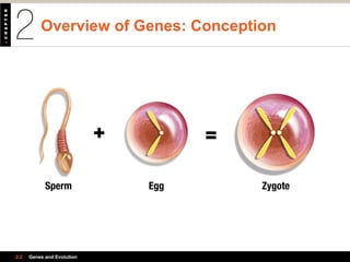
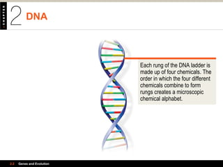
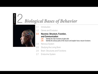
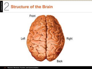
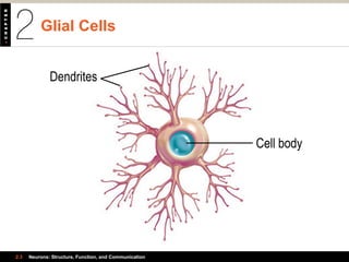
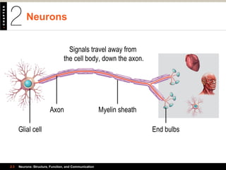
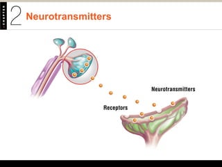
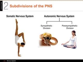
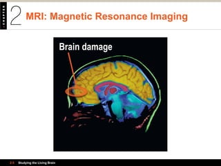
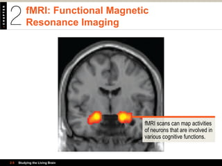
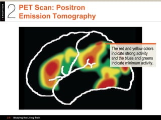
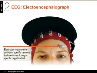
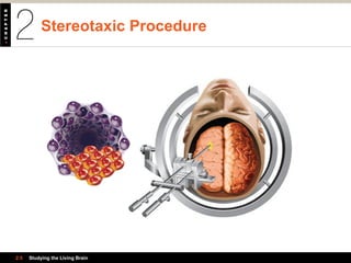
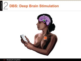
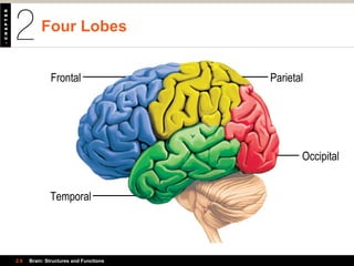
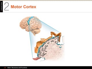
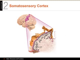
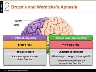
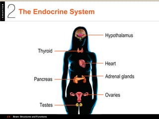
2.1 Introduction
2.2 Genes and Evolution
L02 Describe the structures and processes involved in genetic transmission
L03 Articulate the forces believed to be responsible for the evolution of the human
brain and describe the relevance of the theory of evolution in how psychologists
conduct research today
2.3 Neurons: Structure, Function,
and Communication
2.4 Nervous System
2.5 Studying the Living Brain
2.6 Brain: Structures and Functions
2.7 Endocrine System
Overview of Genes: Conception
2.2 Genes and Evolution
Sperm Egg Zygote
Chromosomes
2.2 Genes and Evolution
Each cell of the human
body contains 46
chromosomes arranged
in 23 pairs.
DNA
2.2 Genes and Evolution
Each rung of the DNA ladder is
made up of four chemicals. The
order in which the four different
chemicals combine to form
rungs creates a microscopic
chemical alphabet.
Genes and Proteins
and Polymorphic Genes
2.2 Genes and Evolution
On each chromosome are
specific segments that
contain particular instructions.
In the chromosome on the
left, each segment represents
the location of a gene.
Dominant and Recessive Genes
2.2 Genes and Evolution
Sperm Egg ZygoteZygote
r
Father Mother
Child
Brown eyesBrown eyesSperm Egg Zygote
D r
Sperm Egg Zygote
D D
Sperm Egg Zygote
rr
Brown eyes
Brown eyes
Blue eyes
DDrDDD rrr
Evolution of the Human Brain
2.2 Genes and Evolution
2.1 Introduction
2.2 Genes and Evolution
2.3 Neurons: Structure, Function,
and Communication
L04 Identify the main functions of glial cells
L05 Identify the various parts of the neuron and explain how a neuron functions
2.4 Nervous System
2.5 Studying the Living Brain
2.6 Brain: Structures and Functions
2.7 Endocrine System
Structure of the Brain
2.3 Neurons: Structure, Function, and Communication
Front
Back
RightLeft
Glial Cells
2.3 Neurons: Structure, Function, and Communication
Cell body
Dendrites
Neurons
2.3 Neurons: Structure, Function, and Communication
Glial cell
Axon Myelin sheath
End bulbs
Signals travel away from
the cell body, down the axon.
Neurotransmitters
Neurotransmitters
Receptors
Reflex Responses
2.3 Neurons: Structure, Function, and Communication
3. Interneuron makes
connections between
neurons, which carry
message to the brain.
Afferent, or
sensory, neuron
4. Efferent, or motor, neuron
carries neural messages
from spinal cord to hand.
2. Afferent, or sensory, neuron
carries neural messages
from hand to spinal cord.
Efferent, or
motor, neuron
1. Sensors
2.1 Introduction
2.2 Genes and Evolution
2.3 Neurons: Structure, Function,
and Communication
2.4 Nervous System
L09 Differentiate between the nerves and neurons
L10 Classify the major divisions and subdivisions of the nervous system
L11 Differentiate the functions of the major divisions and subdivisions
of the nervous system
2.5 Studying the Living Brain
2.6 Brain: Structures and Functions
2.7 Endocrine System
Central Nervous System
2.4 Nervous System
The spinal cord is made up of
neurons and bundles of axons
and dendrites that carry
information back and forth
between the brain and the body.
Peripheral Nervous System
2.4 Nervous System
Nerves carry information from
the senses, skin, muscles,
and the body’s organs to and
from the spinal cord.
Subdivisions of the PNS
2.4 Nervous System
Somatic Nervous System Autonomic Nervous System
Sympathetic
Division
Parasympathetic
Division
2.1 Introduction
2.2 Genes and Evolution
2.3 Neurons: Structure, Function,
and Communication
2.4 Nervous System
2.5 Studying the Living Brain
L12 Describe the different technologies used to investigate the brain
L13 Describe experimental procedures to treat the brain
2.6 Brain: Structures and Functions
2.7 Endocrine System
MRI: Magnetic Resonance Imaging
2.5 Studying the Living Brain
Brain damage
fMRI: Functional Magnetic
Resonance Imaging
2.5 Studying the Living Brain
fMRI scans can map activities
of neurons that are involved in
various cognitive functions.
PET Scan: Positron
Emission Tomography
2.5 Studying the Living Brain
The red and yellow colors
indicate strong activity
and the blues and greens
indicate minimum activity.
EEG: Electoencephalograph
2.5 Studying the Living Brain
Electrodes measure the
activity of specific neurons
that are in use during a
specific cognitive task.
Stem Cells
2.5 Studying the Living Brain
Click on play button
to launch video in
QuickTime player.
Stereotaxic Procedure
2.5 Studying the Living Brain
DBS: Deep Brain Stimulation
2.5 Studying the Living Brain
2.1 Introduction
2.2 Genes and Evolution
2.3 Neurons: Structure, Function,
and Communication
2.4 Nervous System
2.5 Studying the Living Brain
2.6 Brain: Structures and Functions
L14 Identify and locate the major parts of the brain, and state their functions
L15 Identify and locate the four lobes in the cerebral cortex, and state their
key functions
L16 Identify and locate key structures in the limbic system, and state their functions
L17 Identify sex differences in the brain
L18 Describe the lateralization of brain functions
2.7 Endocrine System
Major Parts of the Brain:
The Forebrain and Midbrain
2.6 Brain: Structures and Functions
Forebrain
Midbrain
Pons
Medulla
Cerebellum
Major Parts of the Brain:
The Hindbrain
2.6 Brain: Structures and Functions
Click on play button
to launch video in
QuickTime player.
The Cortex
2.6 Brain: Structures and Functions
Click on play button
to launch video in
QuickTime player.
Four Lobes
2.6 Brain: Structures and Functions
Frontal
Temporal
Occipital
Parietal
Motor Cortex
Toes
Ankle
Knee
Hip
Trunk
Shoulder
Elbow
Wrist
Hand
Little
Ring
MiddleIndexThumbNeck
Brow
Face
Lips
Eyelidand eyeball
Jaw
Tongue
Swallowing
2.6 Brain: Structures and Functions
Toes
Genitals
Foot
Leg
Hip
Trunk
Shoulder
HeadNeck
ElbowArm
Forearm
Wrist
Hand
Little
Ring
MiddleIndexThumbEye
FaceUpper lip
Lower lip
Lips
Nose
Teeth, gums, and jaw
Tongue
Somatosensory Cortex
2.6 Brain: Structures and Functions
Broca’s area Wernicke’s
area
Frontal
lobe
Temporal
lobe
Broca’s and Wernicke’s Aphasia
2.6 Brain: Structures and Functions
Temporal Lobe (understanding)Frontal Lobe (speaking)
Wernicke’s AreaBroca’s Area
Understands sentences
What are you doing in this hospital?
“I have been working as a
nurse in this hospital.”
Produces speech
I am working as a nurse
at this hospital.
Visual Association Area
2.6 Brain: Structures and Functions
Occipital
lobe
The Limbic System
2.6 Brain: Structures and Functions
Click on play button
to launch video in
QuickTime player.
Split Brain
2.6 Brain: Structures and Functions
2.1 Introduction
2.2 Genes and Evolution
2.3 Neurons: Structure, Function,
and Communication
2.4 Nervous System
2.5 Studying the Living Brain
2.6 Brain: Structures and Functions
2.7 Endocrine System
L19 Locate and describe the key elements of the endocrine system
L20 Discuss some ways that hormones regulate behavior
The Endocrine System
2.6 Brain: Structures and Functions
Thyroid
Pancreas
Testes
Hypothalamus
Heart
Adrenal glands
Ovaries

Psychology 101 Plotnik chapter02-v03

  • 2.
    2.1 Introduction 2.2 Genesand Evolution L02 Describe the structures and processes involved in genetic transmission L03 Articulate the forces believed to be responsible for the evolution of the human brain and describe the relevance of the theory of evolution in how psychologists conduct research today 2.3 Neurons: Structure, Function, and Communication 2.4 Nervous System 2.5 Studying the Living Brain 2.6 Brain: Structures and Functions 2.7 Endocrine System
  • 3.
    Overview of Genes:Conception 2.2 Genes and Evolution Sperm Egg Zygote
  • 4.
    Chromosomes 2.2 Genes andEvolution Each cell of the human body contains 46 chromosomes arranged in 23 pairs.
  • 5.
    DNA 2.2 Genes andEvolution Each rung of the DNA ladder is made up of four chemicals. The order in which the four different chemicals combine to form rungs creates a microscopic chemical alphabet.
  • 6.
    Genes and Proteins andPolymorphic Genes 2.2 Genes and Evolution On each chromosome are specific segments that contain particular instructions. In the chromosome on the left, each segment represents the location of a gene.
  • 7.
    Dominant and RecessiveGenes 2.2 Genes and Evolution Sperm Egg ZygoteZygote r Father Mother Child Brown eyesBrown eyesSperm Egg Zygote D r Sperm Egg Zygote D D Sperm Egg Zygote rr Brown eyes Brown eyes Blue eyes DDrDDD rrr
  • 8.
    Evolution of theHuman Brain 2.2 Genes and Evolution
  • 9.
    2.1 Introduction 2.2 Genesand Evolution 2.3 Neurons: Structure, Function, and Communication L04 Identify the main functions of glial cells L05 Identify the various parts of the neuron and explain how a neuron functions 2.4 Nervous System 2.5 Studying the Living Brain 2.6 Brain: Structures and Functions 2.7 Endocrine System
  • 10.
    Structure of theBrain 2.3 Neurons: Structure, Function, and Communication Front Back RightLeft
  • 11.
    Glial Cells 2.3 Neurons:Structure, Function, and Communication Cell body Dendrites
  • 12.
    Neurons 2.3 Neurons: Structure,Function, and Communication Glial cell Axon Myelin sheath End bulbs Signals travel away from the cell body, down the axon.
  • 13.
  • 14.
    Reflex Responses 2.3 Neurons:Structure, Function, and Communication 3. Interneuron makes connections between neurons, which carry message to the brain. Afferent, or sensory, neuron 4. Efferent, or motor, neuron carries neural messages from spinal cord to hand. 2. Afferent, or sensory, neuron carries neural messages from hand to spinal cord. Efferent, or motor, neuron 1. Sensors
  • 15.
    2.1 Introduction 2.2 Genesand Evolution 2.3 Neurons: Structure, Function, and Communication 2.4 Nervous System L09 Differentiate between the nerves and neurons L10 Classify the major divisions and subdivisions of the nervous system L11 Differentiate the functions of the major divisions and subdivisions of the nervous system 2.5 Studying the Living Brain 2.6 Brain: Structures and Functions 2.7 Endocrine System
  • 16.
    Central Nervous System 2.4Nervous System The spinal cord is made up of neurons and bundles of axons and dendrites that carry information back and forth between the brain and the body.
  • 17.
    Peripheral Nervous System 2.4Nervous System Nerves carry information from the senses, skin, muscles, and the body’s organs to and from the spinal cord.
  • 18.
    Subdivisions of thePNS 2.4 Nervous System Somatic Nervous System Autonomic Nervous System Sympathetic Division Parasympathetic Division
  • 19.
    2.1 Introduction 2.2 Genesand Evolution 2.3 Neurons: Structure, Function, and Communication 2.4 Nervous System 2.5 Studying the Living Brain L12 Describe the different technologies used to investigate the brain L13 Describe experimental procedures to treat the brain 2.6 Brain: Structures and Functions 2.7 Endocrine System
  • 20.
    MRI: Magnetic ResonanceImaging 2.5 Studying the Living Brain Brain damage
  • 21.
    fMRI: Functional Magnetic ResonanceImaging 2.5 Studying the Living Brain fMRI scans can map activities of neurons that are involved in various cognitive functions.
  • 22.
    PET Scan: Positron EmissionTomography 2.5 Studying the Living Brain The red and yellow colors indicate strong activity and the blues and greens indicate minimum activity.
  • 23.
    EEG: Electoencephalograph 2.5 Studyingthe Living Brain Electrodes measure the activity of specific neurons that are in use during a specific cognitive task.
  • 24.
    Stem Cells 2.5 Studyingthe Living Brain Click on play button to launch video in QuickTime player.
  • 25.
  • 26.
    DBS: Deep BrainStimulation 2.5 Studying the Living Brain
  • 27.
    2.1 Introduction 2.2 Genesand Evolution 2.3 Neurons: Structure, Function, and Communication 2.4 Nervous System 2.5 Studying the Living Brain 2.6 Brain: Structures and Functions L14 Identify and locate the major parts of the brain, and state their functions L15 Identify and locate the four lobes in the cerebral cortex, and state their key functions L16 Identify and locate key structures in the limbic system, and state their functions L17 Identify sex differences in the brain L18 Describe the lateralization of brain functions 2.7 Endocrine System
  • 28.
    Major Parts ofthe Brain: The Forebrain and Midbrain 2.6 Brain: Structures and Functions Forebrain Midbrain Pons Medulla Cerebellum
  • 29.
    Major Parts ofthe Brain: The Hindbrain 2.6 Brain: Structures and Functions Click on play button to launch video in QuickTime player.
  • 30.
    The Cortex 2.6 Brain:Structures and Functions Click on play button to launch video in QuickTime player.
  • 31.
    Four Lobes 2.6 Brain:Structures and Functions Frontal Temporal Occipital Parietal
  • 32.
  • 33.
  • 34.
    Broca’s area Wernicke’s area Frontal lobe Temporal lobe Broca’sand Wernicke’s Aphasia 2.6 Brain: Structures and Functions Temporal Lobe (understanding)Frontal Lobe (speaking) Wernicke’s AreaBroca’s Area Understands sentences What are you doing in this hospital? “I have been working as a nurse in this hospital.” Produces speech I am working as a nurse at this hospital.
  • 35.
    Visual Association Area 2.6Brain: Structures and Functions Occipital lobe
  • 36.
    The Limbic System 2.6Brain: Structures and Functions Click on play button to launch video in QuickTime player.
  • 37.
    Split Brain 2.6 Brain:Structures and Functions
  • 38.
    2.1 Introduction 2.2 Genesand Evolution 2.3 Neurons: Structure, Function, and Communication 2.4 Nervous System 2.5 Studying the Living Brain 2.6 Brain: Structures and Functions 2.7 Endocrine System L19 Locate and describe the key elements of the endocrine system L20 Discuss some ways that hormones regulate behavior
  • 39.
    The Endocrine System 2.6Brain: Structures and Functions Thyroid Pancreas Testes Hypothalamus Heart Adrenal glands Ovaries

Editor's Notes

  • #4 This Chapter introduces us to the biological bases of behavior. It covers: Genes and Evolution   The structure and function of neurons in the brain and how they communicate   The Nervous System   The Structure of the Brain and its functions   And the Endocrine System
  • #5 The next few slides will be a basic review of the process of conception and the process involved in transmitting genes to the next generation. First is the FERTILIZATION process: The male sperm contains 23 chromosomes and the female egg contains an equal number of 23. When the sperm penetrates the egg at conception the fertilized egg is called a zygote. It contains 46 chromosomes arranged in 23 pairs that contain the basic blueprint for creating a human being.
  • #6 A chromosome is a short, rod-like structure that contains the chemical DNA on tightly coiled structures.
  • #7 These DNA coils are arranged in a long strand that looks like a twisted ladder with rungs.
  • #8 A gene is a segment on the DNA strand that contains the instructions for making proteins that are the chemical building blocks that make build all the parts of a brain and body. Most genes have only one version, but about 1% of them are what we call “Polymorphic Genes” meaning they can carry more than one set of instructions - which helps to explain some of the variations we see in people.
  • #9 A DOMINANT GENE is a polymorphic gene that determines the trait even if paired with a Recessive gene. A RECESSIVE GENE is one that will manifest a trait only if both parents have the gene. When it comes to eye color the dominant gene is for Brown Eyes. BTW…just a little side fact. Blue eyes are actually a genetic mutation of a portion of the gene that controls Melanin – it controls coloration in skin, hair, eyes etc. Only 10,000 years ago everyone had brown eyes… and then there was an anomaly that altered a small part of that gene, resulting in a fading of the iris turning it blue.
  • #10 The Theory of Evolution was based on the revolutionary ideas and research of Charles Darwin in the mid 1800’s. It proposes that we as humans arose as a different species from a common ancestor millions of years ago. According to evolution theory we are the descendant s of a creature that split off from apes. We now have evidence that Darwin was not privileged to at that time…DNA testing actually shows that we are very closely related to Chimpanzees and share 98% of the same DNA. There are two forces at play in this process. 1-genetic mutations – which are accidental error in the genetic instructions (just like the mutation of the gene for eye color we just talked about). And the other is Natural Selection – which means that those organisms that are successful will continue to pass on their genes and insure the success of that species.
  • #12 This is a view of the human brain from the top. It looks sort of like a walnut, with two clear halves, and it is scrunched and wrinkled in appearance. The two halves are connected by nerve fibers called the corpus collosum. It weighs about 3 lbs. and is a pinkish white color with the consistency of Jello. It is fueled by sugar (glucose) and has about one TRILLION cells that come in two forms: Glial cells and Neurons. We’ll look at those two groups individually…
  • #13 The Glial cells are the most numerous – about 900 billion of the trillion cells are Glials (or astrocytes – meaning star shaped). Their main job is to support growing neurons, help insulate the neurons from electrical interference, and release chemicals needed by the neurons to grow and function. And we have recently discovered that some of them may even transmit electrical signals of their own, a function previously thought to be only carried out by Neurons.
  • #14 Neurons account for the remaining 100 thousand or so brain cells. They are smaller in number put play a huge role in making your brain function. They transmit electrical signals at speeds up to 200 MPH – ZOOM! One of the main differences between Glial cells and Neurons is the fact for the most part (there may be a few exceptions) Neurons are not able to reproduce the way Glial cells do. The Neurons you are born with will pretty much have to keep you going for your life. So guard them carefully – your ability to learn and remember new things depends of having ample Neurons in good working order. The research (although highly controversial) being done with Stem Cells that can facilitate the growth of new Neuron cells holds promise for repairing damage from strokes or accidents or diseases such as Alzheimers’ and Parkinsons. This animation defines the parts and the functions of a Neuron and the process called an Action Potential explaining how they “fire”.
  • #16 A REFLEX is an unlearned, involuntary reaction some stimulus. It is “pre-wired” by genetic instructions, but although the reaction is spontaneous and immediate without having to give conscious thought to it, there is still a sequence of neural connections that take place as you can see in this graphic. First comes the sensor…in this case a hot light bulb on the skin. Next the afferent neurons carry the “pain” information to the spinal cord, then it goes to a second neuron called the Interneuron that serves as a sort of relay station before sending it on to the Efferent Neuron, or motor neurons that take the information from the spinal cord to the other parts of the body and causes a response to the pain stimulus…resulting in the reflexive reaction of pulling away. Reflexes have evolved over millions of years to protect our bodies from harm and injury, and they also help regulate heart rate, breathing, and blood pressure.
  • #18 The Brain and Spinal Cord are the Central Nervous System – or CNS. If the spinal cord is severed it cannot be repaired and paralysis is experienced from the point of injury down. A person with lower limb paralysis is known as a paraplegic and if the upper limbs are affected it is called quadriplegia.
  • #19 The Peripheral Nervous System, or the PNS is made up of all the nerves that come out of the spinal cord. If an injury is experienced in the PNS – unlike the one in the spinal cord, it may be able to be reversed. If a severed limb is reattached in time the nerves may regrown and sensation and use of the limb can be restored.
  • #20 The PNS has two subdivisions: the Somatic Nervous System and the Autonomic Nervous System. The Somatic system controls voluntary movements. The Autonomic System has two more subdivisions: The Sympathetic Division is activated by the presence of danger and the “fight or flight” response kicks in, and the Parasympathetic Division restores the body to a calm state once the threat has passed.
  • #22 Modern technology has enabled researchers to view the functions of the brain in ways never before possible. MRI or Magnetic Resonance Imaging passes non-harmful radio waves through the brain, and a computer measures the signals which are transformed into detailed images showing the structure of the brain.
  • #23 A newer version of the MRI is called an fMRI – the f stands for functional. It can measure the activity of specific neurons that are in use during a specific cognitive task.
  • #24 A PET scan, or Positron Emission Tomography is used by injecting a radioactive solution into the blood stream and then measuring the amount that is absorbed by the brain. It shows activity by different colors, the red and yellow colors indicate strong activity and the blues and greens indicate minimum activity.
  • #25 An EEG or electroencephalograph involves attaching electrodes to the scalp to provide information about brain-wave activity.stands for functional. It can measure the activity of specific neurons that are in use during a specific cognitive task.
  • #26 Stem cells are cells found in an embryo at a very early stage of development. At this stage differentiation (dedication to a particular organ) has not yet occurred, and these cells if transplanted have the capacity to become any one of the 220 types of cells in the human body. The possibilities for treating diseases and injuries is extremely promising with this technique, but it is a very controversial and highly politicized topic and funding for research is very limited.
  • #27 It is possible to transplant stem cells into a precise location in the brain by using a Sterotaxic Procedure. The patients head is placed in a holder to keep it stable and a small hole is drilled into to the skull so a long needle can be inserted and the stem cells injected from a syringe.
  • #28 Deep Brain Stimulation - DBS – is a surgical procedure where electrodes are implanted into specific areas of the brain, the electrodes are attached to an electrical stimulator that the patient can operate with a remote control. It can be effective in alleviating symptoms in some neurological diseases such as Parkinson’s.
  • #30 This graphic shows a side view of one hemisphere. The forebrain is the largest part of the brain. It is the executive of the brain in charge of many higher cognitive functions and is especially responsible for judgment and decision-making. The Midbrain houses the reward or pleasure centers. It also contains the reticular formation that arouses the forebrain to process sensory information.
  • #31 The Hindbrain has three structures: the pons, medulla and cerebellum. The video gives us a closer look at their functions.
  • #32 The Cortex as mentioned earlier is very wrinkled into convolutions that enable a large surface area to fit into a small container like the human skull. The video gives us a better view.
  • #33 The Cortex is divided into four separate parts called LOBES. The FRONTAL lobe is involved with personality, some emotions and motor movement. If it is damaged it can result in drastic personality changes. The PARIETAL lobe is involved with sensory experiences and perceptions. The OCCIPITAL lobe is involved in processing visual information. And the TEMPORAL lobe is involved with hearing and speaking. Keep in mind that each hemisphere has these four lobes for a total of eight lobes.
  • #34 The Motor Cortex is a narrow strip located in the frontal lobe. It initiates voluntary movements. The larger the body part, the larger area is assigned to it, and each body part has its own specific location.
  • #35 The somatosensory Cortex is located in the Parietal lobe. It processes sensory information about touch, location of limbs, pain, and body temperature and regulates the sensitivity of all your body parts. This is why your lips are more sensitive than your elbows.
  • #36 Wernicke’s area and Broca’s area are located in the Temporal lobes that you recall are responsible for hearing and speech and also for understanding verbal and written material. If there is damage to Wernecke’s area, called Aphasia it becomes difficult to understand spoken or written words and to speak in meaningful sentences. Broca’s aphasia results in the inability to speak fluently but they can still understand written and spoken words. This animation shows the differences in the two types of Aphasia.
  • #37 The Visual Association Area is located in the Occipital lobe and transforms basic visual sensations into meaningful visual objects. If problems occur in the process it can result in someone not being able to recognize an object as a whole and in the case of Visual Agnosia, the person cannot recognize some object, person, or color, but can still see and even describe the pieces or parts of an object.
  • #38 The Limbic system is often referred to as our primitive or reptilian brain because similar structures are seen the brains of animals that are evolutionarily very old, such as alligators. This video will outline and give us a closer view of the Limbic systems’ parts and functions.
  • #39 The two hemispheres of the brain are joined together by fibers that allow both sides to communicate with each other. The right hemisphere controls the left side of the body and vice versa. Sometimes in cases of extreme Epilepsy the only way to control the violent seizures is to severe the corpus collosum but it leaves each hemisphere to operate on its own which can result in communication problems.
  • #41 The endocrine system is another system the body has for sending signals to help the body operate. It is made up of numerous glands that secrete chemicals called hormones. They have a very strong influence on the body. The Hypothalamus in the brain is the control central for the activity of the glands. The pituitary gland is located just below the hypothalamus and controls the growth rate of an individual as well as activating the other glands. The Thyroid, located in the neck regulates the body’s metabolism. The Adrenal glands regulate sugar and salt balance, and are also involved in development of secondary sexual characteristic. The Gonads produce hormones that regulate the sexual development and growth of the primary sex organs. In males the testes produce testosterone, and in females the ovaries produce estrogen and progesterone.