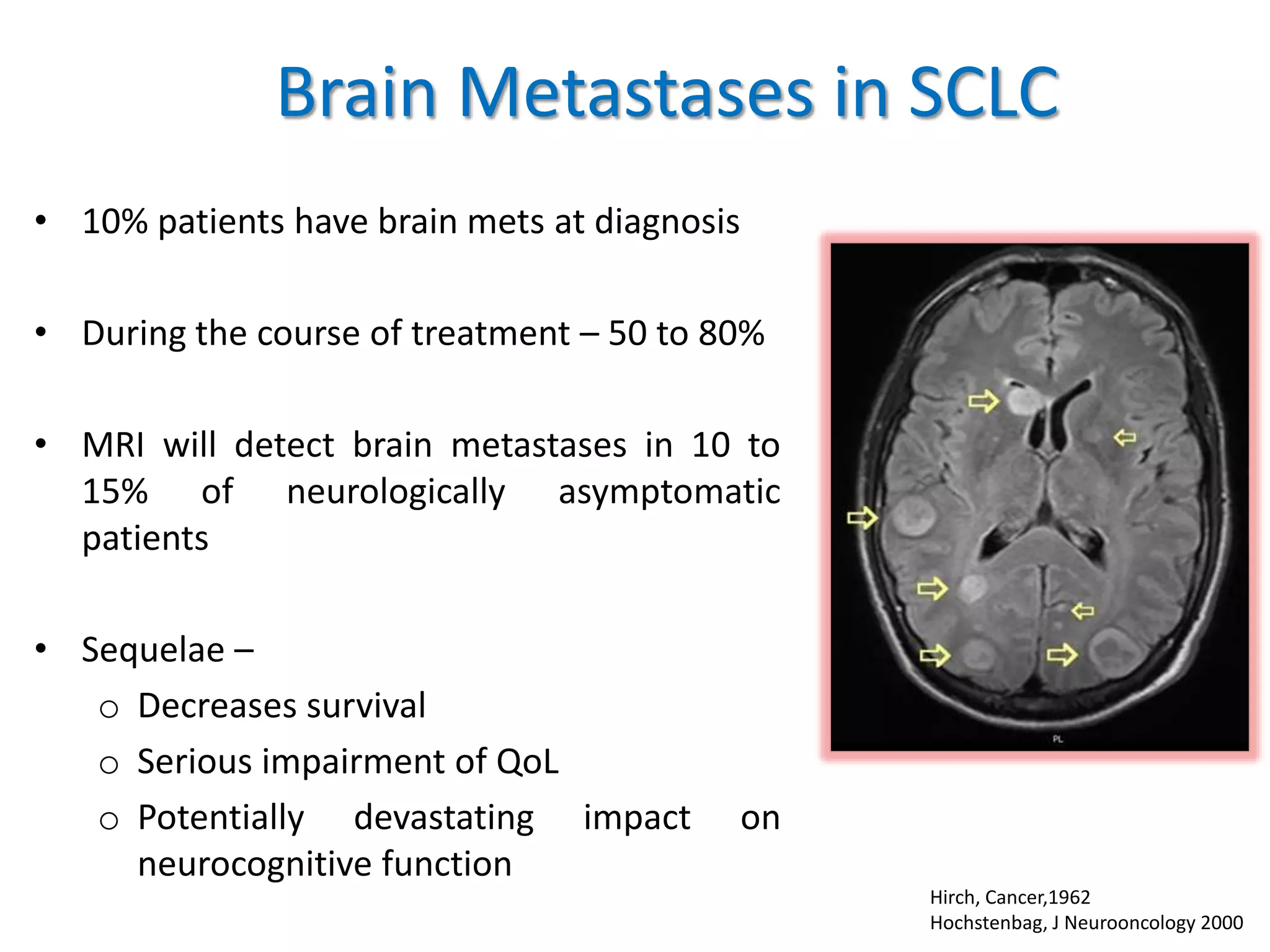
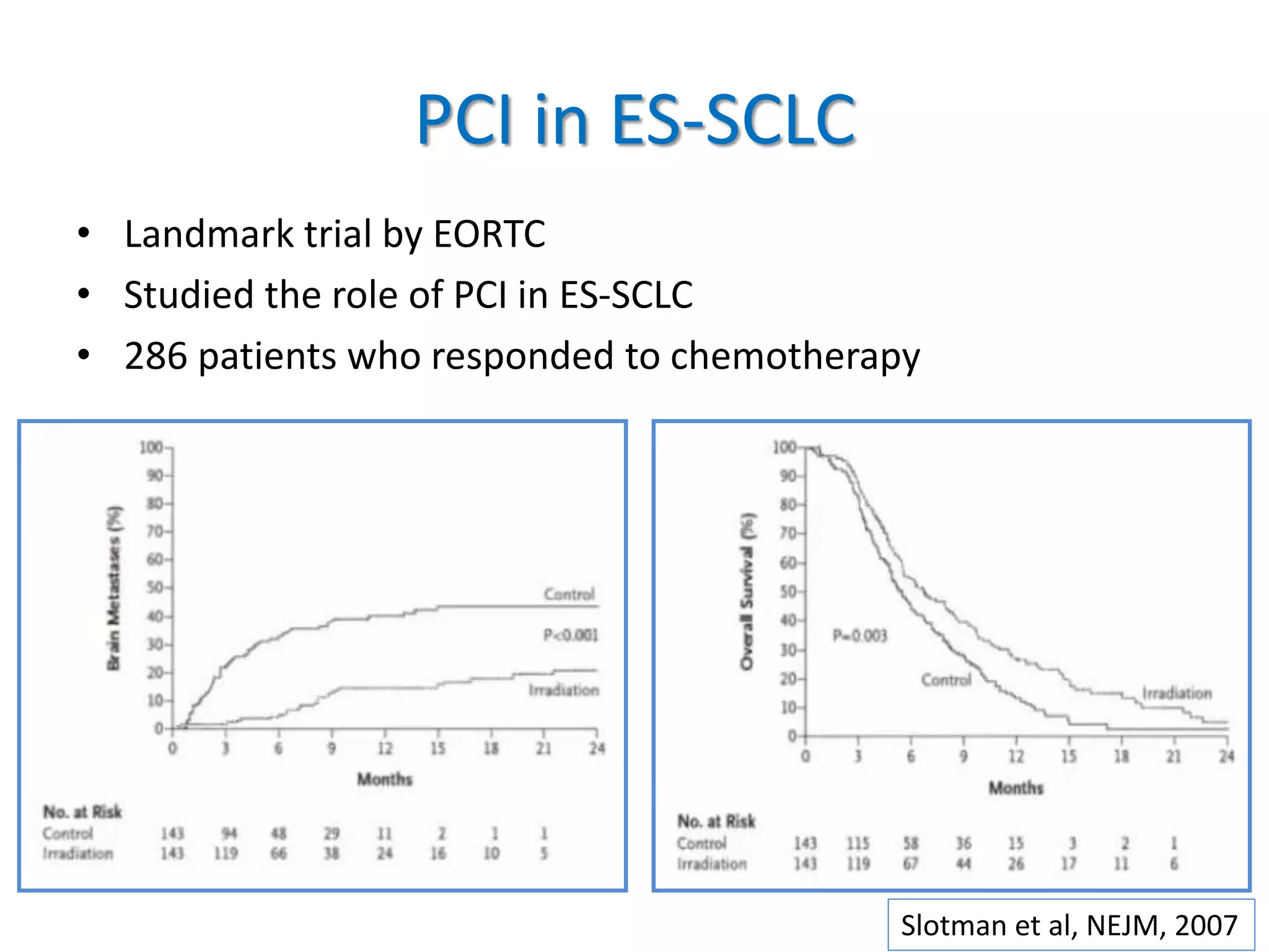
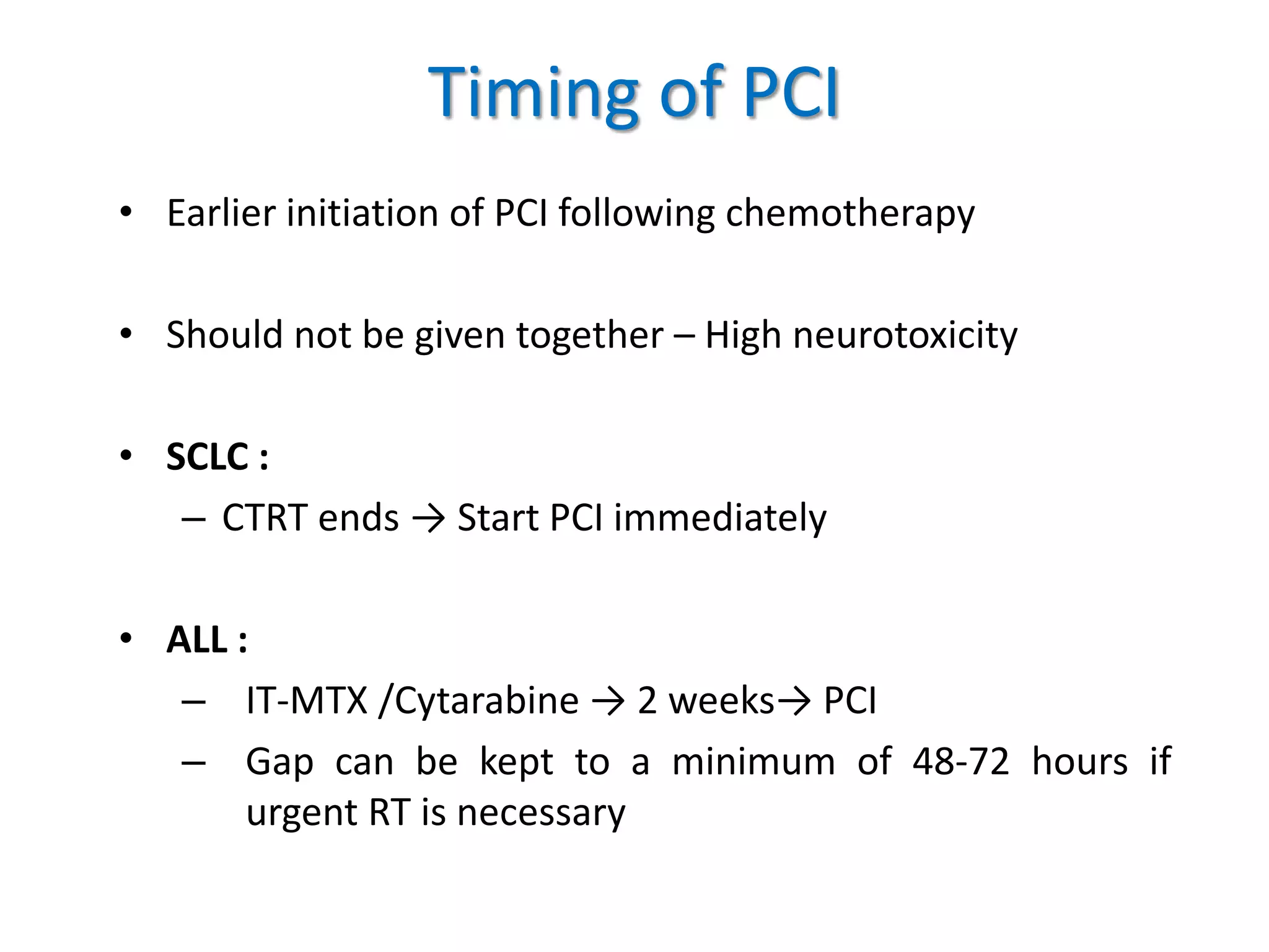
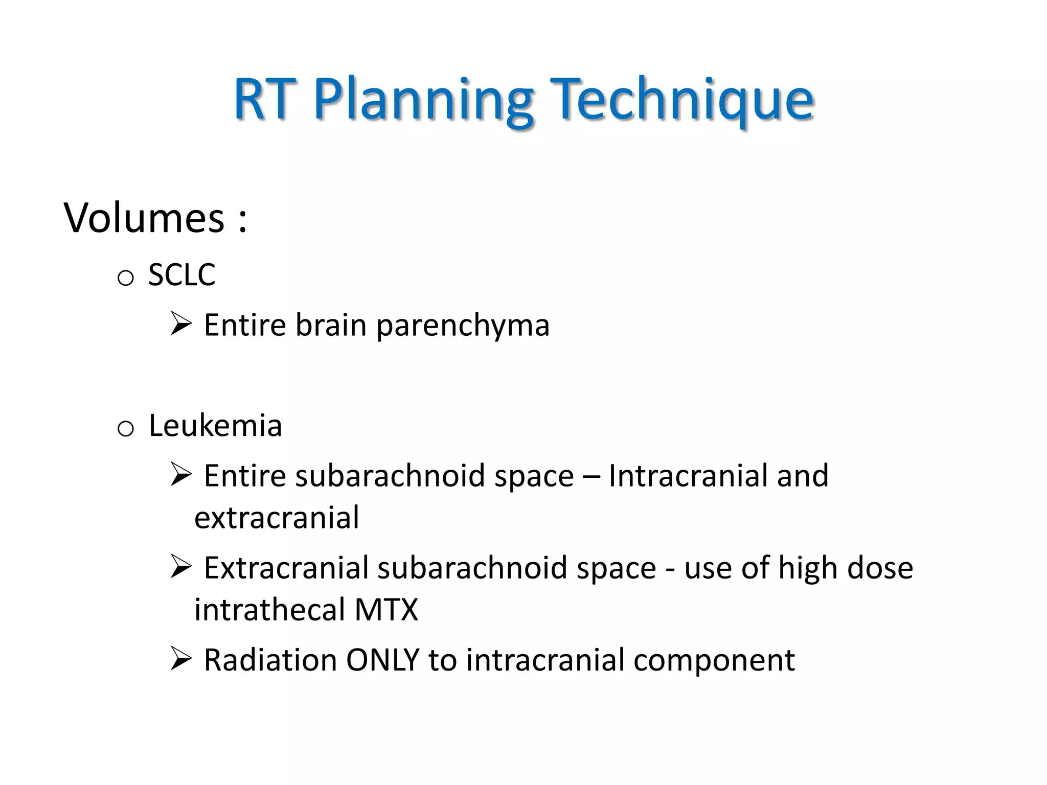
Prophylactic Cranial Irradiation
Dr. Shreya Singh
JR-III
Department of Radiation Oncology
IMS, BHU
Introduction
• Technique used to combat the occurrence of
brain metastasis in highly aggressive cancers with
tendency to metastasize to brain
• PCI is intended as preemptive treatment in
patients with no known current intracranial tumor,
but with high likelihood for harboring occult
microscopic disease and eventual occurrence
No demonstrable evidence of CNS disease
Imaging
CSF Studies
Rationale For PCI
Blood Brain Barrier
Rationale for PCI
• Brain is a sanctuary site
• Poor penetrance for
chemotherapy
• High incidence of CNS
metastasis
Indications
• Small cell lung cancer
• Leukemia
Controversial-
o NSCLC
o Extra-pulmonary small cell cancer
o Uncommon – Testicular lymphoma
Brain Metastases in SCLC
• 10% patients have brain mets at diagnosis
• During the course of treatment – 50 to 80%
• MRI will detect brain metastases in 10 to
15% of neurologically asymptomatic
patients
• Sequelae –
o Decreases survival
o Serious impairment of QoL
o Potentially devastating impact on
neurocognitive function
Hirch, Cancer,1962
Hochstenbag, J Neurooncology 2000
Modified VALSG Staging System
PCI in LS-SCLC
• Arrigada et al,→ Prospective randomised study
• Auperin et al, → Meta-analysis – 987 patients
PCI Observation
2 year rate of brain
mets
40% 67%
Fraction died due
to brain mets alone
19% 45%
PCI Observation
3 year rate of brain
mets
33.3% 58.6%
Increase in OS 20.7% 15.3%
PCI in LS-SCLC
• Auperin Meta Analysis –
o Landmark study
o Higher dose better control
o Early initiation better than late initiation
o All age groups showed benefit
o Caveat- CR assessed by Chest X-Ray only
PCI in ES-SCLC
PCI in ES-SCLC
• Landmark trial by EORTC
• Studied the role of PCI in ES-SCLC
• 286 patients who responded to chemotherapy
Slotman et al, NEJM, 2007
PCI in ES-SCLC
Control PCI
Risk of brain mets 40% 15%
1 year survival rate 13% 27%
Slotman et al, NEJM, 2007
CAVEAT :
NO BRAIN IMAGING DONE
PRIOR TO PCI
PCI in ES-SCLC
• Japanese Group- Randomized 224 patients
• PCI vs no PCI
Takahashi, 2017, Lancet Oncology
PCI in ES-SCLC
• Takahashi et al showed no improvement in OS
despite significant decline in incidence of
brain mets
• Following this study, NCCN has introduced
MRI surveillance (3 monthly) as an alternative
to PCI in ES-SCLC
PCI in NSCLC?
Extra Pulmonary Small Cell Carcinoma
• 2.5% to 4% of all small cell cancers
• 6.4% cases develop brain mets
• Management of EPSCC is modeled on SCLS as no prospective
data exists for this uncommon disease
Naidoo JTO, 2013
• Conclusion :
Brain metastases were uncommon in EPSCC compared with
small-cell lung carcinoma. PCI is thus probably not warranted in
this disease.
PCI Doses
• 720 patients
• 25 Gy in 10 # vs 36 Gy in 18 #
Le Pechoux et al, 2009, Lancet Oncology
Do higher doses offer better control ??
PCI Doses
RESULT:
No significant reduction in incidence of brain metastases in higher dose arm
There was significant increase in chronic neurotoxicity at the end of 1 year
PCI at 25 Gy should
remain standard of care
for LS-SCLC
Le Pechoux et al, 2009, Lancet Oncology
Disease Local Control OS Eviidence
SCLC
LS Yes Yes Indicated
ES Yes Yes PCI vs MRI
surveillance
NSCLC Yes No Not indicated
EPSCC NA NA Not indicated
Leukemia
Evans, Cancer, 1970
Leukemia
• Risk of CNS involvement at diagnosis is low i.e. 3% to 7%
• In absence of CNS directed prophylactic therapy, >50% of
adult patients with leukemia will develop CNS relapse
• CNS prophylaxis can be achieved by
o Radiation (cranial or craniospinal)
o Intrathecal chemotherapy
o High dose systemic chemotherapy, or combinations of
these
AML
• CNS relapse is unusual i.e. 5% to 10 %
• Role of Cranial prophylaxis not well defined for AML
• Some studies show no difference in relapse rate with cranial
irradiation
• High WBC at diagnosis ,monocytic variant – common causes
for CNS relapse
• CNS relapse – poor prognosis
ALL
Role of Radiotherapy in ALL :
• Cranial prophylaxis
• Cranial treatment (all CNS involvement )
• Craniospinal irradiation (overt CNS involvement in adult ALL)
ALL
Backbone to modern leukemic treatment consists of following
steps:
• Induction phase
• Early intensification
• Consolidation → CNS preventive therapy
• Delayed intensification
• Maintenance phase - targeted at eliminating residual disease
Risk Factors for CNS involvement in
ALL
• T-cell immunophenotype
• High leukemic cell proliferation index
• Mature B-cell subtype
• Elevated LDH levels
• Others –
– Age : <1 year, >10 years
– WBC > 50,000
– Philadelphia chromosome
CNS Prophylaxis in ALL
• In most regimens, CNS prophylaxis for patients at lower risk is
achieved with systemic and intrathecal chemotherapy without
cranial irradiation
• Children with high-risk features are at an increased risk of CNS
relapse and, historically, have received prophylactic cranial
irradiation
• Infants younger than age 12 months with 11q23 abnormalities
are at high risk of CNS relapse but because of their young age
are usually treated without cranial irradiation, using
intensified systemic and intrathecal chemotherapy
Current Status in ALL
• Does not recommend PCI in children / adults with
CNS1 and CNS2
• Cranial RT in :
o CNS 3
o CNS relapse
o Pre HCT (Hematopoietic cell transplant)
Dose
• Historically, the standard dose for high-risk ALL cranial prophylaxis
had been 1800 cGy
• Berlin-Frankfurt-Munich (BFM) group used 1200 cGy in patients
with CNS-1 disease with good results
• The standard dose for prophylactic cranial irradiation in those
patients with high-risk disease still treated with irradiation is now
1200 cGy
• For patients with an isolated CNS relapse occurring 18 months or
more after initial diagnosis, a dose of 1800 cGy to the cranial field
has been shown to be effective
Dose Overview
BFM-95
Timing of PCI
• Earlier initiation of PCI following chemotherapy
• Should not be given together – High neurotoxicity
• SCLC :
– CTRT ends → Start PCI immediately
• ALL :
– IT-MTX /Cytarabine → 2 weeks→ PCI
– Gap can be kept to a minimum of 48-72 hours if
urgent RT is necessary
RT Planning Technique
Volumes :
o SCLC
 Entire brain parenchyma
o Leukemia
 Entire subarachnoid space – Intracranial and
extracranial
 Extracranial subarachnoid space - use of high dose
intrathecal MTX
 Radiation ONLY to intracranial component
RT Planning Technique
The technique for cranial
irradiation must ensure
coverage of the cranial
meninges as well as other
areas of potential access to
the CNS such as :
 Cribriform plate
 Posterior retina and
posterior globe of eye
 Inferior extent of temporal
meninges
RT Planning Technique
• To ensure adequate dose to the meninges, the entire calvaria
is covered and the energy should be 4 MV or 6 MV photons
• Custom blocks can be used to shield normal tissues
• Parallel opposed lateral fields are used
• Immobilisation is critical to ensure precise treatment and
reproducibility and use of immobilisation devices is
recommended when possible
RT Planning Technique
• Sedation may be required for younger patients who are
unable to fully co-operate
• Every field should be treated in every sessions
• Daily single dose is administered in 5 sessions per week until
the total dose has been applied
• Angulation of the beam (3-5 deg posterior) - To avoid
ophthalmological complications
Recent Advances
• Results extrapolated from studies for brain mets
• Hippocampal Sparing PCI :
o RTOG – 0933
o HA-WBRT resulted in a mean decline of 7% in HVLT-DR scores from
baseline to 4 months, which was significantly lower
o Dutch Study
o No mets in avoidance region
o Decline by ≥ 5 points on HVLT-total recall score in 28% patients
o No significant difference in two arms
• Memantine / Donepezil
o RTOG – 0614
o Less decline in delayed recall at 24 weeks - NS
Radiation Induced Brain Injury
• Leads to an acute increase in BBB leakiness and edema
• White matter necrosis often occurs following brain
irradiation
• Brain irradiation leads to an acute burst in apoptotic
cell death
• Structural alterations – demyelination and vasculopathy
Toxicity
• Acute :
o Fatigue
o Alopecia
o Pharyngitis
• Chronic :
o Neurocognitive defects
o Endocrine disorder – growth retardation
o Secondary malignancy
o Delayed dentition
o Cataract formation
Prophylactic cranial irradiation

Prophylactic cranial irradiation

  • 1.
    Prophylactic Cranial Irradiation Dr.Shreya Singh JR-III Department of Radiation Oncology IMS, BHU
  • 2.
    Introduction • Technique usedto combat the occurrence of brain metastasis in highly aggressive cancers with tendency to metastasize to brain • PCI is intended as preemptive treatment in patients with no known current intracranial tumor, but with high likelihood for harboring occult microscopic disease and eventual occurrence
  • 3.
    No demonstrable evidenceof CNS disease Imaging CSF Studies
  • 4.
  • 5.
    Rationale for PCI •Brain is a sanctuary site • Poor penetrance for chemotherapy • High incidence of CNS metastasis
  • 6.
    Indications • Small celllung cancer • Leukemia Controversial- o NSCLC o Extra-pulmonary small cell cancer o Uncommon – Testicular lymphoma
  • 7.
    Brain Metastases inSCLC • 10% patients have brain mets at diagnosis • During the course of treatment – 50 to 80% • MRI will detect brain metastases in 10 to 15% of neurologically asymptomatic patients • Sequelae – o Decreases survival o Serious impairment of QoL o Potentially devastating impact on neurocognitive function Hirch, Cancer,1962 Hochstenbag, J Neurooncology 2000
  • 8.
  • 9.
    PCI in LS-SCLC •Arrigada et al,→ Prospective randomised study • Auperin et al, → Meta-analysis – 987 patients PCI Observation 2 year rate of brain mets 40% 67% Fraction died due to brain mets alone 19% 45% PCI Observation 3 year rate of brain mets 33.3% 58.6% Increase in OS 20.7% 15.3%
  • 10.
    PCI in LS-SCLC •Auperin Meta Analysis – o Landmark study o Higher dose better control o Early initiation better than late initiation o All age groups showed benefit o Caveat- CR assessed by Chest X-Ray only
  • 11.
  • 12.
    PCI in ES-SCLC •Landmark trial by EORTC • Studied the role of PCI in ES-SCLC • 286 patients who responded to chemotherapy Slotman et al, NEJM, 2007
  • 13.
    PCI in ES-SCLC ControlPCI Risk of brain mets 40% 15% 1 year survival rate 13% 27% Slotman et al, NEJM, 2007 CAVEAT : NO BRAIN IMAGING DONE PRIOR TO PCI
  • 14.
    PCI in ES-SCLC •Japanese Group- Randomized 224 patients • PCI vs no PCI Takahashi, 2017, Lancet Oncology
  • 15.
    PCI in ES-SCLC •Takahashi et al showed no improvement in OS despite significant decline in incidence of brain mets • Following this study, NCCN has introduced MRI surveillance (3 monthly) as an alternative to PCI in ES-SCLC
  • 16.
  • 17.
    Extra Pulmonary SmallCell Carcinoma • 2.5% to 4% of all small cell cancers • 6.4% cases develop brain mets • Management of EPSCC is modeled on SCLS as no prospective data exists for this uncommon disease Naidoo JTO, 2013 • Conclusion : Brain metastases were uncommon in EPSCC compared with small-cell lung carcinoma. PCI is thus probably not warranted in this disease.
  • 18.
    PCI Doses • 720patients • 25 Gy in 10 # vs 36 Gy in 18 # Le Pechoux et al, 2009, Lancet Oncology Do higher doses offer better control ??
  • 19.
    PCI Doses RESULT: No significantreduction in incidence of brain metastases in higher dose arm There was significant increase in chronic neurotoxicity at the end of 1 year PCI at 25 Gy should remain standard of care for LS-SCLC Le Pechoux et al, 2009, Lancet Oncology
  • 20.
    Disease Local ControlOS Eviidence SCLC LS Yes Yes Indicated ES Yes Yes PCI vs MRI surveillance NSCLC Yes No Not indicated EPSCC NA NA Not indicated
  • 21.
  • 22.
    Leukemia • Risk ofCNS involvement at diagnosis is low i.e. 3% to 7% • In absence of CNS directed prophylactic therapy, >50% of adult patients with leukemia will develop CNS relapse • CNS prophylaxis can be achieved by o Radiation (cranial or craniospinal) o Intrathecal chemotherapy o High dose systemic chemotherapy, or combinations of these
  • 23.
    AML • CNS relapseis unusual i.e. 5% to 10 % • Role of Cranial prophylaxis not well defined for AML • Some studies show no difference in relapse rate with cranial irradiation • High WBC at diagnosis ,monocytic variant – common causes for CNS relapse • CNS relapse – poor prognosis
  • 24.
    ALL Role of Radiotherapyin ALL : • Cranial prophylaxis • Cranial treatment (all CNS involvement ) • Craniospinal irradiation (overt CNS involvement in adult ALL)
  • 25.
    ALL Backbone to modernleukemic treatment consists of following steps: • Induction phase • Early intensification • Consolidation → CNS preventive therapy • Delayed intensification • Maintenance phase - targeted at eliminating residual disease
  • 26.
    Risk Factors forCNS involvement in ALL • T-cell immunophenotype • High leukemic cell proliferation index • Mature B-cell subtype • Elevated LDH levels • Others – – Age : <1 year, >10 years – WBC > 50,000 – Philadelphia chromosome
  • 28.
    CNS Prophylaxis inALL • In most regimens, CNS prophylaxis for patients at lower risk is achieved with systemic and intrathecal chemotherapy without cranial irradiation • Children with high-risk features are at an increased risk of CNS relapse and, historically, have received prophylactic cranial irradiation • Infants younger than age 12 months with 11q23 abnormalities are at high risk of CNS relapse but because of their young age are usually treated without cranial irradiation, using intensified systemic and intrathecal chemotherapy
  • 29.
  • 30.
    • Does notrecommend PCI in children / adults with CNS1 and CNS2 • Cranial RT in : o CNS 3 o CNS relapse o Pre HCT (Hematopoietic cell transplant)
  • 31.
    Dose • Historically, thestandard dose for high-risk ALL cranial prophylaxis had been 1800 cGy • Berlin-Frankfurt-Munich (BFM) group used 1200 cGy in patients with CNS-1 disease with good results • The standard dose for prophylactic cranial irradiation in those patients with high-risk disease still treated with irradiation is now 1200 cGy • For patients with an isolated CNS relapse occurring 18 months or more after initial diagnosis, a dose of 1800 cGy to the cranial field has been shown to be effective
  • 32.
  • 33.
    Timing of PCI •Earlier initiation of PCI following chemotherapy • Should not be given together – High neurotoxicity • SCLC : – CTRT ends → Start PCI immediately • ALL : – IT-MTX /Cytarabine → 2 weeks→ PCI – Gap can be kept to a minimum of 48-72 hours if urgent RT is necessary
  • 34.
    RT Planning Technique Volumes: o SCLC  Entire brain parenchyma o Leukemia  Entire subarachnoid space – Intracranial and extracranial  Extracranial subarachnoid space - use of high dose intrathecal MTX  Radiation ONLY to intracranial component
  • 35.
    RT Planning Technique Thetechnique for cranial irradiation must ensure coverage of the cranial meninges as well as other areas of potential access to the CNS such as :  Cribriform plate  Posterior retina and posterior globe of eye  Inferior extent of temporal meninges
  • 36.
  • 37.
  • 38.
  • 39.
    RT Planning Technique •To ensure adequate dose to the meninges, the entire calvaria is covered and the energy should be 4 MV or 6 MV photons • Custom blocks can be used to shield normal tissues • Parallel opposed lateral fields are used • Immobilisation is critical to ensure precise treatment and reproducibility and use of immobilisation devices is recommended when possible
  • 40.
    RT Planning Technique •Sedation may be required for younger patients who are unable to fully co-operate • Every field should be treated in every sessions • Daily single dose is administered in 5 sessions per week until the total dose has been applied • Angulation of the beam (3-5 deg posterior) - To avoid ophthalmological complications
  • 41.
    Recent Advances • Resultsextrapolated from studies for brain mets • Hippocampal Sparing PCI : o RTOG – 0933 o HA-WBRT resulted in a mean decline of 7% in HVLT-DR scores from baseline to 4 months, which was significantly lower o Dutch Study o No mets in avoidance region o Decline by ≥ 5 points on HVLT-total recall score in 28% patients o No significant difference in two arms • Memantine / Donepezil o RTOG – 0614 o Less decline in delayed recall at 24 weeks - NS
  • 42.
    Radiation Induced BrainInjury • Leads to an acute increase in BBB leakiness and edema • White matter necrosis often occurs following brain irradiation • Brain irradiation leads to an acute burst in apoptotic cell death • Structural alterations – demyelination and vasculopathy
  • 43.
    Toxicity • Acute : oFatigue o Alopecia o Pharyngitis • Chronic : o Neurocognitive defects o Endocrine disorder – growth retardation o Secondary malignancy o Delayed dentition o Cataract formation

Editor's Notes

  • #18 Naidoo - The number of patients who developed brain metastases, their survival, and treatment data were documented. Patients who received PCI were investigated.