Introduction to CPR
•CPR = Cardiopulmonary Resuscitation
• Emergency procedure for cardiac arrest
• Maintains circulation & oxygenation
• Goal: Prevent brain damage & death
4.
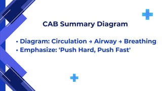
What is CABin
CPR?
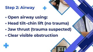
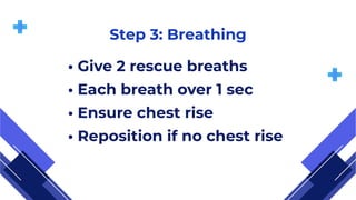
• C = Circulation
• A = Airway
• B = Breathing
• Ensures early chest compressions
5.
Integrity and ethics
Innovationand continuous improvement
Why CAB Instead of ABC?
• Focus on early compressions = higher survival
• Oxygen in blood is enough initially
• Circulation must start immediately
6.
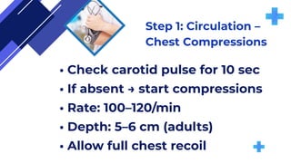
Step 1: Circulation–
Chest Compressions
• Check carotid pulse for 10 sec
• If absent → start compressions
• Rate: 100–120/min
• Depth: 5–6 cm (adults)
• Allow full chest recoil
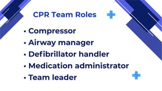
CPR Team Roles
•Compressor
• Airway manager
• Defibrillator handler
• Medication administrator
• Team leader
25.
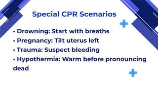
Special CPR Scenarios
•Drowning: Start with breaths
• Pregnancy: Tilt uterus left
• Trauma: Suspect bleeding
• Hypothermia: Warm before pronouncing
dead


![ONFH[AVN HIP] -TRIPLE REGIME -A NOVAL SURGICAL CONCEPT .pptx](https://cdn.slidesharecdn.com/ss_thumbnails/onfhavnhip2026koaconcalicutdrgokuldevdrmashraf-260210064517-213ec005-thumbnail.jpg?width=640&height=640&fit=bounds)


