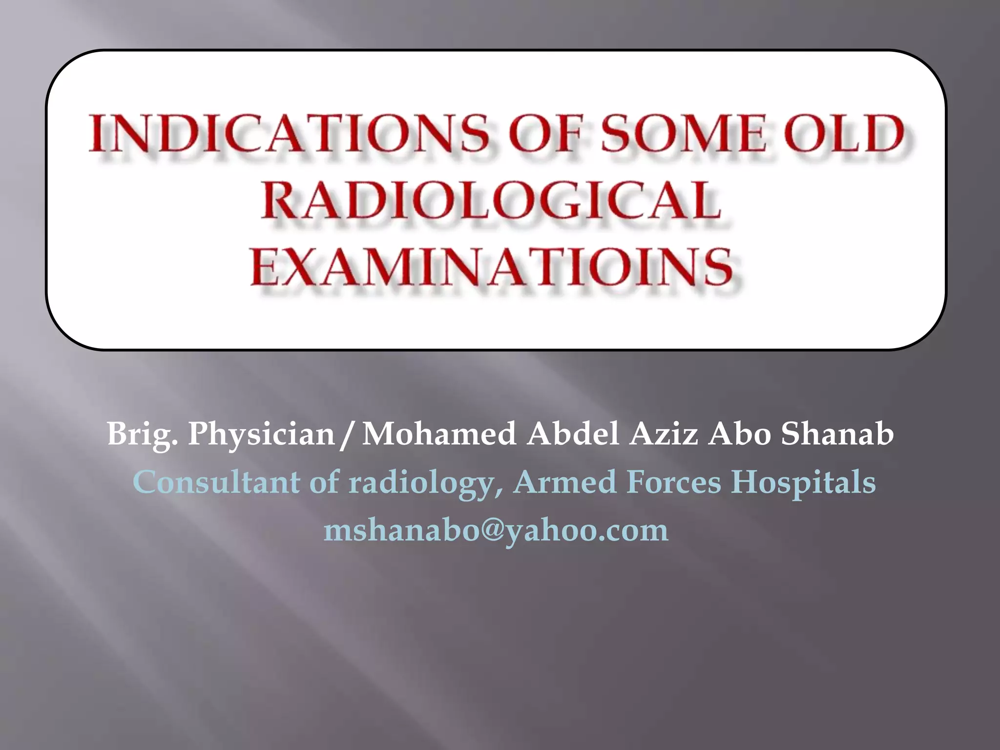
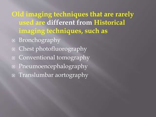
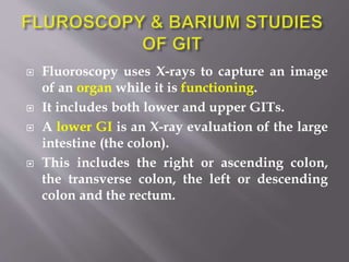
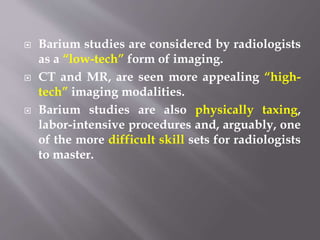
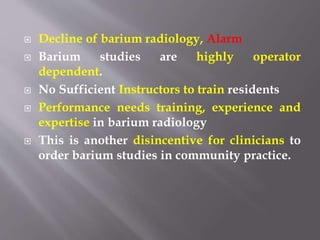
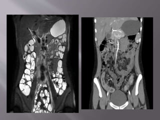
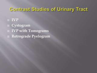
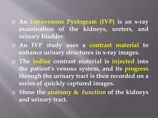
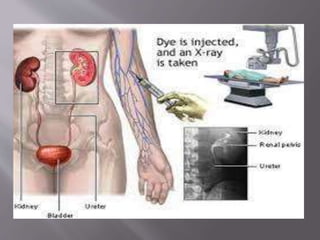
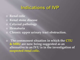
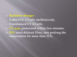
Brig. Physician / Mohamed Abdel Aziz Abo Shanab
Hospitals
Consultant of radiology, Armed Forces
mshanabo@yahoo.com
Old imaging techniques that are rarely
used are different from Historical
imaging techniques, such as
 Bronchography
 Chest photofluorography
 Conventional tomography
 Pneumoencephalography
 Translumbar aortography
 The field of diagnostic and therapeutic
radiology has always been characterized by
constant innovation & creativity to evolve to
its current form.
 There are numerous imaging techniques that
were once prevalent but have become
outdated & replaced by the current
examinations and modalities, which improve
diagnostic accuracy and patient outcomes.
 Knowledge of these old radiologic
examinations and procedures is important to
understand how we have arrived at the
current practice of radiology & how our field
can continue to evolve to improve our
diagnostic and therapeutic abilities to fit the
changing needs of our patients.
 Fluoroscopy uses X-rays to capture an image
of an organ while it is functioning.
 It includes both lower and upper GITs.
 A lower GI is an X-ray evaluation of the large
intestine (the colon).
 This includes the right or ascending colon,
the transverse colon, the left or descending
colon and the rectum.
 An upper GI Study is an X-ray examination of
the esophagus, stomach and first part of the
small intestine.
 Upper GIT must be coated or filled with a
contrast material called barium, an element
that appears bright white on radiographs.
 The barium is given to the patient to drink.
 Some patients are asked to swallow baking-
soda crystals to create gas and further
improve the images; this procedure has the
modified name of air-contrast or double-
contrast upper GI.
 Esophogram
 Barium Meal, UGI Series
 Barium Follow through, Small Bowel and
UGI
 Barium Enema
 The dedicated small bowel follow-through
has been replaced to a lesser degree by CT
Abdomen, CT and MR Enterography &
capsule endoscopy.
 For lower GI symptoms and colon cancer
screening, the double-contrast barium enema
has increasingly been discarded in favor of
Colonoscopy, CT Abdomen, CT
Colonography.
 Barium studies are considered by radiologists
as a “low-tech” form of imaging.
 CT and MR, are seen more appealing “high-
tech” imaging modalities.
 Barium studies are also physically taxing,
labor-intensive procedures and, arguably, one
of the more difficult skill sets for radiologists
to master.
 Decline of barium radiology, Alarm
 Barium studies are highly operator
dependent.
 No Sufficient Instructors to train residents
 Performance needs training, experience and
expertise in barium radiology
 This is another disincentive for clinicians to
order barium studies in community practice.
 IVP
 Cystogram
 IVP with Tomograms
 Retrograde Pyelogram
 An Intravenous Pyelogram (IVP) is an x-ray
examination of the kidneys, ureters, and
urinary bladder.
 An IVP study uses a contrast material to
enhance urinary structures in x-ray images.
 The iodine contrast material is injected into
the patient's venous system, and its progress
through the urinary tract is then recorded on a
series of quickly captured images.
 Show the anatomy & function of the kidneys
and urinary tract.
 Renal colic
 Renal stone disease
 Calyceal pathology
 Hematuria
 Chronic upper urinary tract obstruction.
 The commonest situation in which the CTU
& MRU are now being suggested as an
alternative to an IVU is in the investigation of
suspected renal colic.
 Radiation dosage:-
- 3-shot IVU 1.5 mSv (millisievert).
- Unenhanced CT 4.5 mSv
 CT scan, performed within few minutes.
 IVU need delayed films, may prolong the
examination for more than 12 h.
IVP vs CT
 CTU detection for the cause of haematuria is high
(sensitivity 92 - 100 %, specificity 89 - 97 %) &
significantly better than IVP (sensitivity 61 %,
specificity 97 %).
 Radiation doses of IVP < CT but varies with the
number of films
 Many practices no longer have tomographic
equipment.
 Many " younger " radiologists have never
performed or interpreted an IVP
 IVP may be inconvenient to the patient as its
procedures include, puncture by cannula
(pain), allergy to contrast medium,
 It is interesting to speculate about which of
the imaging modalities and techniques we
rely on today will become the antiquated
examinations of the future!!
 AI may replace jobs, personnel as radiologists
in the future!
THANK YOU

PRESENTATION. old rad techniques.pptx

  • 1.
    Brig. Physician /Mohamed Abdel Aziz Abo Shanab Hospitals Consultant of radiology, Armed Forces mshanabo@yahoo.com
  • 2.
    Old imaging techniquesthat are rarely used are different from Historical imaging techniques, such as  Bronchography  Chest photofluorography  Conventional tomography  Pneumoencephalography  Translumbar aortography
  • 3.
     The fieldof diagnostic and therapeutic radiology has always been characterized by constant innovation & creativity to evolve to its current form.  There are numerous imaging techniques that were once prevalent but have become outdated & replaced by the current examinations and modalities, which improve diagnostic accuracy and patient outcomes.
  • 4.
     Knowledge ofthese old radiologic examinations and procedures is important to understand how we have arrived at the current practice of radiology & how our field can continue to evolve to improve our diagnostic and therapeutic abilities to fit the changing needs of our patients.
  • 5.
     Fluoroscopy usesX-rays to capture an image of an organ while it is functioning.  It includes both lower and upper GITs.  A lower GI is an X-ray evaluation of the large intestine (the colon).  This includes the right or ascending colon, the transverse colon, the left or descending colon and the rectum.
  • 6.
     An upperGI Study is an X-ray examination of the esophagus, stomach and first part of the small intestine.  Upper GIT must be coated or filled with a contrast material called barium, an element that appears bright white on radiographs.
  • 7.
     The bariumis given to the patient to drink.  Some patients are asked to swallow baking- soda crystals to create gas and further improve the images; this procedure has the modified name of air-contrast or double- contrast upper GI.
  • 9.
     Esophogram  BariumMeal, UGI Series  Barium Follow through, Small Bowel and UGI  Barium Enema
  • 11.
     The dedicatedsmall bowel follow-through has been replaced to a lesser degree by CT Abdomen, CT and MR Enterography & capsule endoscopy.  For lower GI symptoms and colon cancer screening, the double-contrast barium enema has increasingly been discarded in favor of Colonoscopy, CT Abdomen, CT Colonography.
  • 12.
     Barium studiesare considered by radiologists as a “low-tech” form of imaging.  CT and MR, are seen more appealing “high- tech” imaging modalities.  Barium studies are also physically taxing, labor-intensive procedures and, arguably, one of the more difficult skill sets for radiologists to master.
  • 13.
     Decline ofbarium radiology, Alarm  Barium studies are highly operator dependent.  No Sufficient Instructors to train residents  Performance needs training, experience and expertise in barium radiology  This is another disincentive for clinicians to order barium studies in community practice.
  • 15.
     IVP  Cystogram IVP with Tomograms  Retrograde Pyelogram
  • 16.
     An IntravenousPyelogram (IVP) is an x-ray examination of the kidneys, ureters, and urinary bladder.  An IVP study uses a contrast material to enhance urinary structures in x-ray images.  The iodine contrast material is injected into the patient's venous system, and its progress through the urinary tract is then recorded on a series of quickly captured images.  Show the anatomy & function of the kidneys and urinary tract.
  • 18.
     Renal colic Renal stone disease  Calyceal pathology  Hematuria  Chronic upper urinary tract obstruction.  The commonest situation in which the CTU & MRU are now being suggested as an alternative to an IVU is in the investigation of suspected renal colic.
  • 19.
     Radiation dosage:- -3-shot IVU 1.5 mSv (millisievert). - Unenhanced CT 4.5 mSv  CT scan, performed within few minutes.  IVU need delayed films, may prolong the examination for more than 12 h.
  • 20.
    IVP vs CT CTU detection for the cause of haematuria is high (sensitivity 92 - 100 %, specificity 89 - 97 %) & significantly better than IVP (sensitivity 61 %, specificity 97 %).  Radiation doses of IVP < CT but varies with the number of films  Many practices no longer have tomographic equipment.  Many " younger " radiologists have never performed or interpreted an IVP
  • 23.
     IVP maybe inconvenient to the patient as its procedures include, puncture by cannula (pain), allergy to contrast medium,
  • 24.
     It isinteresting to speculate about which of the imaging modalities and techniques we rely on today will become the antiquated examinations of the future!!  AI may replace jobs, personnel as radiologists in the future!
  • 25.