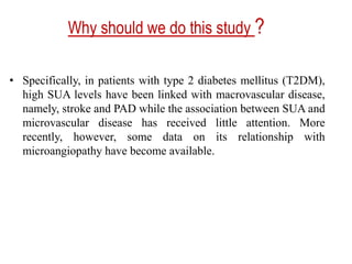
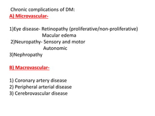
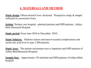
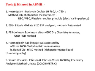
This document outlines a thesis protocol on examining the relationship between serum uric acid level and microvascular and macrovascular complications in diabetes mellitus. The candidate is Dr. Sachinkumar Pandey from Aditya Birla Memorial Hospital in Pune, India. The study will examine 139 type 2 diabetes patients to determine if increased serum uric acid levels are associated with chronic complications like retinopathy, nephropathy, neuropathy, and dyslipidemia. Laboratory tests, medical history, and clinical examinations will be used to analyze the relationship between hyperuricemia and microvascular and macrovascular complication risk. Statistical analysis will then be performed on the collected data.

![INTRODUCTION
• Diabetes mellitus (DM) is a metabolic disease that affects many people
across the world but more so in India. India is known as the "diabetes
capital of the world". From 51 million people in 2010, the number of
persons with type 2 diabetes mellitus in India is estimated to register a
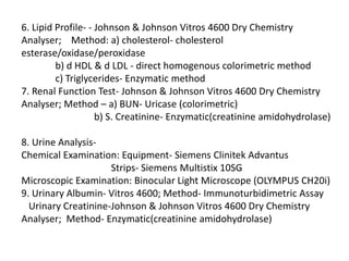
58% increase to reach an alarming level of 87 million by the year 2030. In
a recent study from India, the prevalence of microvascular complications
in newly diagnosed type 2 diabetics was reported 1,2,3as 32.55% (one in
three patients) .[1]
• Diabetes mellitus (DM) refers to a group of common metabolic disorders
that share the phenotype of hyperglycemia. The two broad categories of
DM designated as type 1 and type 2. Type 1 diabetes is the result of
complete or near total insulin deficiency. Type 2 diabetes is a
heterogenous group of disorders characterized by variable degrees of
insulin resistance, impaired insulin secretion, and increased glucose
production. In accordance with the criteria of the American Diabetes
Association and World Health Organization, DM is diagnosed when 1)
fasting blood glucose ≥126 mg/dl or 2)two hour post glucose >200 mg/dl.
The diabetic duration is defined as the duration from the first diagnosis of
type 2 DM to the time of blood sampling.[25]](https://image.slidesharecdn.com/bcww9whr9od7921qaerg-ppt-230528123146-3a8b8e6e/85/PPT-THESIS_PROTOCOL-pptx-5-320.jpg)
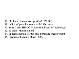
![Methodology
In study total 139 patients of known case of type 2 DM will be included. We will take
full medical history of all individuals and detailed clinical examinations with laboratory
investigations will be undertaken to exclude any other systemic and/or local diseases
that may affect the parameters examined in this study.
For this study, microvascular complications were defined as follows:
Nephropathy: Measurement of a spot urine sample for albumin alone (whether by
immunoassay or by using a sensitive dipstick test specific for albuminuria) was done.
Values ≥ 30μg/mg creatinine is taken as increased urinary albumin excretion as given
by ADA 6guidelines .[1,13]
Neuropathy: Patients are screened using tests such as pinprick sensation, vibration
perception (using a 128-Hz tuning fork), and 10 g monofilament pressure sensation at
the distal plantar aspect of both great toes and metatarsal joints, and assessment of
ankle reflexes. The presence of neuropathy is confirmed by physician if diagnosed with
one or more abnormal finding of 10 gram monofilament, pinprick sensations and
ankle reflexes.[1,9]](https://image.slidesharecdn.com/bcww9whr9od7921qaerg-ppt-230528123146-3a8b8e6e/85/PPT-THESIS_PROTOCOL-pptx-14-320.jpg)
![Methodology
Coronary artery disease (CAD) will be diagnosed on the basis of
electrocardiographic findings.
Peripheral Arterial Disease will be diagnosed by on the basis of Arterial
Doppler study.
Cerebrovascular disease will be diagnosed on the basis of previous history of
stroke, cerebral small vessel disease and acute cerebral vascular disease.
Haemoglobin A1c (HbA1c) by high performance liquid chromatography and
serum uric acid level will be assessed for each and every patient of our study.
Hyperuricemia is diagnosed when the serum uric acid concentration is > 416
micro mol/L (>7.0 mg/dl) in men or >386 micro mol/L(>6.5 mg/dl) in women,
or when patients were taking allopurinol. [7]](https://image.slidesharecdn.com/bcww9whr9od7921qaerg-ppt-230528123146-3a8b8e6e/85/PPT-THESIS_PROTOCOL-pptx-16-320.jpg)
![Methodology
Arbitrary Criteria for Education :
1] Able to read or write any single language is taken as ‘educated’.
2] Not able to read or write even a single language is taken as ‘uneducated’.
Arbitrary Criteria for Awareness : It is based on following two questions :
1] Do you know about DM ?
2] Whether good DM control is essential or not ?
On the basis of knowledge of the subjects of these two questions, they are
classified as Aware ,Partial aware and Not aware.](https://image.slidesharecdn.com/bcww9whr9od7921qaerg-ppt-230528123146-3a8b8e6e/85/PPT-THESIS_PROTOCOL-pptx-17-320.jpg)
![REVIEW OF LITERATURE
3.In 2013, Alberico Catapano, University of Milan, Italy ,et al.
conducted a search which yielded 9 eligible articles including
20,891 T2DM patients. Pooled estimates for the relationship
suggested that each 0.1 mmol/l increase in SUA resulted in a
28% increase in the risk of diabetic vascular complications and a
9% increase in the risk of diabetic mortality. In stratification-
analysis, the positive relationship between SUA and vascular
complications remained significant irrespective of mean age,
adjustment for metabolic variables and medications. However, it
was inconsistent in different populations (significantly positive in
the Asian but not in Australian and Italian population) and
sample sizes (significantly positive in the relatively large sample
size [≥1000] but non-significant in the small sample size [<1000]).](https://image.slidesharecdn.com/bcww9whr9od7921qaerg-ppt-230528123146-3a8b8e6e/85/PPT-THESIS_PROTOCOL-pptx-20-320.jpg)


![Wilm's tumour - The most common kidney tumor in children - Dr Vishnu A [VCR],...](https://cdn.slidesharecdn.com/ss_thumbnails/vishnu-wilmstumour-210312145616-thumbnail.jpg?width=640&height=640&fit=bounds)


