Downloaded 1,145 times
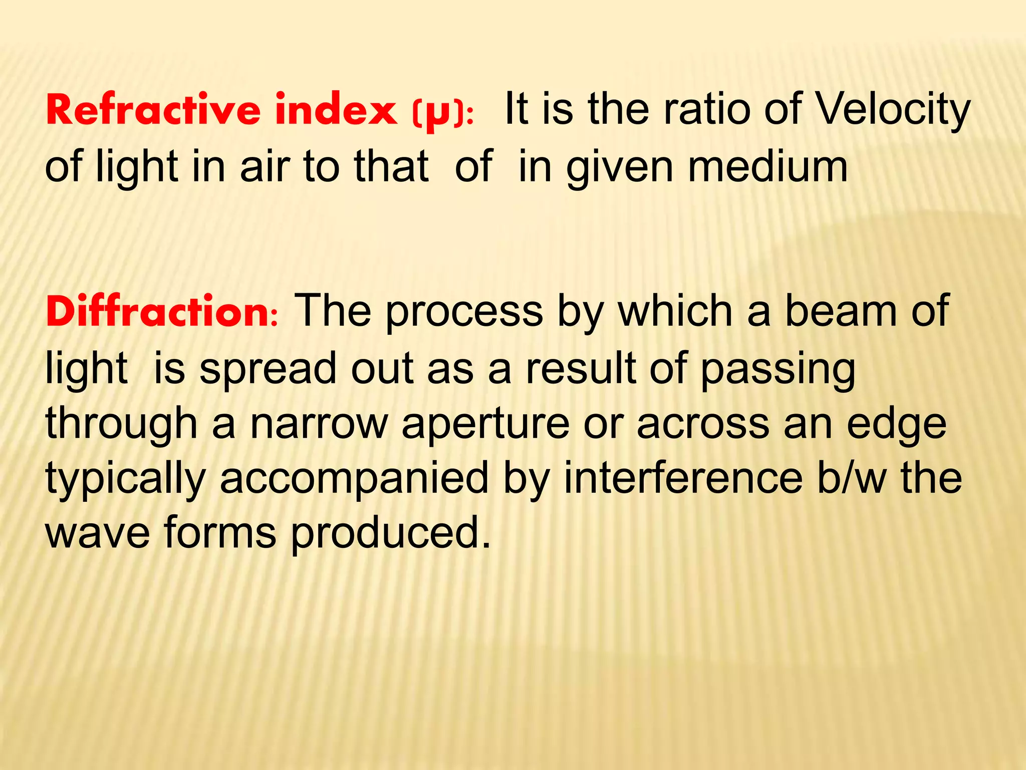
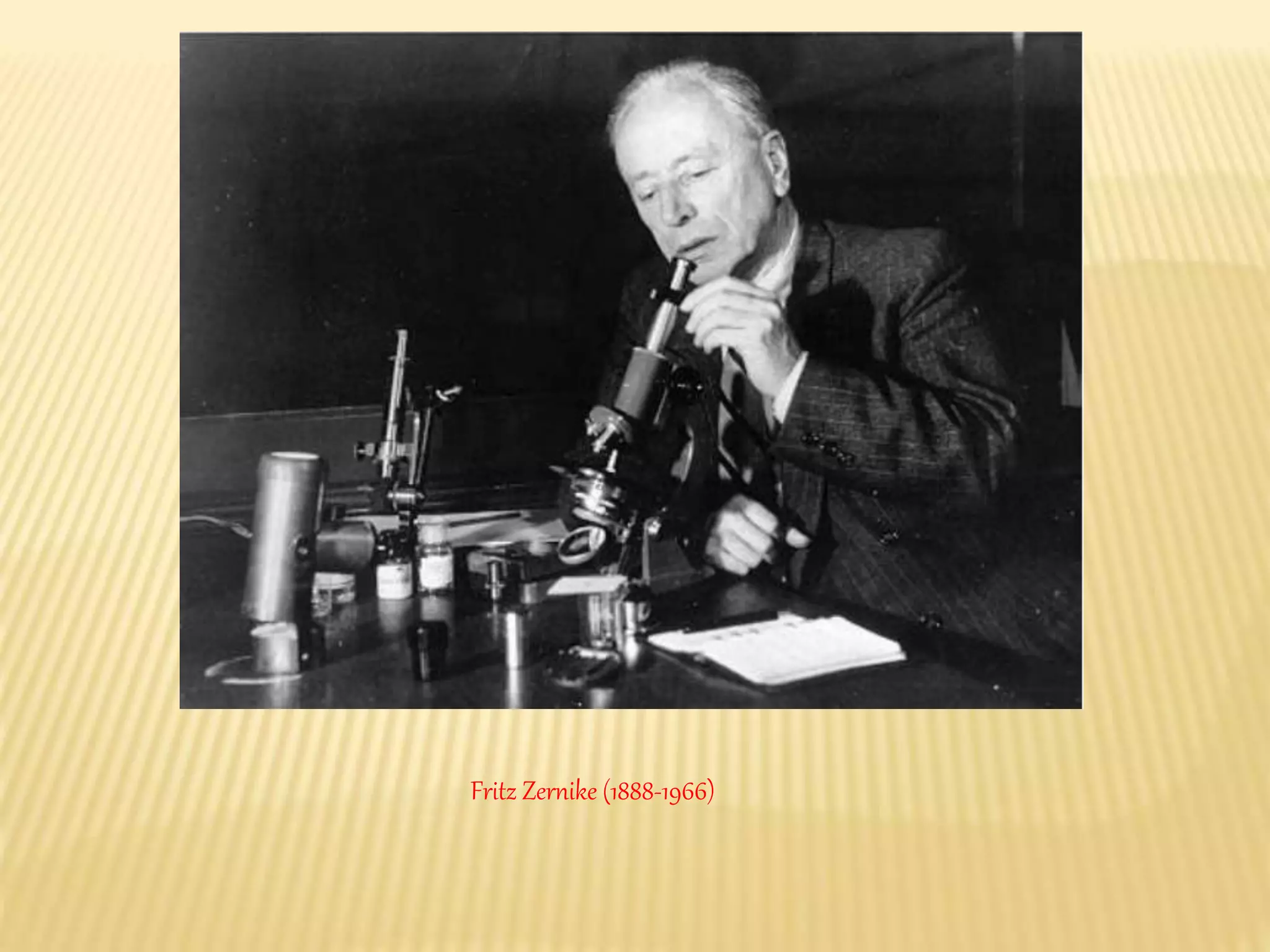
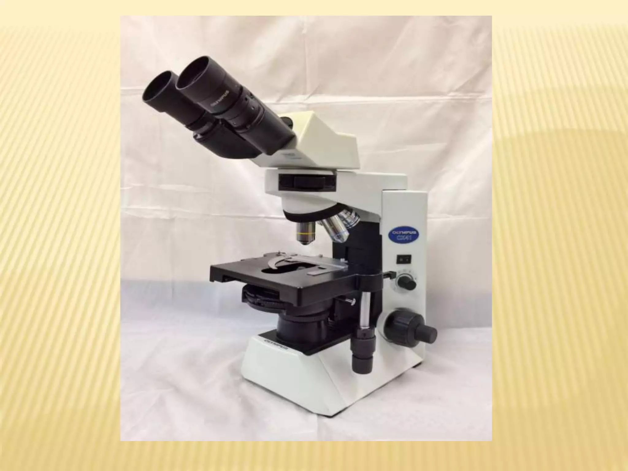

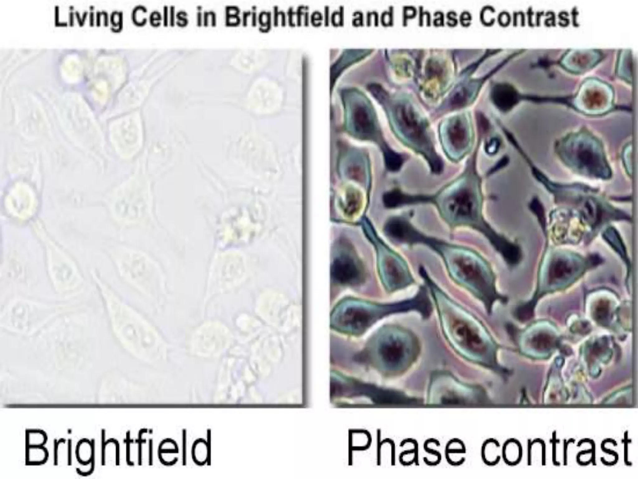
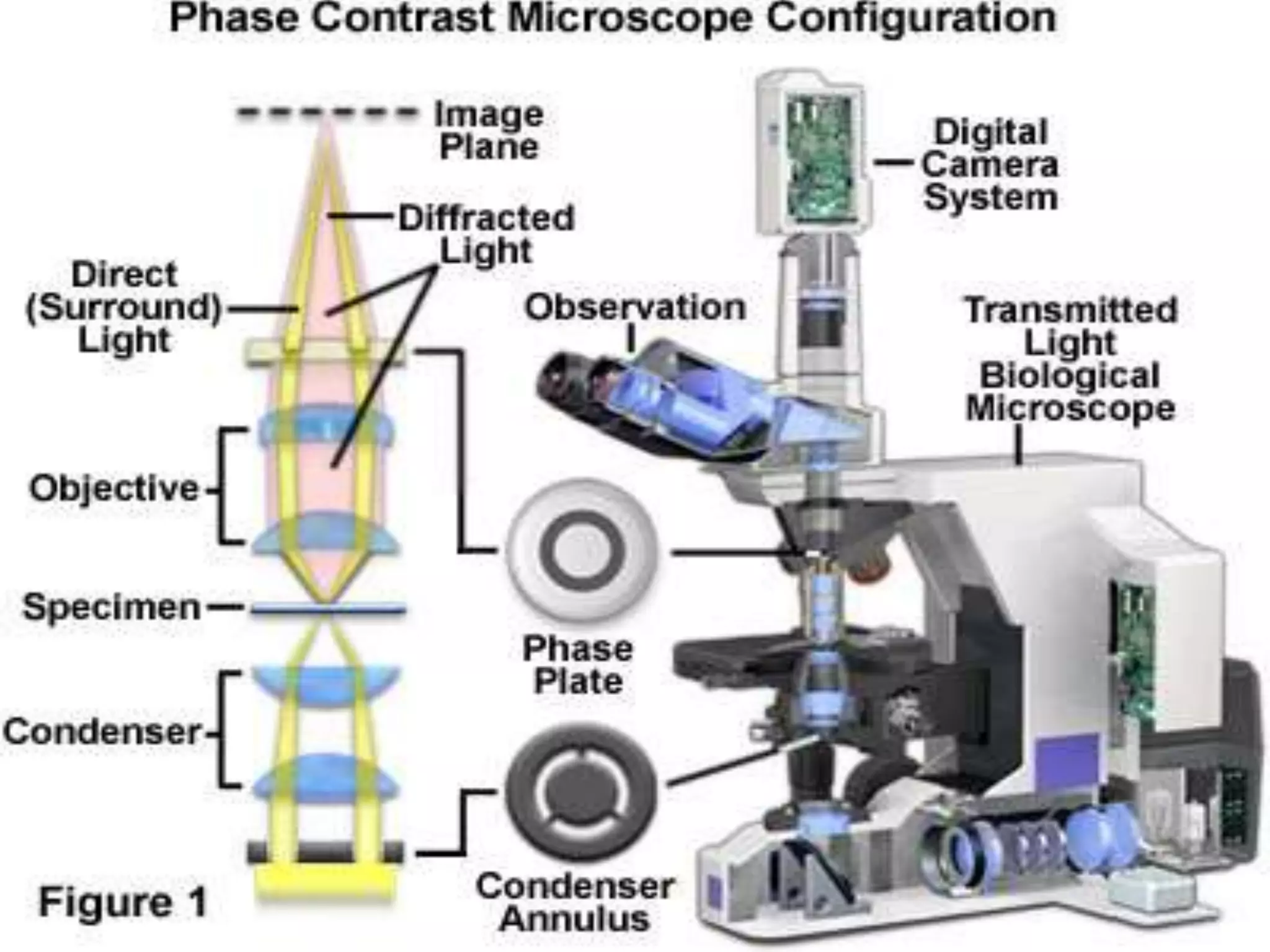

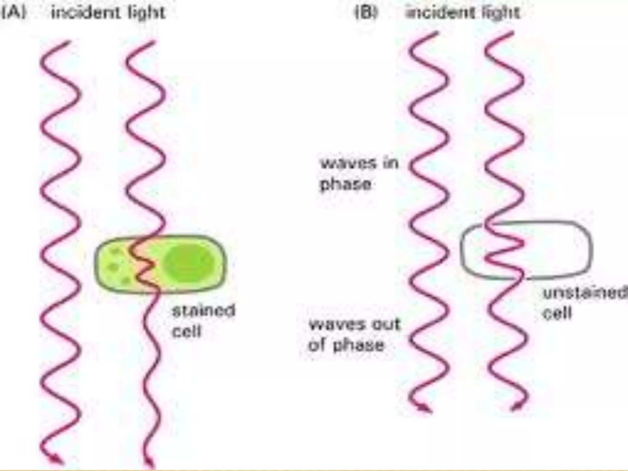
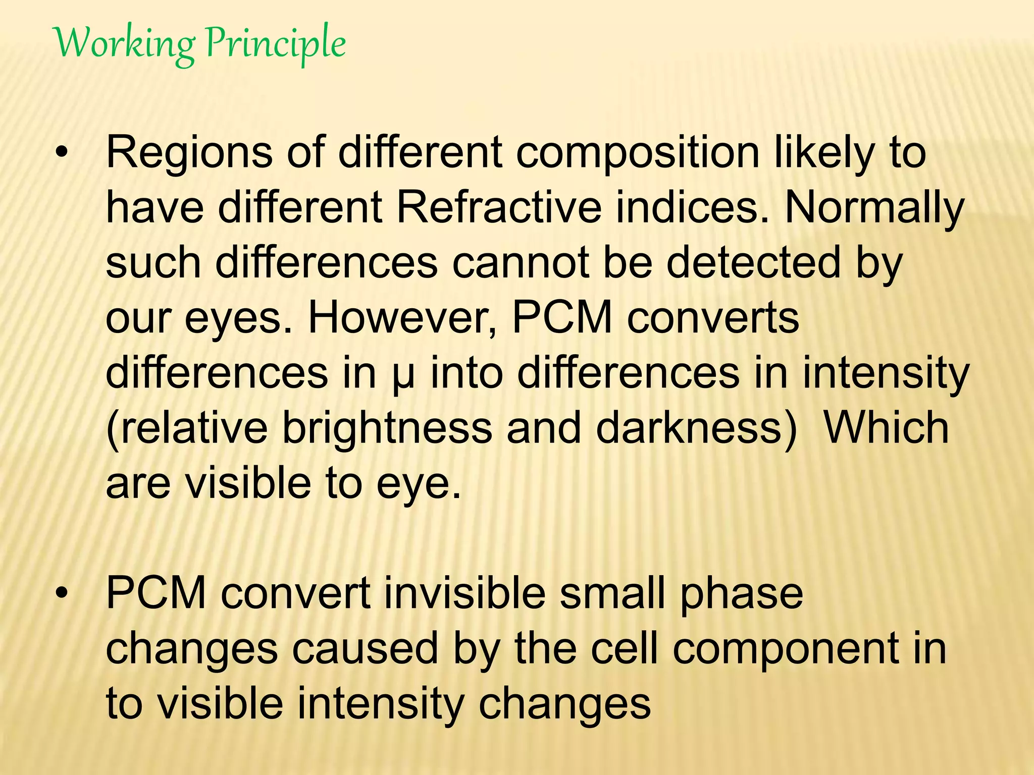
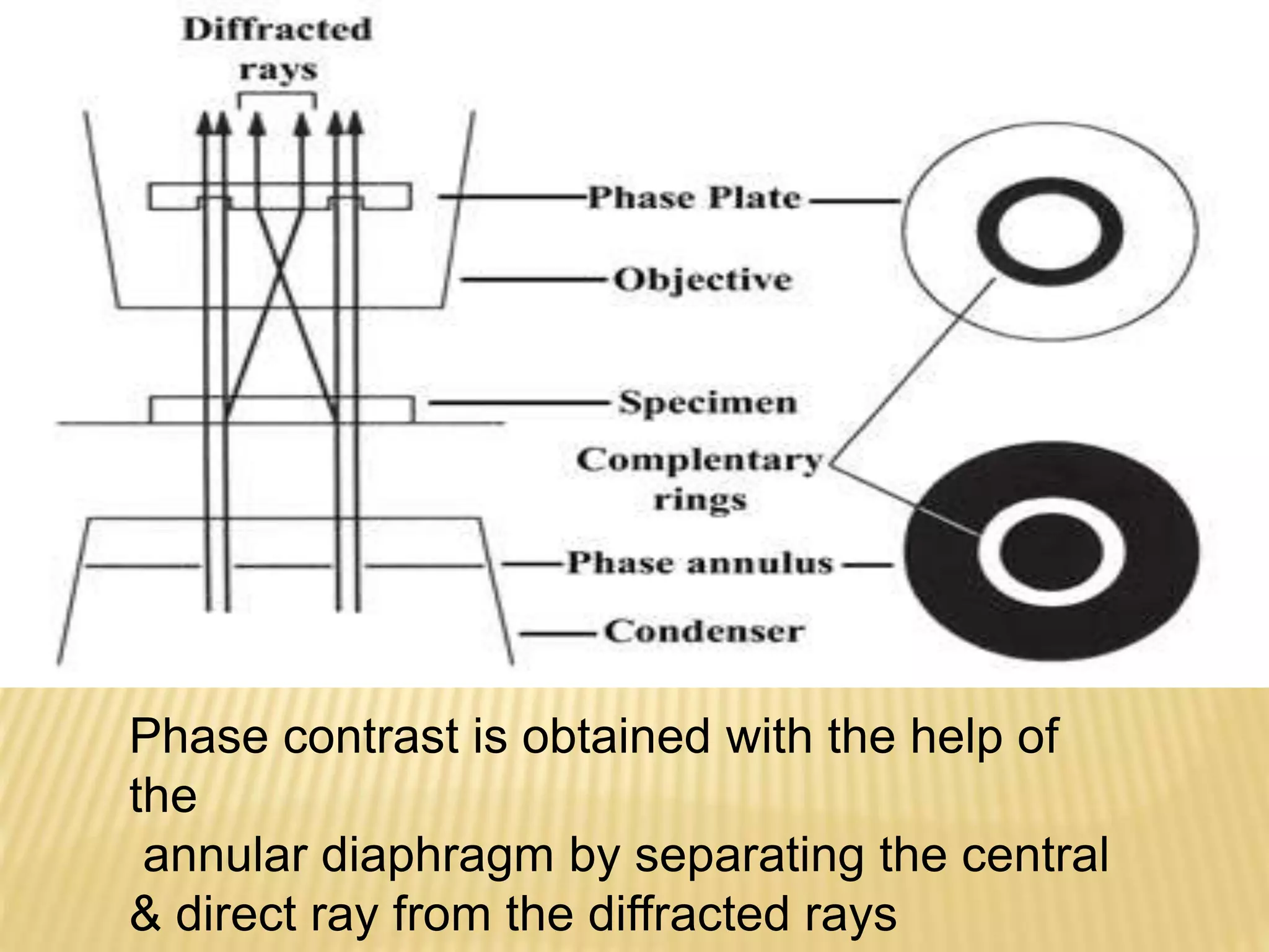


The document discusses phase contrast microscopy, which was developed by Fritz Zernike in the 1930s. It allows living or unstained cells and intracellular components to be visible under a microscope. The phase contrast microscope works by converting small phase changes caused by differences in refractive index of cell structures into visible brightness and darkness differences. This makes organelles and other structures visible without using staining. The phase contrast is achieved using an annular diaphragm and phase rings or filters to shift the phase of light passing through or around the specimen.


