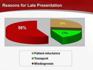
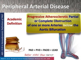
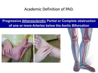
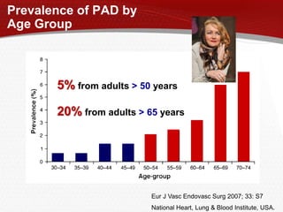
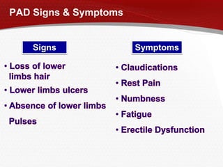
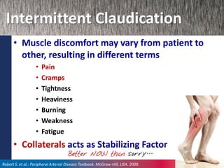
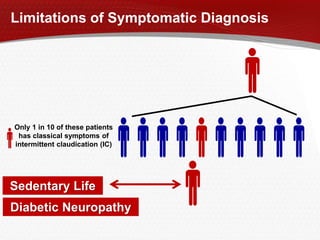
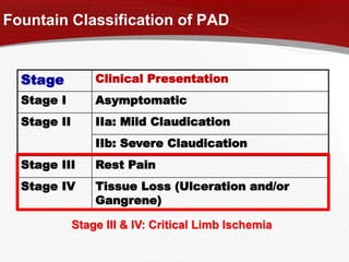
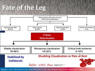
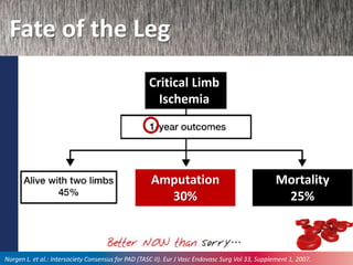
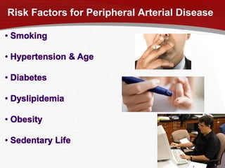
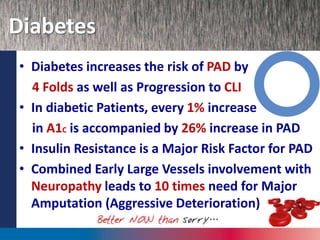
Peripheral Arterial Disease
Check the Pedal Pulse
Alaa Wafa . MD
Associate Professor of Internal Medicine
PGDIP DM Cardiff University UK
Diabetes & Endocrine Unit.
Mansoura University
2014
Amputation every 30 Seconds
Every 30 Sec. there is an Amputation
somewhere in the World
The global burden of diabetic foot disease. Lancet. Vol 366 November 12, 2005
Reasons for Late Presentation
Peripheral Arterial Disease
Progressive Atherosclerotic Partial
or Complete Obstruction
of one or more Arteries below the
Aortic Bifurcation
Academic
Definition
Robert S. et al.: Peripheral Arterial Disease Textbook. McGraw-Hill, USA. 2009.
PAD = PVD = PAOD = LEAD
ABI<0.9isHemodynamicDefinitionofPAD
Academic Definition of PAD.
Progressive Atherosclerotic Partial or Complete obstruction
of one or more Arteries below the Aortic Bifurcation
Prevalence of PAD by
Age Group
Eur J Vasc Endovasc Surg 2007; 33: S7
National Heart, Lung & Blood Institute, USA.
5% from adults > 50 years
20% from adults > 65 years
PAD Signs & Symptoms
Signs Symptoms
Intermittent Claudication
• Muscle discomfort may vary from patient to
other, resulting in different terms
• Pain
• Cramps
• Tightness
• Heaviness
• Burning
• Weakness
• Fatigue
• Collaterals acts as Stabilizing Factor
Robert S. et al.: Peripheral Arterial Disease Textbook. McGraw-Hill, USA. 2009.
Limitations of Symptomatic Diagnosis
Only 1 in 10 of these patients
has classical symptoms of
intermittent claudication (IC)
Diabetic Neuropathy
Sedentary Life
Stage Clinical Presentation
Stage I Asymptomatic
Stage II IIa: Mild Claudication
IIb: Severe Claudication
Stage III Rest Pain
Stage IV Tissue Loss (Ulceration and/or
Gangrene)
Fountain Classification of PAD
Stage III & IV: Critical Limb Ischemia
Fate of the Leg
Disabling Claudication or Pain at RestStabilized by
Collaterals
Norgen L. et al.: Intersociety Consensus for PAD (TASC II). Eur J Vasc Endovasc Surg Vol 33, Supplement 1, 2007.
5 Years
Deterioration
Fate of the Leg
Critical Limb
Ischemia
Norgen L. et al.: Intersociety Consensus for PAD (TASC II). Eur J Vasc Endovasc Surg Vol 33, Supplement 1, 2007.
Amputation
30%
Mortality
25%
Risk Factors for Peripheral Arterial Disease
Factors Magnifying Critical
Limb Ischemia incidence
Norgen L. et al.: Intersociety Consensus for PAD (TASC II). Eur J Vasc Endovasc Surg Vol 33, Supplement 1, 2007.
Diabetes
• Diabetes increases the risk of PAD by
4 Folds as well as Progression to CLI
• In diabetic Patients, every 1% increase
in A1C is accompanied by 26% increase in PAD
• Insulin Resistance is a Major Risk Factor for PAD
• Combined Early Large Vessels involvement with
Neuropathy leads to 10 times need for Major
Amputation (Aggressive Deterioration)
Diabetes
• More Severe & Longstanding Diabetes
is associated with more incidence of PAD
• Major Contributor are Advanced Glycated
End Products (AGEs)
– Combined excess Glucose with Collagen Fibers
– AGEs encourage Plaque Formation, Atherosclerosis &
increased trapping of LDL
Diabetic Vascular Complications
 All patients with diabetes are at risk of developing several chronic
complications, categorized as microvascular and macrovascular1,2
Diabetic
Retinopathy
Microvascular Complications Macrovascular Complications
Diabetic
Nephropathy
Diabetic
Neuropathy
Stroke
Peripheral
Vascular Disease
Heart
Disease
1. Fowler. Clinical Diabetes 2008;26(2):77-82.
2. http://www.who.int/diabetesactiononline/diabetes/basics/en/index3.html
DPNP
Diabetic Peripheral Neuropathic Pain:
A Frequent and Debilitating Complication
 10%-20% of patients with diabetic peripheral neuropathy
develop pain1
 This pain broadly interferes with daily functioning and quality
of life1-4
• General activity
• Walking
• Energy level
• Social activities
• Ability to sleep
• Change in mood, feelings of depression and anxiety
• Overall enjoyment of life
1. Argoff et al. Mayo Clin Proc 2006;81(4 Suppl):S3-11.
2. Boulton. Clin Diabetes 2005;23:9-15.
DPNP 3. Galer et al. Diabetes Res Clin Pract 2000;47(2):123-8.
4. Gore et al. J Pain Symptom Manage 2005;30(4):374-85.
Diabetic Neuropathy: Clinical Presentation
Large-fiber
Neuropathy
Small-fiber
Neuropathy
Proximal Motor
Neuropathy
Acute
Mononeuropathies Pressure Palsies
Sensory loss: 0+++
(touch, vibration)
Pain: ++++
Tendon reflex:
N
Motor deficit: 0+++
Sensory loss: 0+
(thermal, allodynia)
Pain: ++++
Tendon reflex: N
Motor deficit: 0
Sensory loss: 0+
Pain: ++++
Tendon reflex:
Proximal motor deficit:
++++
Sensory loss: 0+
Pain: ++++
Tendon reflex: N
Motor deficit:
++++
Sensory loss in nerve
distribution: ++++
Pain: +++
Tendon reflex: N
Motor deficit: ++++
III VI
Truncal
Ulnar
Lateral
popliteal
Median
Reprinted from: Vinik et al. Diabetologia 2000;43(8):957-73.DPNP
DIABETIC FOOT
1. HISTORY
HISTORY
Duration of
diabetes
Quality of
glycemic
control
Complications
and
comorbidities
Patient foot specific
medical history
Initial
wounding
event
History of
recurrent
wounds
Previous
wound
healing
Prior
diagnostic
testing
Prior treatment
and response
Social
history
2. EXAMINATION
Assessment
of PAD
Assessment of
Neuropathy
Foot and
Ulcer
examination
Infection
Evaluation
FOOT &ULCER EXAMINATION
• Evaluate etiology:neuropathic,ischemic,neuro-ischemic
• Evaluation of perfusion
• Ischemic skin changes
• Dermatological changes-callus, muscle wasting
• Ulcer characteristics-location,site,shape,size
• Wound edges,wound bed,wound base,paeriwound skin
• and exudates
• Presence of necrosis and wound associated pain
• Biomechanical status-forefoot deformities,weakness,gait
abnormalities
• Complications-cellulitis,gangrene,osteomyelitis
• Charcot deformity
INFECTION EVALUATION
• Red,hot,tender
• Swelling
• Exudates
• Delayed healing
• Friable and discolored granulation tissue
• Foul odor
• Wound breakdown
• Increased ESR & CRP
 Signs-
loss of vibratory and position sense
loss of deep tendon reflexes
trophic ulceration
foot drop
muscle atrophy
excessive callus formation
 Semmes-Weinstein filament
 Biothesiometer
 Pulsation of dorsalis pedis artery and
posterior tibial artery
 Bruit at iliac or femoral arteries
 Skin atrophy
 Loss of pedal hair growth
 Toe cyanosis
 Ulceration or ischemic necrosis
 Pallor and rubor after 1-2 minutes of
elevation above heart level
 History of claudication
ASSESSMENT OF
NEUROPATHY
ASSESSMENT OF PERIPHERAL
ARTERIAL DISEASE
CLASSIFICATION OF
DIABETIC FOOT ULCER
 Texas Classification
 Wagner Classification
3. INVESTIGATIONS
 Complete Blood Count
 Pulse-Volume Recording
 Doppler Ultrasound
 Ankle-Brachial Index
 Plain Radiography
 CT and MRI
 Bone scan
 Angiography
PREVENTION
PREVENTION
Patient
Education
Foot
examination
Optimizing
glycemic
control
Smoking
cessation
Custom
Footwear
and
Orthotics
Debridement
of calluses
Multidisciplinary
team
Prophylactic
foot surgery
Revascularization
CUSTOM FOOTWEAR
TREATMENT
Vascular
perfusion
Debridement
Infection
Control
Off-
loading
Wound
coverage
Treatment of
Charcot foot
Hyperbaric
Oxygen
Treatment
Dietary
Changes
Activity
restriction
Amputation
Recent
Management
options for PAD
Cilostazol
Naftidrofuryl
Role of Serotonin in PAD
Serotonin
5-HT2
5 Hydroxytryptamine-2
100% stored in Platelets
•Promotes Platelets Aggregation
•Thrombus Formation
•Augments Aggregating Factors
•RBCs Aggregation (Rouleaux)
•Increase Blood Viscosity
•Direct Vasoconstriction
•Collaterals: Serotonin Sensitive
•Serotonin Sensitivity is defined by
Hyper-reactivity to Serotonin
(increased by Age, Hypertension, ischemia
Atherosclerosis Hypercholesterolemia)
Naftidrofuryl Multi Modes of Action
5-HT2
↑ Platelets aggregation
↑ Vasoconstriction
↑ RBCs Rigidity
Guidelines
Recommendations
Global Guideline for the Management of
PAD (TASC II).
Inter-Society Consensus for the Management of
Peripheral Arterial Disease
Pharmacotherapy
A. Drugs with evidence of clinical utility in claudication
(Grade A)
Blood
Platelets
RBCsV. Smooth
Muscles
5-HT
Naftidrofuryl is the only
available specific Serotonin
S2 - receptor blocker on
blood platelets and
vascular smooth muscles
Global Guideline for the Management of
PAD (TASC II).
10
dralaawafa@hotmail.com

Peipheral arterial dse

  • 1.
    Peripheral Arterial Disease Checkthe Pedal Pulse Alaa Wafa . MD Associate Professor of Internal Medicine PGDIP DM Cardiff University UK Diabetes & Endocrine Unit. Mansoura University 2014
  • 2.
    Amputation every 30Seconds Every 30 Sec. there is an Amputation somewhere in the World The global burden of diabetic foot disease. Lancet. Vol 366 November 12, 2005
  • 3.
    Reasons for LatePresentation
  • 4.
    Peripheral Arterial Disease ProgressiveAtherosclerotic Partial or Complete Obstruction of one or more Arteries below the Aortic Bifurcation Academic Definition Robert S. et al.: Peripheral Arterial Disease Textbook. McGraw-Hill, USA. 2009. PAD = PVD = PAOD = LEAD ABI<0.9isHemodynamicDefinitionofPAD
  • 5.
    Academic Definition ofPAD. Progressive Atherosclerotic Partial or Complete obstruction of one or more Arteries below the Aortic Bifurcation
  • 6.
    Prevalence of PADby Age Group Eur J Vasc Endovasc Surg 2007; 33: S7 National Heart, Lung & Blood Institute, USA. 5% from adults > 50 years 20% from adults > 65 years
  • 7.
    PAD Signs &Symptoms Signs Symptoms
  • 8.
    Intermittent Claudication • Musclediscomfort may vary from patient to other, resulting in different terms • Pain • Cramps • Tightness • Heaviness • Burning • Weakness • Fatigue • Collaterals acts as Stabilizing Factor Robert S. et al.: Peripheral Arterial Disease Textbook. McGraw-Hill, USA. 2009.
  • 9.
    Limitations of SymptomaticDiagnosis Only 1 in 10 of these patients has classical symptoms of intermittent claudication (IC) Diabetic Neuropathy Sedentary Life
  • 10.
    Stage Clinical Presentation StageI Asymptomatic Stage II IIa: Mild Claudication IIb: Severe Claudication Stage III Rest Pain Stage IV Tissue Loss (Ulceration and/or Gangrene) Fountain Classification of PAD Stage III & IV: Critical Limb Ischemia
  • 11.
    Fate of theLeg Disabling Claudication or Pain at RestStabilized by Collaterals Norgen L. et al.: Intersociety Consensus for PAD (TASC II). Eur J Vasc Endovasc Surg Vol 33, Supplement 1, 2007. 5 Years Deterioration
  • 12.
    Fate of theLeg Critical Limb Ischemia Norgen L. et al.: Intersociety Consensus for PAD (TASC II). Eur J Vasc Endovasc Surg Vol 33, Supplement 1, 2007. Amputation 30% Mortality 25%
  • 13.
    Risk Factors forPeripheral Arterial Disease
  • 14.
    Factors Magnifying Critical LimbIschemia incidence Norgen L. et al.: Intersociety Consensus for PAD (TASC II). Eur J Vasc Endovasc Surg Vol 33, Supplement 1, 2007.
  • 15.
    Diabetes • Diabetes increasesthe risk of PAD by 4 Folds as well as Progression to CLI • In diabetic Patients, every 1% increase in A1C is accompanied by 26% increase in PAD • Insulin Resistance is a Major Risk Factor for PAD • Combined Early Large Vessels involvement with Neuropathy leads to 10 times need for Major Amputation (Aggressive Deterioration)
  • 16.
    Diabetes • More Severe& Longstanding Diabetes is associated with more incidence of PAD • Major Contributor are Advanced Glycated End Products (AGEs) – Combined excess Glucose with Collagen Fibers – AGEs encourage Plaque Formation, Atherosclerosis & increased trapping of LDL
  • 18.
    Diabetic Vascular Complications All patients with diabetes are at risk of developing several chronic complications, categorized as microvascular and macrovascular1,2 Diabetic Retinopathy Microvascular Complications Macrovascular Complications Diabetic Nephropathy Diabetic Neuropathy Stroke Peripheral Vascular Disease Heart Disease 1. Fowler. Clinical Diabetes 2008;26(2):77-82. 2. http://www.who.int/diabetesactiononline/diabetes/basics/en/index3.html DPNP
  • 19.
    Diabetic Peripheral NeuropathicPain: A Frequent and Debilitating Complication  10%-20% of patients with diabetic peripheral neuropathy develop pain1  This pain broadly interferes with daily functioning and quality of life1-4 • General activity • Walking • Energy level • Social activities • Ability to sleep • Change in mood, feelings of depression and anxiety • Overall enjoyment of life 1. Argoff et al. Mayo Clin Proc 2006;81(4 Suppl):S3-11. 2. Boulton. Clin Diabetes 2005;23:9-15. DPNP 3. Galer et al. Diabetes Res Clin Pract 2000;47(2):123-8. 4. Gore et al. J Pain Symptom Manage 2005;30(4):374-85.
  • 20.
    Diabetic Neuropathy: ClinicalPresentation Large-fiber Neuropathy Small-fiber Neuropathy Proximal Motor Neuropathy Acute Mononeuropathies Pressure Palsies Sensory loss: 0+++ (touch, vibration) Pain: ++++ Tendon reflex: N Motor deficit: 0+++ Sensory loss: 0+ (thermal, allodynia) Pain: ++++ Tendon reflex: N Motor deficit: 0 Sensory loss: 0+ Pain: ++++ Tendon reflex: Proximal motor deficit: ++++ Sensory loss: 0+ Pain: ++++ Tendon reflex: N Motor deficit: ++++ Sensory loss in nerve distribution: ++++ Pain: +++ Tendon reflex: N Motor deficit: ++++ III VI Truncal Ulnar Lateral popliteal Median Reprinted from: Vinik et al. Diabetologia 2000;43(8):957-73.DPNP
  • 21.
  • 23.
    1. HISTORY HISTORY Duration of diabetes Qualityof glycemic control Complications and comorbidities Patient foot specific medical history Initial wounding event History of recurrent wounds Previous wound healing Prior diagnostic testing Prior treatment and response Social history
  • 24.
    2. EXAMINATION Assessment of PAD Assessmentof Neuropathy Foot and Ulcer examination Infection Evaluation
  • 25.
    FOOT &ULCER EXAMINATION •Evaluate etiology:neuropathic,ischemic,neuro-ischemic • Evaluation of perfusion • Ischemic skin changes • Dermatological changes-callus, muscle wasting • Ulcer characteristics-location,site,shape,size • Wound edges,wound bed,wound base,paeriwound skin • and exudates • Presence of necrosis and wound associated pain • Biomechanical status-forefoot deformities,weakness,gait abnormalities • Complications-cellulitis,gangrene,osteomyelitis • Charcot deformity INFECTION EVALUATION • Red,hot,tender • Swelling • Exudates • Delayed healing • Friable and discolored granulation tissue • Foul odor • Wound breakdown • Increased ESR & CRP
  • 26.
     Signs- loss ofvibratory and position sense loss of deep tendon reflexes trophic ulceration foot drop muscle atrophy excessive callus formation  Semmes-Weinstein filament  Biothesiometer  Pulsation of dorsalis pedis artery and posterior tibial artery  Bruit at iliac or femoral arteries  Skin atrophy  Loss of pedal hair growth  Toe cyanosis  Ulceration or ischemic necrosis  Pallor and rubor after 1-2 minutes of elevation above heart level  History of claudication ASSESSMENT OF NEUROPATHY ASSESSMENT OF PERIPHERAL ARTERIAL DISEASE
  • 27.
    CLASSIFICATION OF DIABETIC FOOTULCER  Texas Classification  Wagner Classification
  • 28.
    3. INVESTIGATIONS  CompleteBlood Count  Pulse-Volume Recording  Doppler Ultrasound  Ankle-Brachial Index  Plain Radiography  CT and MRI  Bone scan  Angiography
  • 31.
  • 32.
  • 33.
  • 34.
  • 35.
  • 36.
  • 37.
  • 38.
    Role of Serotoninin PAD Serotonin 5-HT2 5 Hydroxytryptamine-2 100% stored in Platelets •Promotes Platelets Aggregation •Thrombus Formation •Augments Aggregating Factors •RBCs Aggregation (Rouleaux) •Increase Blood Viscosity •Direct Vasoconstriction •Collaterals: Serotonin Sensitive •Serotonin Sensitivity is defined by Hyper-reactivity to Serotonin (increased by Age, Hypertension, ischemia Atherosclerosis Hypercholesterolemia)
  • 39.
    Naftidrofuryl Multi Modesof Action 5-HT2 ↑ Platelets aggregation ↑ Vasoconstriction ↑ RBCs Rigidity
  • 40.
  • 41.
    Global Guideline forthe Management of PAD (TASC II). Inter-Society Consensus for the Management of Peripheral Arterial Disease
  • 42.
    Pharmacotherapy A. Drugs withevidence of clinical utility in claudication (Grade A) Blood Platelets RBCsV. Smooth Muscles 5-HT Naftidrofuryl is the only available specific Serotonin S2 - receptor blocker on blood platelets and vascular smooth muscles Global Guideline for the Management of PAD (TASC II). 10
  • 43.