A sedentary male of 70 kg (154 lbs) requires about 8400 kJ
(2000 kcal) for a day’s worth of activity. To provide this much energy
requires 83 kg of ATP. However, human beings possess only about 250 g
of ATP at any given moment. The disparity between the amount of ATP
that we have and the amount that we require is compensated by
recycling ADP back to ATP. Each ATP molecule is recycled
approximately 300 times per day. This recycling takes place primarily
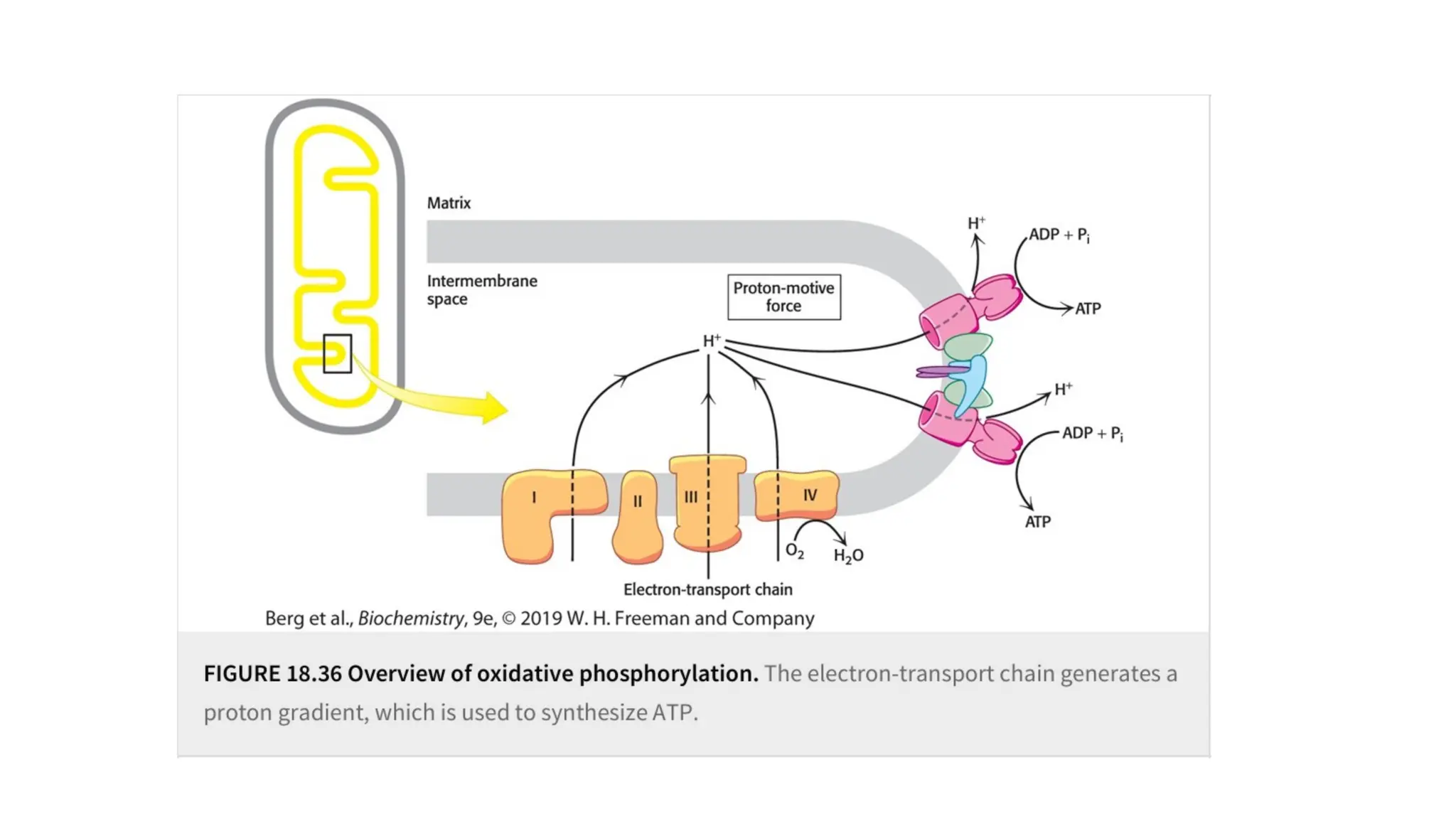
through oxidative phosphorylation.


![Bioenergetics [autosaved]](https://cdn.slidesharecdn.com/ss_thumbnails/bioenergeticsautosaved-210315111006-thumbnail.jpg?width=640&height=640&fit=bounds)


