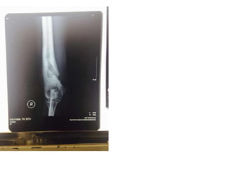
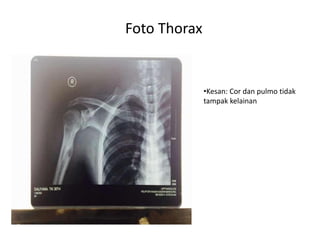
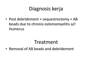
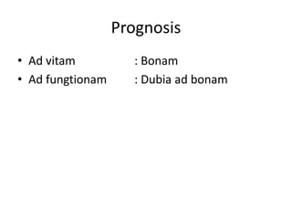
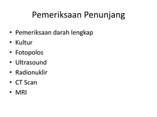
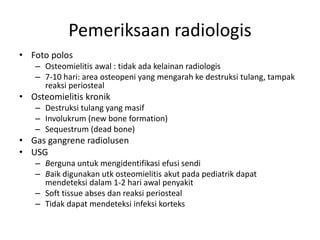
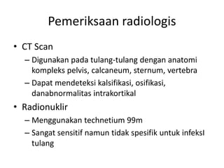
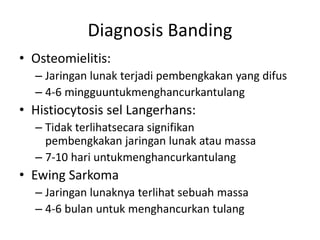
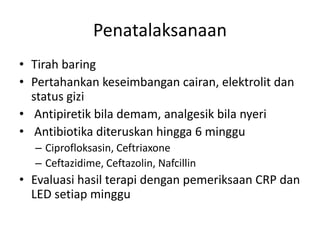
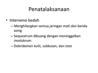
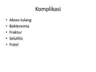
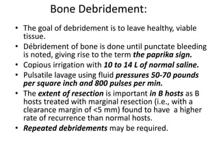
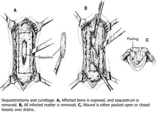
Case Report Session 
Osteomyelitis 
Pembimbing: 
Dr. Yoyos Dias Ismiranto, dr. SpOT., M.Kes FICS 
Penyusun: Devyashini Prabhakaran 
Bagian Orthopedi dan Traumatologi 
Fakultas Kedokteran 
Universitas Padjadjaran 
Bandung 
2014
1. Identitas Pasien 
• Nama :Tn. DA 
• Umur :30 thn 
• Jenis kelamin : Laki-laki 
• Agama : Islam 
• Alamat : Kg. Babakan,RT 01, RW 
06,Citapen, Cihampelas, 
Bandung 
• Status Pernikahan : Bernikah 
• Pekerjaan : Buruh
• Anamnesa 
KU: Keluar nanah dari lengan atas kanan 
• Anamnesa khusus: 
– 2 tahun SMRS, os mengeluh timbul bisul di 
lengan atas kanan. Bisul pecah dengan 
mengeluarkan nanah. 1 bulan yang lalu, os 
dilakukan operasi pembersihan tulang dan 
pemasangan selang antibiotik. Sekarang nanah 
berkurang dan selang antibiotik nyeri 
dilepaskan.
• Riwayat penyakit dahulu: Post op 
debridement + sequestrectomy + AB beads 
• Riwayat penyakit pada keluarga: tidak ada.
Pemeriksaan Fisik 
Keadaan umum : Compos Mentis 
Tanda vital 
 Tensi : 100/70 mmHg 
 Nadi : 80 x/menit 
 Respirasi : 20 x/menit 
 Suhu : 36,6 oC 
Status Generalis 
Kepala : Konjungtiva tidak anemis, Sklera tidak ikterik 
Leher : KGB tidak teraba membesar 
Dada : Bentuk dan gerak simetris, VBS ki=ka, rhonki (-/-) 
wheezing (-/-),Bunyi jantung murni reguler 
Abdomen : Datar dan lembut, Bising usus (+) normal, Hepar dan 
lien tidak teraba, Ruang traube kosong 
Ekstremitas : Lihat status lokalis
Status Lokalis: 
a/r humerus 
L : Scar (+),Swelling (+), pus (+), fistel (+) 
F : NT (+), sensitibilitas dalam batas normal, 
kapiler refil (+ <2 detik) 
M : elbow stiffness 90°
Pemeriksaan Penunjang 
• Lab Darah 
• X-ray
• Tampak lesi litik dan 
sklerotik pada 1/3 distal os 
humerus dan 1/3 proximal 
os radius. 
• Tampak lesi periosteal pada 
1/3 distal os humerus 
kanan dan 1/3 proksimal os 
radius kanan 
• Kesan: menyokong suatu 
osteomielitis kronis 
Foto elbow
Foto Thorax 
•Kesan: Cor dan pulmo tidak 
tampak kelainan
Diagnosis kerja 
• Post debridement + sequestrectomy + AB 
beads due to chronis ostemomyelitis a/r 
Humerus 
Treatment 
• Removal of AB beads and debridement
Prognosis 
• Ad vitam : Bonam 
• Ad fungtionam : Dubia ad bonam
Pembahasan Osteomyelitis
Definisi 
• Suatu proses inflamasi akut ataupun kronis 
daritulang dan struktur-struktur disekitarnya 
akibatinfeksi dari kuman-kuman piogenik
Patogenesis 
• Infeksi dapat berkembang melalui 
beberapacara, yaitu : 
– Penyebaran hematogen dari infeksi di bagian 
tubuh lain (osteomielitis hematogen) 
– Pembedahan jaringan tubuh terpapar 
denganlingkungan sekitarnya (osteomielitis 
esogenik) 
– Luka penetrasi langsung (osteomielitis esogenik)
Insiden 
• Osteomielitis akut hematogen : anak-anak 
• Osteomielitis dari trauma direk /contiguous: 
usiaremaja dan dewasa muda 
• Spinal osteomielitis : usia > 45 tahun 
• dapat pula ditemukan pada bayi dan ‘infant’ 
• Anak laki-laki > anak perempuan (4:1) 
• Lokasi yang tersering : tulang-tulang panjang
Klasifikasi 
• Menurut durasi (tradisional) 
– Akut (7-14 hari) 
– Subakut (14 hari -3 bulan) 
– Kronik (> 3 bulan) 
• Menurut etiologi dan kronisitas (Waldvogel) 
– Hematogen 
– Contiguous 
– Kronik 
• Menurut penyebaran anatomis (Cierny-Mader ) 
– Stadium I -Medular 
– Stadium II -Cortex superfisial 
– Stadium III- medular dan korteks terlokalisasi 
– Stadium IV- Medular dan korteks difus
Presentasi klinis 
• Osteomielitis hematogen 
• Sesuai gejala dan tanda inflamasi akut 
• Nyeri terlokalisasi 
• Penderita menghindari menggunakan bagian 
tubuhyang sakit 
• PF : nyeri tekan lokal, pergerakan sendi terbatas,oedem 
dan kemerahan jarang ditemukan 
• Gejala sistemik : demam, malaise, menggigil, nafsu 
makan menurun 
• Lab : peningkatan CRP ,LED, dan leukosit
Presentasi klinis 
• Osteomyelitis subakut 
• Sering pada anak-anak 
• Akibat kuman bervirulensi rendah tanpa gejala 
• Gambaran radiologis : 
– Seperti osteomielitis akut osteolisis dan elevasi 
periosteal 
– Seperti osteomielitis kronik zona sirkumferensial 
tulangyang sklerotik
Presentasi klinis 
• Osteomielitis kronik 
• Akibat infeksi akut atau subakut yang tidak 
diobati 
• Ulkus persisten 
• Fistel atau drainase pus 
• Fatigue 
• Malaise
Pemeriksaan Penunjang 
• Pemeriksaan darah lengkap 
• Kultur 
• Fotopolos 
• Ultrasound 
• Radionuklir 
• CT Scan 
• MRI
Pemeriksaan radiologis 
• Foto polos 
– Osteomielitis awal : tidak ada kelainan radiologis 
– 7-10 hari: area osteopeni yang mengarah ke destruksi tulang, tampak 
reaksi periosteal 
• Osteomielitis kronik 
– Destruksi tulang yang masif 
– Involukrum (new bone formation) 
– Sequestrum (dead bone) 
• Gas gangrene radiolusen 
• USG 
– Berguna untuk mengidentifikasi efusi sendi 
– Baik digunakan utk osteomielitis akut pada pediatrik dapat 
mendeteksi dalam 1-2 hari awal penyakit 
– Soft tissue abses dan reaksi periosteal 
– Tidak dapat mendeteksi infeksi korteks
Pemeriksaan radiologis 
• CT Scan 
– Digunakan pada tulang-tulang dengan anatomi 
kompleks pelvis, calcaneum, sternum, vertebra 
– Dapat mendeteksi kalsifikasi, osifikasi, 
danabnormalitas intrakortikal 
• Radionuklir 
– Menggunakan technetium 99m 
– Sangat sensitif namun tidak spesifik untuk infeksI 
tulang
Diagnosis Banding 
• Osteomielitis: 
– Jaringan lunak terjadi pembengkakan yang difus 
– 4-6 mingguuntukmenghancurkantulang 
• Histiocytosis sel Langerhans: 
– Tidak terlihatsecara signifikan 
pembengkakan jaringan lunak atau massa 
– 7-10 hari untukmenghancurkantulang 
• Ewing Sarkoma 
– Jaringan lunaknya terlihat sebuah massa 
– 4-6 bulan untuk menghancurkan tulang
Penatalaksanaan 
• Tirah baring 
• Pertahankan keseimbangan cairan, elektrolit dan 
status gizi 
• Antipiretik bila demam, analgesik bila nyeri 
• Antibiotika diteruskan hingga 6 minggu 
– Ciprofloksasin, Ceftriaxone 
– Ceftazidime, Ceftazolin, Nafcillin 
• Evaluasi hasil terapi dengan pemeriksaan CRP dan 
LED setiap minggu
Penatalaksanaan 
• Intervensi bedah 
– Menghilangkan semua jaringan mati dan benda 
asing 
– Sequestrum dibuang dengan meninggalkan 
involukrum 
– Debridemen kulit, subkutan, dan otot
Komplikasi 
• Abses tulang 
• Bakteremia 
• Fraktur 
• Selulitis 
• Fistel
Surgical treatment
Bone Debridement: 
• The goal of debridement is to leave healthy, viable 
tissue. 
• Débridement of bone is done until punctate bleeding 
is noted, giving rise to the term the paprika sign. 
• Copious irrigation with 10 to 14 L of normal saline. 
• Pulsatile lavage using fluid pressures 50-70 pounds 
per square inch and 800 pulses per min. 
• The extent of resection is important in B hosts as B 
hosts treated with marginal resection (i.e., with a 
clearance margin of <5 mm) found to have a higher 
rate of recurrence than normal hosts. 
• Repeated debridements may be required.
Sequestrectomy and curettage. A, Affected bone is exposed, and sequestrum is 
removed. B, All infected matter is removed. C, Wound is either packed open or closed 
loosely over drains.
When to do sequestrectomy? 
Early sequestrectomy 
- Eradicate infection 
-Better environment for periosteum to respond 
Delayed sequestrectomy 
-Wait till sufficient involucrum has formed before 
doing a sequestrectomy to mimimize the risk of 
fracture, deformity & segmental loss
Prerequisites for Sequestrectomy 
Radiological 
• Well formed 
involucrum 
surrounding the 
discretely visible 
sequestrum 
adequately at least 
2/3rd diameter of 
bone (3 intact walls 
on two views ensure 
3/4th intact walls) 
Clinical 
• Symptomatic patient 
with pus discharge or 
chronic unreleaved 
disabling pain due to 
osteomyelitis per se 
and type A/B host.
Management of Dead Space: 
Antibiotic Beads 
 May be used to sterilize and temporarily maintain a 
dead space. 
 Beads are made with PMMA+ab 
 Cement -40 gm. 
 Genta- 1-2 gm. or vanco 1-2 gm. 
 Other antibiotics that can be used are Tobramycin, 
Penicillin, cephalosporins, amikacin, vancomycin. 
 The gentamycin concentration remain for 30 days 
after implantation. 
 The shape and type of methylmethacrylate has a 
significant effect on the amount of antibiotic 
delivery, as well as duration. 
 The best delivery profile was with PMMA beads 
impregnated with gentamicin. 
 Usually removed within two to four weeks and are 
replaced with a cancellous bone graft.
Antibiotic beads: 
• Can act as a biomaterial surface to which bacteria 
preferentially adhere. 
• To avoid such a problem, biodegradable antibiotic-impregnated 
(calcium sulfate) beads have been 
employed recently and have shown favorable antibiotic 
release kinetics 
– Elution testing of 4% by weight loaded calcium 
sulfate pellets revealed a maximum concentration 
of 828 μg/ml and undetectable levels by day 15. 
• Antibiotic-impregnated cancellous bone grafts were 
recently used in a clinical trial of forty-six patients, and 
the osteomyelitis was arrested in 95% of them
Management of Dead Space: 
• Antibiotics (clindamycin and amikacin) have 
also been delivered directly into dead spaces 
with an implantable pump. 
• Very high local and low systemic levels of 
antibiotics have been achieved.
Soft-Tissue Coverage: 
• Three methods commonly used: 
– Primary closure- if no infection 
– Let tissue heal by secondary intention 
– Small soft-tissue defects may be covered with a split-thickness 
skin graft. 
– Local muscle flaps and free vascularized muscle flaps in 
the presence of a large soft-tissue defect or an 
inadequate soft-tissue envelope. 
• Healing by so-called secondary intention should be 
discouraged, since the scar tissue that fills the defect may 
later become avascular.
Bone Stabilization: 
• If skeletal instability is present at the site of an infection, 
measures must be taken to achieve stability with 
– Plates 
– Screws 
– Rods 
– An external fixator 
• External fixation is preferred over internal fixation because 
– of the tendency of the sites of medullary rods to become 
secondarily infected and to spread the extent of the 
infection. 
• Rigid fixation helpful in union of fracture sites.
Limb reconstruction: 
• Ilizarov external fixation 
– Is used for reconstruction of segmental defects and difficult infected 
nonunions. 
– Based on the technique of distraction osteogenesis whereby an 
osteotomy created in the metaphyseal region of the bone is gradually 
distracted to fill in the defect. 
– Used for difficult cases of osteomyelitis when stabilization and bone-lengthening 
are necessary. 
– May also be used to compress nonunions and to correct malunions.
AMPUTATION: 
• Infrequently performed 
• INDICATIONS 
1. Malignancy 
2. Arterial insufficiency 
3. Nerve paralysis 
4. Jt. Contracture & stiffness making limb 
nonfunctional
Osteomyelitis Case Report Session

Osteomyelitis Case Report Session

  • 1.
    Case Report Session Osteomyelitis Pembimbing: Dr. Yoyos Dias Ismiranto, dr. SpOT., M.Kes FICS Penyusun: Devyashini Prabhakaran Bagian Orthopedi dan Traumatologi Fakultas Kedokteran Universitas Padjadjaran Bandung 2014
  • 2.
    1. Identitas Pasien • Nama :Tn. DA • Umur :30 thn • Jenis kelamin : Laki-laki • Agama : Islam • Alamat : Kg. Babakan,RT 01, RW 06,Citapen, Cihampelas, Bandung • Status Pernikahan : Bernikah • Pekerjaan : Buruh
  • 3.
    • Anamnesa KU:Keluar nanah dari lengan atas kanan • Anamnesa khusus: – 2 tahun SMRS, os mengeluh timbul bisul di lengan atas kanan. Bisul pecah dengan mengeluarkan nanah. 1 bulan yang lalu, os dilakukan operasi pembersihan tulang dan pemasangan selang antibiotik. Sekarang nanah berkurang dan selang antibiotik nyeri dilepaskan.
  • 4.
    • Riwayat penyakitdahulu: Post op debridement + sequestrectomy + AB beads • Riwayat penyakit pada keluarga: tidak ada.
  • 5.
    Pemeriksaan Fisik Keadaanumum : Compos Mentis Tanda vital  Tensi : 100/70 mmHg  Nadi : 80 x/menit  Respirasi : 20 x/menit  Suhu : 36,6 oC Status Generalis Kepala : Konjungtiva tidak anemis, Sklera tidak ikterik Leher : KGB tidak teraba membesar Dada : Bentuk dan gerak simetris, VBS ki=ka, rhonki (-/-) wheezing (-/-),Bunyi jantung murni reguler Abdomen : Datar dan lembut, Bising usus (+) normal, Hepar dan lien tidak teraba, Ruang traube kosong Ekstremitas : Lihat status lokalis
  • 6.
    Status Lokalis: a/rhumerus L : Scar (+),Swelling (+), pus (+), fistel (+) F : NT (+), sensitibilitas dalam batas normal, kapiler refil (+ <2 detik) M : elbow stiffness 90°
  • 7.
    Pemeriksaan Penunjang •Lab Darah • X-ray
  • 8.
    • Tampak lesilitik dan sklerotik pada 1/3 distal os humerus dan 1/3 proximal os radius. • Tampak lesi periosteal pada 1/3 distal os humerus kanan dan 1/3 proksimal os radius kanan • Kesan: menyokong suatu osteomielitis kronis Foto elbow
  • 10.
    Foto Thorax •Kesan:Cor dan pulmo tidak tampak kelainan
  • 11.
    Diagnosis kerja •Post debridement + sequestrectomy + AB beads due to chronis ostemomyelitis a/r Humerus Treatment • Removal of AB beads and debridement
  • 12.
    Prognosis • Advitam : Bonam • Ad fungtionam : Dubia ad bonam
  • 13.
  • 14.
    Definisi • Suatuproses inflamasi akut ataupun kronis daritulang dan struktur-struktur disekitarnya akibatinfeksi dari kuman-kuman piogenik
  • 15.
    Patogenesis • Infeksidapat berkembang melalui beberapacara, yaitu : – Penyebaran hematogen dari infeksi di bagian tubuh lain (osteomielitis hematogen) – Pembedahan jaringan tubuh terpapar denganlingkungan sekitarnya (osteomielitis esogenik) – Luka penetrasi langsung (osteomielitis esogenik)
  • 16.
    Insiden • Osteomielitisakut hematogen : anak-anak • Osteomielitis dari trauma direk /contiguous: usiaremaja dan dewasa muda • Spinal osteomielitis : usia > 45 tahun • dapat pula ditemukan pada bayi dan ‘infant’ • Anak laki-laki > anak perempuan (4:1) • Lokasi yang tersering : tulang-tulang panjang
  • 17.
    Klasifikasi • Menurutdurasi (tradisional) – Akut (7-14 hari) – Subakut (14 hari -3 bulan) – Kronik (> 3 bulan) • Menurut etiologi dan kronisitas (Waldvogel) – Hematogen – Contiguous – Kronik • Menurut penyebaran anatomis (Cierny-Mader ) – Stadium I -Medular – Stadium II -Cortex superfisial – Stadium III- medular dan korteks terlokalisasi – Stadium IV- Medular dan korteks difus
  • 18.
    Presentasi klinis •Osteomielitis hematogen • Sesuai gejala dan tanda inflamasi akut • Nyeri terlokalisasi • Penderita menghindari menggunakan bagian tubuhyang sakit • PF : nyeri tekan lokal, pergerakan sendi terbatas,oedem dan kemerahan jarang ditemukan • Gejala sistemik : demam, malaise, menggigil, nafsu makan menurun • Lab : peningkatan CRP ,LED, dan leukosit
  • 19.
    Presentasi klinis •Osteomyelitis subakut • Sering pada anak-anak • Akibat kuman bervirulensi rendah tanpa gejala • Gambaran radiologis : – Seperti osteomielitis akut osteolisis dan elevasi periosteal – Seperti osteomielitis kronik zona sirkumferensial tulangyang sklerotik
  • 20.
    Presentasi klinis •Osteomielitis kronik • Akibat infeksi akut atau subakut yang tidak diobati • Ulkus persisten • Fistel atau drainase pus • Fatigue • Malaise
  • 21.
    Pemeriksaan Penunjang •Pemeriksaan darah lengkap • Kultur • Fotopolos • Ultrasound • Radionuklir • CT Scan • MRI
  • 22.
    Pemeriksaan radiologis •Foto polos – Osteomielitis awal : tidak ada kelainan radiologis – 7-10 hari: area osteopeni yang mengarah ke destruksi tulang, tampak reaksi periosteal • Osteomielitis kronik – Destruksi tulang yang masif – Involukrum (new bone formation) – Sequestrum (dead bone) • Gas gangrene radiolusen • USG – Berguna untuk mengidentifikasi efusi sendi – Baik digunakan utk osteomielitis akut pada pediatrik dapat mendeteksi dalam 1-2 hari awal penyakit – Soft tissue abses dan reaksi periosteal – Tidak dapat mendeteksi infeksi korteks
  • 23.
    Pemeriksaan radiologis •CT Scan – Digunakan pada tulang-tulang dengan anatomi kompleks pelvis, calcaneum, sternum, vertebra – Dapat mendeteksi kalsifikasi, osifikasi, danabnormalitas intrakortikal • Radionuklir – Menggunakan technetium 99m – Sangat sensitif namun tidak spesifik untuk infeksI tulang
  • 24.
    Diagnosis Banding •Osteomielitis: – Jaringan lunak terjadi pembengkakan yang difus – 4-6 mingguuntukmenghancurkantulang • Histiocytosis sel Langerhans: – Tidak terlihatsecara signifikan pembengkakan jaringan lunak atau massa – 7-10 hari untukmenghancurkantulang • Ewing Sarkoma – Jaringan lunaknya terlihat sebuah massa – 4-6 bulan untuk menghancurkan tulang
  • 25.
    Penatalaksanaan • Tirahbaring • Pertahankan keseimbangan cairan, elektrolit dan status gizi • Antipiretik bila demam, analgesik bila nyeri • Antibiotika diteruskan hingga 6 minggu – Ciprofloksasin, Ceftriaxone – Ceftazidime, Ceftazolin, Nafcillin • Evaluasi hasil terapi dengan pemeriksaan CRP dan LED setiap minggu
  • 26.
    Penatalaksanaan • Intervensibedah – Menghilangkan semua jaringan mati dan benda asing – Sequestrum dibuang dengan meninggalkan involukrum – Debridemen kulit, subkutan, dan otot
  • 27.
    Komplikasi • Absestulang • Bakteremia • Fraktur • Selulitis • Fistel
  • 28.
  • 29.
    Bone Debridement: •The goal of debridement is to leave healthy, viable tissue. • Débridement of bone is done until punctate bleeding is noted, giving rise to the term the paprika sign. • Copious irrigation with 10 to 14 L of normal saline. • Pulsatile lavage using fluid pressures 50-70 pounds per square inch and 800 pulses per min. • The extent of resection is important in B hosts as B hosts treated with marginal resection (i.e., with a clearance margin of <5 mm) found to have a higher rate of recurrence than normal hosts. • Repeated debridements may be required.
  • 30.
    Sequestrectomy and curettage.A, Affected bone is exposed, and sequestrum is removed. B, All infected matter is removed. C, Wound is either packed open or closed loosely over drains.
  • 31.
    When to dosequestrectomy? Early sequestrectomy - Eradicate infection -Better environment for periosteum to respond Delayed sequestrectomy -Wait till sufficient involucrum has formed before doing a sequestrectomy to mimimize the risk of fracture, deformity & segmental loss
  • 32.
    Prerequisites for Sequestrectomy Radiological • Well formed involucrum surrounding the discretely visible sequestrum adequately at least 2/3rd diameter of bone (3 intact walls on two views ensure 3/4th intact walls) Clinical • Symptomatic patient with pus discharge or chronic unreleaved disabling pain due to osteomyelitis per se and type A/B host.
  • 33.
    Management of DeadSpace: Antibiotic Beads  May be used to sterilize and temporarily maintain a dead space.  Beads are made with PMMA+ab  Cement -40 gm.  Genta- 1-2 gm. or vanco 1-2 gm.  Other antibiotics that can be used are Tobramycin, Penicillin, cephalosporins, amikacin, vancomycin.  The gentamycin concentration remain for 30 days after implantation.  The shape and type of methylmethacrylate has a significant effect on the amount of antibiotic delivery, as well as duration.  The best delivery profile was with PMMA beads impregnated with gentamicin.  Usually removed within two to four weeks and are replaced with a cancellous bone graft.
  • 34.
    Antibiotic beads: •Can act as a biomaterial surface to which bacteria preferentially adhere. • To avoid such a problem, biodegradable antibiotic-impregnated (calcium sulfate) beads have been employed recently and have shown favorable antibiotic release kinetics – Elution testing of 4% by weight loaded calcium sulfate pellets revealed a maximum concentration of 828 μg/ml and undetectable levels by day 15. • Antibiotic-impregnated cancellous bone grafts were recently used in a clinical trial of forty-six patients, and the osteomyelitis was arrested in 95% of them
  • 35.
    Management of DeadSpace: • Antibiotics (clindamycin and amikacin) have also been delivered directly into dead spaces with an implantable pump. • Very high local and low systemic levels of antibiotics have been achieved.
  • 36.
    Soft-Tissue Coverage: •Three methods commonly used: – Primary closure- if no infection – Let tissue heal by secondary intention – Small soft-tissue defects may be covered with a split-thickness skin graft. – Local muscle flaps and free vascularized muscle flaps in the presence of a large soft-tissue defect or an inadequate soft-tissue envelope. • Healing by so-called secondary intention should be discouraged, since the scar tissue that fills the defect may later become avascular.
  • 37.
    Bone Stabilization: •If skeletal instability is present at the site of an infection, measures must be taken to achieve stability with – Plates – Screws – Rods – An external fixator • External fixation is preferred over internal fixation because – of the tendency of the sites of medullary rods to become secondarily infected and to spread the extent of the infection. • Rigid fixation helpful in union of fracture sites.
  • 38.
    Limb reconstruction: •Ilizarov external fixation – Is used for reconstruction of segmental defects and difficult infected nonunions. – Based on the technique of distraction osteogenesis whereby an osteotomy created in the metaphyseal region of the bone is gradually distracted to fill in the defect. – Used for difficult cases of osteomyelitis when stabilization and bone-lengthening are necessary. – May also be used to compress nonunions and to correct malunions.
  • 39.
    AMPUTATION: • Infrequentlyperformed • INDICATIONS 1. Malignancy 2. Arterial insufficiency 3. Nerve paralysis 4. Jt. Contracture & stiffness making limb nonfunctional