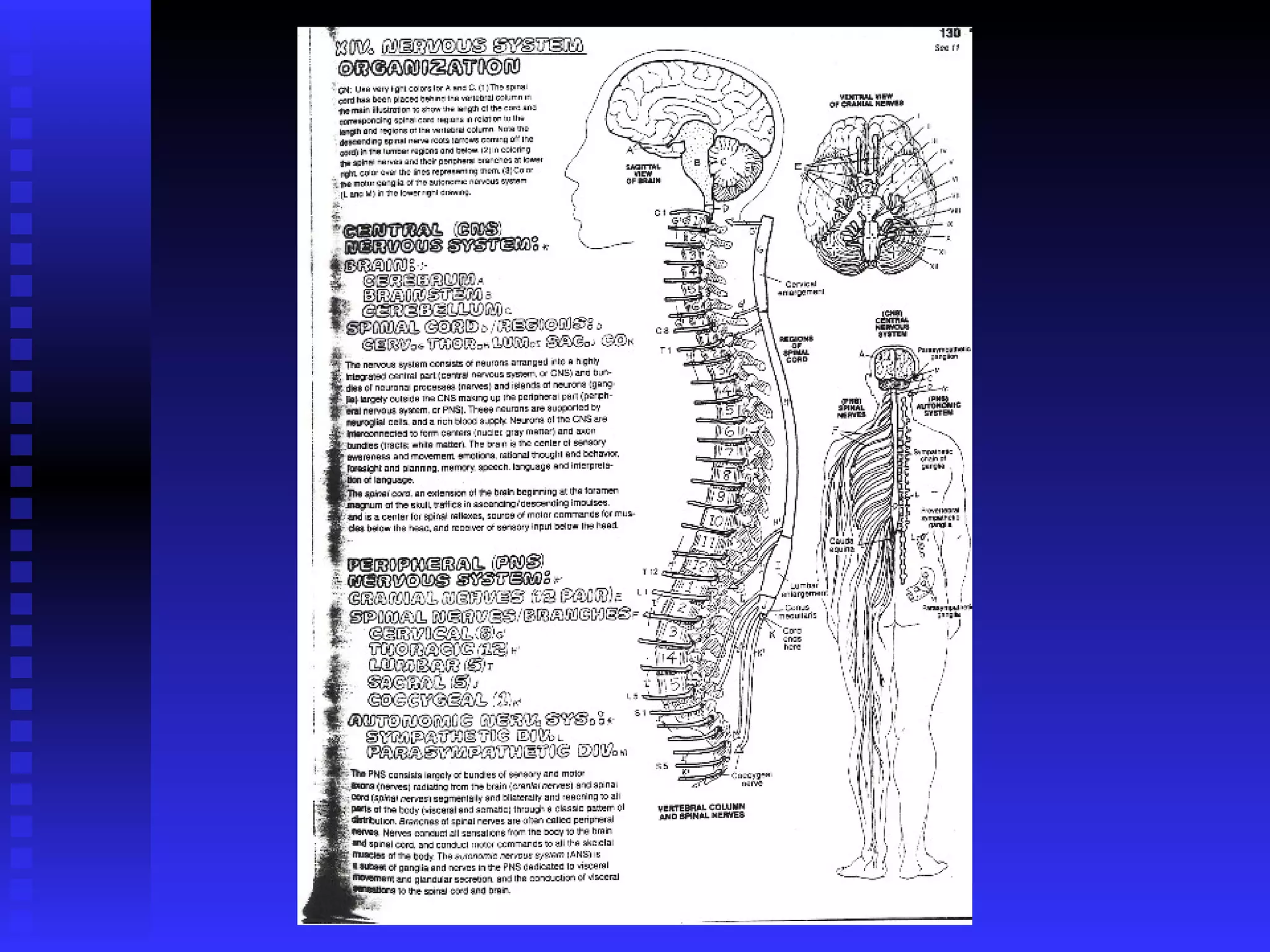
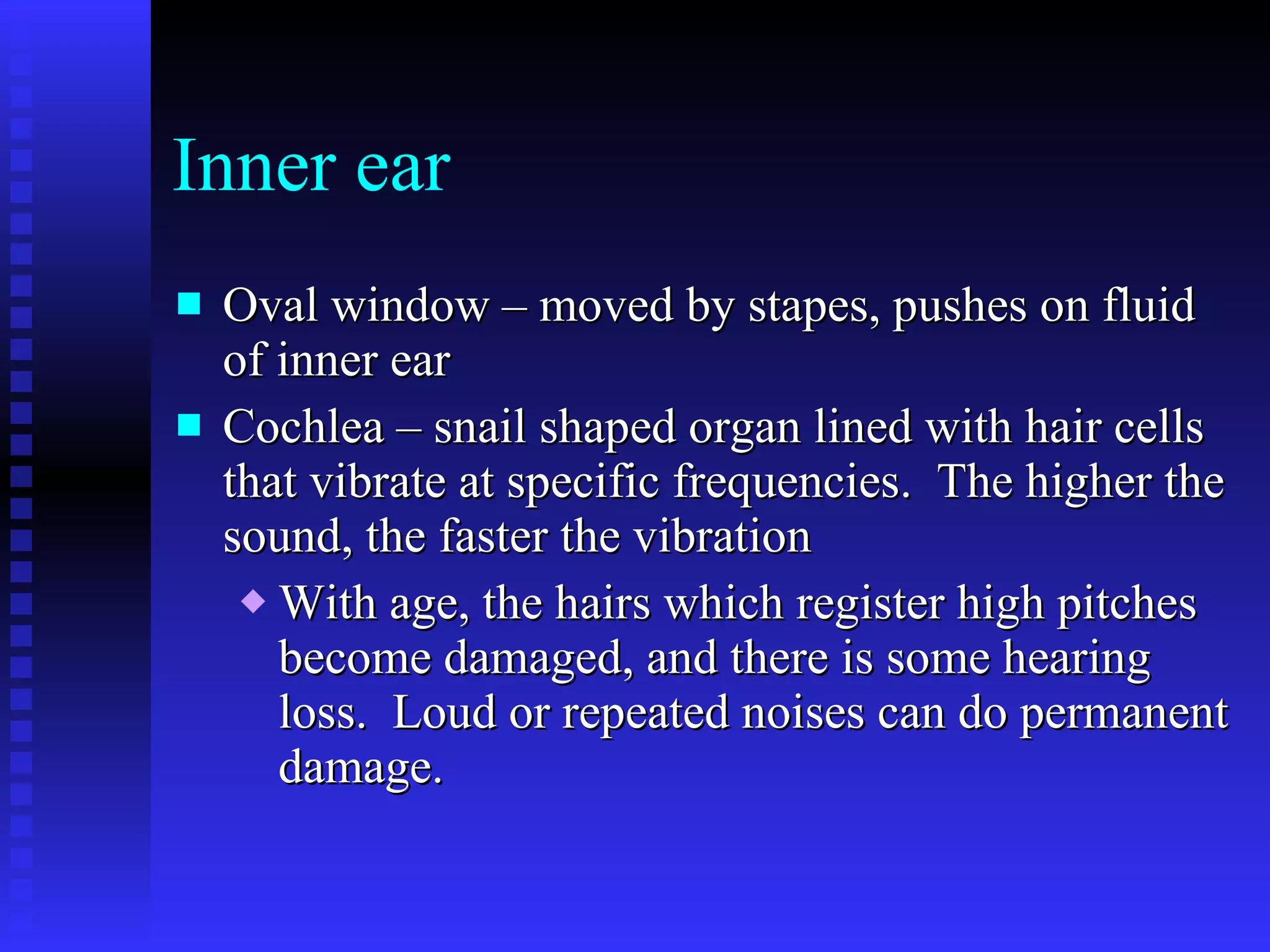
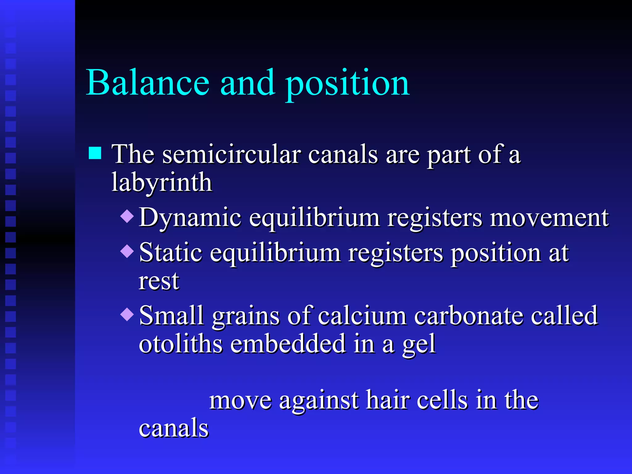
Nervous system Sensory – detect changes Integrative – make decisions Responsive - take action
Types of nerves Sensory – gather information; take it to the brain Associative/interneurons – connect other neurons, process information Motor – carry impulses to  effectors  (muscles or glands) where action is taken
Divisions CNS – brain and spinal cord PNS – nerves running to and from the CNS Somatic NS – all sensory, and motor nerves to muscles Autonomic NS – to visceral organs and skin Sympathetic – originate in the thoracic and lumbar areas Parasympathetic – originate in the brain, cervical or sacral areas
The sympathetic and parasympathetic often have opposite actions on organs.  One stimulates an increase while the other causes a decrease.  Homeostasis. Neurotransmitters can be  Excitatory – trigger nerve impulses Inhibitory – prevent nerve impulses
Structures found along nerves in the PNS Ganglion – enlarged spot along a nerve that houses somas for sensory neurons outside the CNS Plexus – a spot where many nerves (or blood vessels) connect to one another
 
Nerve pathway  A route traveled by an impulse as it travels through the nervous system The more often this path is traveled, the more stable it becomes, new connections are memories; reinforcing them makes a memory stronger, able to be recalled easier
Reflexes  Automatic, unconscious responses to changes Help maintain homeostasis (heart rate, breathing rate, digestion, blood pressure) Automatic actions (coughing, sneezing, swallowing, vomiting)
Reflex Arc -  Consists of sensory neuron, reflex center, motor neuron, effector
Reflex Arc cont. 1. A sensory neuron is stimulated – ex. Hot stove, rubber hammer on tendon stimulates stretch receptor in muscle 2. Impulse travels along sensory (afferent) neuron to the spinal cord 3. In the spinal cord the sensory neuron stimulates several neurons
Reflex Arc cont. 4. An impulse is immediately sent down an efferent motor neuron to the muscle.  Ex: bicep jerks hand back, leg kicks Simultaneously, a message is sent to the brain registering what happened * brain is NOT involved in the decision to move
Types of reflexes Monosynaptic – sensory neuron connects directly to motor neuron – knee jerk Polysynaptic – sensory and motor neurons are connected by associative neurons – hot stove reaction
The Brain – 3 major parts Cerebrum – largest part of brain, has sensory areas, motor areas and is involved in higher thinking Folded surface allows more cells; grey matter on the surface is somas, deeper white matter is axons Gyri are hills, sulci are valleys Corpus Callosum made of axons connects the 2 sides of the brain
Cerebellum  Smaller, posterior and inferior to cerebrum Consists of smaller folds Coordinates movements Operates below the level of consciousness Ventricles – spaces within the brain filled with cerebrospinal fluid
Brain Stem Anterior to cerebellum, connects to spinal cord Midbrain – most superior portion, connects lower brain to cerebrum Pons – inferior to midbrain, links cerebellum and cerebrum Medulla Oblongata – most inferior part of brain, controls breathing, heart rate and blood pressure
 
Cranial nerves There are 12 pairs of cranial nerves that come from the brain.  Pair I are the Olfactory nerves lead to the nose.  (smell) Pair II are the optic nerves that lead to the eyes.  (vision) Pair VIII are the vestibular-cochlear nerves which lead to the ear.  (balance and hearing) The other pairs are a combination of sensory and motor to the head, neck and trunk, including the vagus nerve (X) to the heart.
Spinal cord Slender nerve column that passes downward from the brain into the vertebral column Consists of 31 segments, each having a pair of spinal nerves that branch out to various body parts and connect them with the CNS A core of gray matter arranged in “horns” surrounded by white matter that has ascending and descending tracts of axons. Conduct nerve impulses and serve as a center for reflexes.
Meninges -  the coverings of the CNS   Pia mater  – a delicate covering on the surface of the brain which contains blood vessels   Arachnoid mater  – spiderweblike covering which cushions   Dura mater  – a much tougher almost leathery covering
Blood-brain barrier There is no blood actually within the brain or spinal cord Nutrients are carried by cerebrospinal fluid
Sight and the Eye – the orbit Eyelids – protect eye from debris, clean and lubricate with each blink Extrinsic muscles – 3 pairs move eyeball up, down; side to side; and twist slightly Lacrimal glands – located above eye, lateral;  make tears that wash across surface, contain lysozyme;  are collected in Lacrimal ducts – medial lower corner of eyelid; lead to nasal passages
Structure of eye Sclera – white of the eye, has blood vessels Conjunctiva – on surface of sclera Cornea – clear, smooth surface, light passes through Aqueous humor – clear fluid posterior to cornea Iris – colored part of eye, actually a muscle that controls the amount of light coming in
Structure of eye cont. Pupil – hole in the center of the iris  Lens – posterior to pupil; hard but flexible, bends light as it passes through to focus it on the back of the eye;  loses flexibility as you age Suspensory ligaments/ciliary muscles – pull on the lens to slightly change its shape, focus for far or near,
Structure of eye cont. Vitreous humor – clear gel posterior to lens that fills the cavity inside the eyeball Choroid coat – pigmented layer just deep to the sclera at the back of the eye.  Absorbs extra light.  This is reflective in many nocturnal species
Retina – at the back of the eye Layers in order that light passes through Nerve fibers Connecting neurons Receptor cells Rods – detect light and dark, rhodopsin Cones – detect color; red, green, blue Choroid coat – light stops here
Retina cont. Fovea centralis– area where most of light hits, pit with densely packed cones Blind spot – the area where the optic nerve leaves the eye, no receptor cells
Optic nerve Nerves from each eye come together at the chiasma where info from the left side of each eye goes one way, and from the right goes the other. Travels to the visual center of the cerebrum at the rear of the brain
The Ear – hearing and balance External Ear –  auricle  – funnels sound into ear canal Tympanic membrane – eardrum that vibrates from sound waves Middle ear Ossicles – 3 small bones that amplify sound waves Malleus (hammer), incus (anvil) and stapes (stirrup)
Inner ear Oval window – moved by stapes, pushes on fluid of inner ear Cochlea – snail shaped organ lined with hair cells that vibrate at specific frequencies.  The higher the sound, the faster the vibration With age, the hairs which register high pitches become damaged, and there is some hearing loss.  Loud or repeated noises can do permanent damage.
Balance and position The semicircular canals are part of a labyrinth  Dynamic equilibrium registers movement  Static equilibrium registers position at rest Small grains of calcium carbonate called otoliths embedded in a gel  move against hair cells in the canals

Nervous system

  • 1.
    Nervous system Sensory– detect changes Integrative – make decisions Responsive - take action
  • 2.
    Types of nervesSensory – gather information; take it to the brain Associative/interneurons – connect other neurons, process information Motor – carry impulses to effectors (muscles or glands) where action is taken
  • 3.
    Divisions CNS –brain and spinal cord PNS – nerves running to and from the CNS Somatic NS – all sensory, and motor nerves to muscles Autonomic NS – to visceral organs and skin Sympathetic – originate in the thoracic and lumbar areas Parasympathetic – originate in the brain, cervical or sacral areas
  • 4.
    The sympathetic andparasympathetic often have opposite actions on organs. One stimulates an increase while the other causes a decrease. Homeostasis. Neurotransmitters can be Excitatory – trigger nerve impulses Inhibitory – prevent nerve impulses
  • 5.
    Structures found alongnerves in the PNS Ganglion – enlarged spot along a nerve that houses somas for sensory neurons outside the CNS Plexus – a spot where many nerves (or blood vessels) connect to one another
  • 6.
  • 7.
    Nerve pathway A route traveled by an impulse as it travels through the nervous system The more often this path is traveled, the more stable it becomes, new connections are memories; reinforcing them makes a memory stronger, able to be recalled easier
  • 8.
    Reflexes Automatic,unconscious responses to changes Help maintain homeostasis (heart rate, breathing rate, digestion, blood pressure) Automatic actions (coughing, sneezing, swallowing, vomiting)
  • 9.
    Reflex Arc - Consists of sensory neuron, reflex center, motor neuron, effector
  • 10.
    Reflex Arc cont.1. A sensory neuron is stimulated – ex. Hot stove, rubber hammer on tendon stimulates stretch receptor in muscle 2. Impulse travels along sensory (afferent) neuron to the spinal cord 3. In the spinal cord the sensory neuron stimulates several neurons
  • 11.
    Reflex Arc cont.4. An impulse is immediately sent down an efferent motor neuron to the muscle. Ex: bicep jerks hand back, leg kicks Simultaneously, a message is sent to the brain registering what happened * brain is NOT involved in the decision to move
  • 12.
    Types of reflexesMonosynaptic – sensory neuron connects directly to motor neuron – knee jerk Polysynaptic – sensory and motor neurons are connected by associative neurons – hot stove reaction
  • 13.
    The Brain –3 major parts Cerebrum – largest part of brain, has sensory areas, motor areas and is involved in higher thinking Folded surface allows more cells; grey matter on the surface is somas, deeper white matter is axons Gyri are hills, sulci are valleys Corpus Callosum made of axons connects the 2 sides of the brain
  • 14.
    Cerebellum Smaller,posterior and inferior to cerebrum Consists of smaller folds Coordinates movements Operates below the level of consciousness Ventricles – spaces within the brain filled with cerebrospinal fluid
  • 15.
    Brain Stem Anteriorto cerebellum, connects to spinal cord Midbrain – most superior portion, connects lower brain to cerebrum Pons – inferior to midbrain, links cerebellum and cerebrum Medulla Oblongata – most inferior part of brain, controls breathing, heart rate and blood pressure
  • 16.
  • 17.
    Cranial nerves Thereare 12 pairs of cranial nerves that come from the brain. Pair I are the Olfactory nerves lead to the nose. (smell) Pair II are the optic nerves that lead to the eyes. (vision) Pair VIII are the vestibular-cochlear nerves which lead to the ear. (balance and hearing) The other pairs are a combination of sensory and motor to the head, neck and trunk, including the vagus nerve (X) to the heart.
  • 18.
    Spinal cord Slendernerve column that passes downward from the brain into the vertebral column Consists of 31 segments, each having a pair of spinal nerves that branch out to various body parts and connect them with the CNS A core of gray matter arranged in “horns” surrounded by white matter that has ascending and descending tracts of axons. Conduct nerve impulses and serve as a center for reflexes.
  • 19.
    Meninges - the coverings of the CNS Pia mater – a delicate covering on the surface of the brain which contains blood vessels Arachnoid mater – spiderweblike covering which cushions Dura mater – a much tougher almost leathery covering
  • 20.
    Blood-brain barrier Thereis no blood actually within the brain or spinal cord Nutrients are carried by cerebrospinal fluid
  • 21.
    Sight and theEye – the orbit Eyelids – protect eye from debris, clean and lubricate with each blink Extrinsic muscles – 3 pairs move eyeball up, down; side to side; and twist slightly Lacrimal glands – located above eye, lateral; make tears that wash across surface, contain lysozyme; are collected in Lacrimal ducts – medial lower corner of eyelid; lead to nasal passages
  • 22.
    Structure of eyeSclera – white of the eye, has blood vessels Conjunctiva – on surface of sclera Cornea – clear, smooth surface, light passes through Aqueous humor – clear fluid posterior to cornea Iris – colored part of eye, actually a muscle that controls the amount of light coming in
  • 23.
    Structure of eyecont. Pupil – hole in the center of the iris Lens – posterior to pupil; hard but flexible, bends light as it passes through to focus it on the back of the eye; loses flexibility as you age Suspensory ligaments/ciliary muscles – pull on the lens to slightly change its shape, focus for far or near,
  • 24.
    Structure of eyecont. Vitreous humor – clear gel posterior to lens that fills the cavity inside the eyeball Choroid coat – pigmented layer just deep to the sclera at the back of the eye. Absorbs extra light. This is reflective in many nocturnal species
  • 25.
    Retina – atthe back of the eye Layers in order that light passes through Nerve fibers Connecting neurons Receptor cells Rods – detect light and dark, rhodopsin Cones – detect color; red, green, blue Choroid coat – light stops here
  • 26.
    Retina cont. Foveacentralis– area where most of light hits, pit with densely packed cones Blind spot – the area where the optic nerve leaves the eye, no receptor cells
  • 27.
    Optic nerve Nervesfrom each eye come together at the chiasma where info from the left side of each eye goes one way, and from the right goes the other. Travels to the visual center of the cerebrum at the rear of the brain
  • 28.
    The Ear –hearing and balance External Ear – auricle – funnels sound into ear canal Tympanic membrane – eardrum that vibrates from sound waves Middle ear Ossicles – 3 small bones that amplify sound waves Malleus (hammer), incus (anvil) and stapes (stirrup)
  • 29.
    Inner ear Ovalwindow – moved by stapes, pushes on fluid of inner ear Cochlea – snail shaped organ lined with hair cells that vibrate at specific frequencies. The higher the sound, the faster the vibration With age, the hairs which register high pitches become damaged, and there is some hearing loss. Loud or repeated noises can do permanent damage.
  • 30.
    Balance and positionThe semicircular canals are part of a labyrinth Dynamic equilibrium registers movement Static equilibrium registers position at rest Small grains of calcium carbonate called otoliths embedded in a gel move against hair cells in the canals