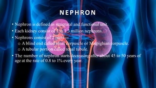
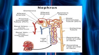
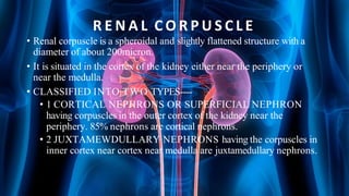
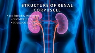
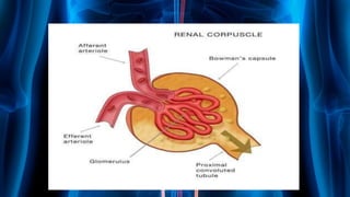
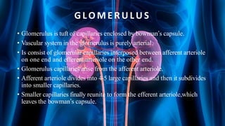
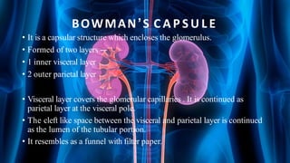
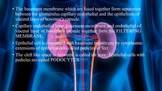
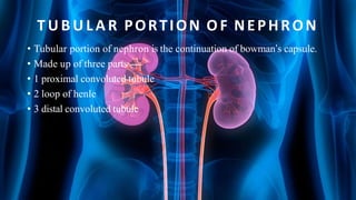
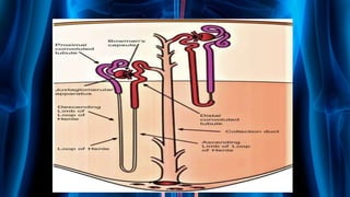
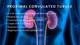
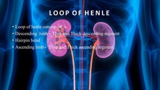
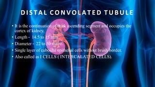
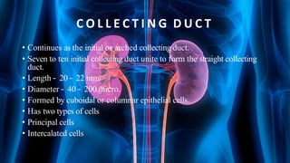
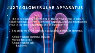
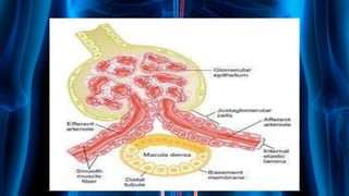
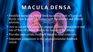
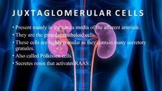
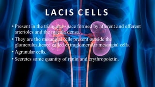
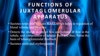
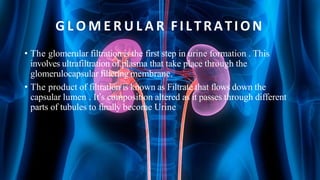
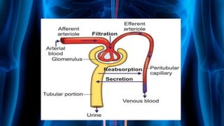
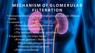
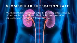
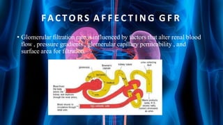
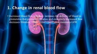
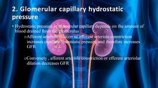
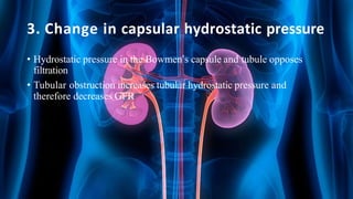
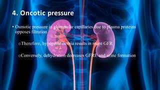
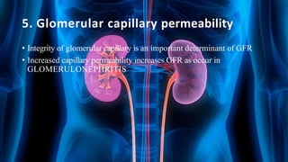
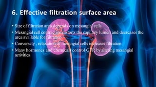
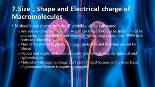
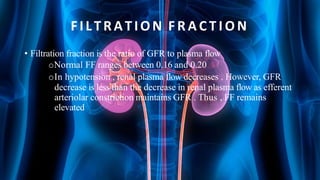
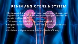
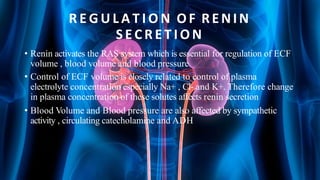
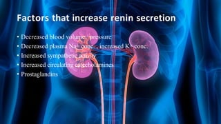
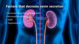
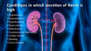
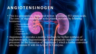
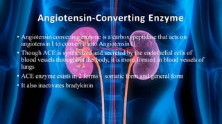
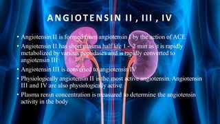
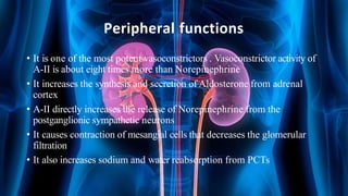
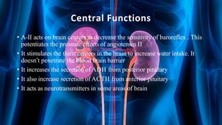
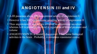
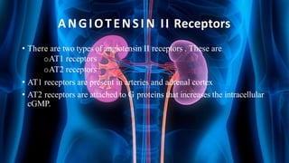
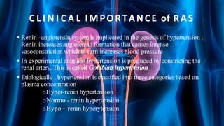
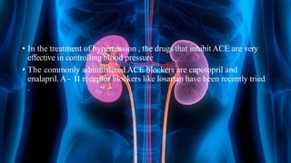
SlideShare a Scribd company logo
1 of 68
NEPHRON U L T R A ST R U C T U R E ,
G L O M E R U L A R FILTRATION A N D
R E G U L A T I O N OF G F R , R O L E OF
J G A A N D RENIN-ANGIOTENSIN
A L D O S T E R O N E SYSTEM
Presented by-
Shreyash Sinha
Simran Kaur Khanuja
Sneha Srivastav
Soumya Nirala
IN D E X
Nephron
Juxtaglomerular apparatus
Role of JGA
Glomerular Filtration Rate
Factors affecting GFR
Renin – Angiotensin Aldosterone System
NEPHRON
• Nephron is defined as structural and functional unit.
• Each kidney consist of 1 to 1.3 million nephrons.
• Nephrons consist of 2 parts ---
o A blind end called renal corpuscle or Malpighian corpuscle.
o A tubular portion called renal tubule.
• The number of nephron starts decreasing after about 45 to 50 years of
age at the rate of 0.8 to 1% every year.
R E N A L C O R P U S C L E
• Renal corpuscle is a spheroidal and slightly flattened structure with a
diameter of about 200micron.
• It is situated in the cortex of the kidney either near the periphery or
near the medulla.
• CLASSIFIED INTO TWO TYPES----
• 1 CORTICAL NEPHRONS OR SUPERFICIAL NEPHRON
having corpuscles in the outer cortex of the kidney near the
periphery. 85% nephrons are cortical nephrons.
• 2 JUXTAMEWDULLARY NEPHRONS having the corpuscles in
inner cortex near cortex near medulla are juxtamedullary nephrons.
S T R U C T U R E OF R E N A L
C O R P U S C L E
• It is formed by two portions
• GLOMERULUS
• BOWMAN’S CAPSULE
G L O M E R U L U S
• Glomerulus is tuft of capillaries enclosed by bowman’s capsule.
• Vascular system in the glomerulus is purely arterial.
• Is consist of glomerular capillaries interposed between afferent arteriole
on one end and efferent arteriole on the other end.
• Glomerulus capillaries arise from the afferent arteriole.
• Afferent arteriole divides into 4-5 large capillaries and then it subdivides
into smaller capillaries.
• Smaller capillaries finally reunite to form the efferent arteriole,which
leaves the bowman’s capsule.
BOWMAN’S C A P S U L E
• It is a capsular structure which encloses the glomerulus.
• Formed of two layers ---
• 1 inner visceral layer
• 2 outer parietal layer
• Visceral layer covers the glomerular capillaries . It is continued as
parietal layer at the visceral pole.
• The cleft like space between the visceral and parietal layer is continued
as the lumen of the tubular portion.
• It resembles as a funnel with filter paper.
• The basement membrane which are fused together form separation
between the glomerulus capillary endothelial and the epithelium of
visceral layer of bowman’s capsule.
• Capillary endothelial layer, basement membrane and endothelial of
visceral layer of bowman’s capsule together form the FILTERING
MEMBRANE.
• Epithelial cell is connected with basement membrane by cytoplasmic
extensions of epithelial cells called pedicles or feet.
• The cleft like space in between is called slit pore. Epithelial cells with
pedicles are called PODOCYTES.
T U B U L A R PORTION OF NEPHRON
• Tubular portion of nephron is the continuation of bowman’s capsule.
• Made up of three parts ----
• 1 proximal convoluted tubule
• 2 loop of henle
• 3 distal convoluted tubule
PROXIMAL C O N V U L A T E D T U B U L E
• It is the coiled portion arising from bowmans capsule.
• Situated near the cortex.
• Continued as descending limb of henle.
• Length of PCT is 14mm
• Diameter is 55micro.
• Single layer of cuboidal epithelial cells.
LOOP OF H E N L E
• Loop of henle consists of
• Descending limb – Thin and Thick descending segment
• Hairpin bend
• Ascending limb – Thin and Thick ascending segment
D I S T A L C O N V O L A T E D T U B U L E
• It is the continuation of thick ascending segment and occupies the
cortex of kidney.
• Length – 14.5 to 15 mm
• Diameter – 22 to 50 micro.
• Single layer of cuboidal epithelial cells without brush border.
• Also called as I CELLS ( INTERCALATED CELLS).
C O L L E C T I N G D U C T
• Continues as the initial or arched collecting duct.
• Seven to ten initial collecting duct unite to form the straight collecting
duct.
• Length – 20 – 22 mm
• Diameter – 40 – 200 micro.
• Formed by cuboidal or columnar epithelial cells.
• Has two types of cells
• Principal cells
• Intercalated cells
J U X T A G L O M E R U L A R APPARATUS
•
1.The thick ascending limb of loop of Henle when comes in contact
with the glomerulus of the same renal corpuscle,structural modifications
occur in the tubule and afferent and efferent arterioles.
2. The entire modified structure is called juxtaglomerular apparatus.
3. Juxtaglomerular apparatus includes
Macula densa cells
Juxtaglomerular cells
Lacis cells
M A C U L A D E NSA
• Modified epithelial cells of thick ascending limb of Loop of
Henle when it comes in contact with the afferent and efferent
arteriole.
• Acts as sensor that monitors changes in ionic composition and
rate of flow of tubular fluid in the lumen of the tubule.
• Provides appropriate feedback signal to renal corpuscle.
• Important component in the tubuloglomerular feedback
circuit.
J U X T A G L O M E R U L A R C E L L S
• Present mainly in the tunica media of the afferent arteriole.
• They are the granular epitheloid cells.
• These cells are highly granular as they contain many secretory
granules.
• Also called Polkissen cells.
• Secretes renin that activates RAAS .
L A C I S C E L L S
• Present in the triangular space formed by afferent and efferent
arterioles and the macula densa.
• They are the mesangial cells present outside the
glomerulus,hence called extraglomerular mesangial cells.
• Agranular cells.
• Secretes some quantity of renin and erythropoietin.
FUNCTIONS OF
J U X TAG L OMERULAR
APPARATUS
• Secretes renin that activates RAAS which helps in regulation of
blood volume and pressure.
• Detects the change in rate of flow and volume of flow in the
tubule, and composition of tubular fluid and thus, forms the
basis of tubuloglomerular feedback.
• Secretes renin and erythropoietin.
G L O M E R U L A R FILTRATION
• The glomerular filtration is the first step in urine formation . This
involves ultrafiltration of plasma that take place through the
glomerulocapsular filtering membrane.
• The product of filtration is known as Filtrate that flows down the
capsular lumen . It’s composition altered as it passes through different
parts of tubules to finally become Urine
MECHANISM OF G L O M E R U L A R
FILTERATION
• Glomerular filtration occurs through glomerulocapsular filtration
barrier.
• It consists of
oFenestrated capillary endothelium
oThe basement membrane
oThe podocytes of capsular epithelium
• It is governed by two major factors
oPressure gradients ( Starling forces)
Hydrostatic pressure gradient
Osmotic pressure gradient
oFiltration coeffecient
G L O M E R U L A R FILTERATION R A T E
• Glomerular filtration rate is defined as the amount of filtrate formed by
glomerular filtering membrane of both the kidneys in a unit time
• Normally, it is 125ml/min or 7.5 l/h or 180 l/day
FACTORS A F F E C T I N G G F R
• Glomerular filtration rate is influenced by factors that alter renal blood
flow , pressure gradients , glomerular capillary permeability , and
surface area for filtration.
1. Change in renal blood flow
• Increased blood flow to kidney increases the delivery of blood to
glomerulus that promotes filtration and conversely decreased flow
decreases filtration . Thus , renal vasodilation maintains GFR
2. Glomerular capillary hydrostatic
pressure
• Hydrostatic pressure in glomerular capillary depends on the amount of
blood drained from the glomerulus
oAfferent arteriole dilation or efferent arteriole constriction
increases capillary hydrostatic pressure and therefore increases
GFR
oConversely , afferent arteriole constriction or efferent arteriolar
dilation decreases GFR
3. Change in capsular hydrostatic pressure
• Hydrostatic pressure in the Bowmen’s capsule and tubule opposes
filtration
• Tubular obstruction increases tubular hydrostatic pressure and
therefore decreases GFR
4. Oncotic pressure
• Osmotic pressure in glomerular capillaries due to plasma proteins
opposes filtration
oTherefore, hypoproteinemia results in more GFR
oConversely, dehydration decreases GFRE and urine formation
5. Glomerular capillary permeability
• Integrity of glomerular capillary is an important determinant of GFR
• Increased capillary permeability increases GFR as occur in
GLOMERULONEPHRITIS
6. Effective filtration surface area
• Size of filtration area depends on mesangial cells
• Mesangial cell contraction distorts the capillary lumen and decreases the
area available for filtration
• Conversely , relaxation of mesangial cells increases filtration
• Many hormones and chemicals control GFR by altering mesangial
activities
7.Size , Shape and Electrical charge of
Macromolecules
• Molecule size determines the filterability of the substance
o Any substance having molecular weight less than 10000 can be freely filtered by
glomerular filtration barrier and molecules having weight more than 10000 have
restricted filterability
o Most of the protein in plasma are larger molecules and therefore can not be
filtered
o Slender and supple molecules can easily pass through than the spherical and
rigid molecules
o Molecules with negative charge can’t easily filtered because all the three layers
of glomerular filtration is negatively charged
FILTRATION F R A C T I O N
• Filtration fraction is the ratio of GFR to plasma flow
oNormal FF ranges between 0.16 and 0.20
oIn hypotension , renal plasma flow decreases . However, GFR
decrease is less than the decrease in renal plasma flow as efferent
arteriolar constriction maintains GFR . Thus , FF remains
elevated
RENIN ANGIOTENSIN SYSTEM
• Renin secreted from JG cells of kidney activates angiotensinogen to
angiotensin I, which on further enzymatic action gets converted into
angiotensin II and III
• This system is called renin – angiotensin system (RAS)
• It is primarily involved in the control of blood volume and blood
pressure
• Renin is an acid protease secreted from JG cells of Kidney
R E G U L A T I O N OF RENIN
SE C R E T I O N
• Renin activates the RAS system which is essential for regulation of ECF
volume , blood volume and blood pressure
• Control of ECF volume is closely related to control of plasma
electrolyte concentration especially Na+ , Cl- and K+. Therefore change
in plasma concentration of these solutes affects renin secretion
• Blood Volume and Blood pressure are also affected by sympathetic
activity , circulating catecholamine and ADH
Factors that increase renin secretion
• Decreased blood volume / pressure
• Decreased plasma Na+ conc. , increased K+ conc.
• Increased sympathetic activity
• Increased circulating catecholamines
• Prostaglandins
Factors that decrease renin secretion
• Increased blood Na+ concentration
• Increased blood pressure
• ADH
• Angiotensin II
Conditions in which secretion of Renin is
high
• Hypotension
• Hyponatremia
• Hyperkalemia
• Hypovolemia
• Dehydration
• Constriction of renal artery
• Cardiac failure
• Standing
• Liver cirrhosis
ANGIOTENSINOGEN
• This is a glycoprotein synthesized in liver. It contains 453 amino acids.
The conc. of angiotensinogen in the plasma increases by following
hormones
o Glucocorticoids
o Thyroid hormone
o Estrogen
o Cytokines
o Angiotensin II
• Angiotensin II provides a positive feedback for further synthesis of
angiotensinogen. Angiotensinogen is physiologically inactive and act as
precursor for the formation of Angiotensin I which is further converted
into Angiotensin II with the help of ACE enzyme.
Angiotensin-Converting Enzyme
• Angiotensin converting enzyme is a carboxypeptidase that acts on
angiotensin I to convert it into Angiotensin II
• Though ACE is synthesized and secreted by the endothelial cells of
blood vessels throughout the body, it is more formed in blood vessels of
lungs
• ACE enzyme exists in 2 forms – somatic form and general form
• It also inactivates bradykinin
ANGIOTENSIN II , III , IV
• Angiotensin II is formed from angiotensin I by the action of ACE
• Angiotensin II has short plasma half life 1 – 2 min as it is rapidly
metabolized by various peptidases and is rapidly converted to
angiotensin III
• Angiotensin III is converted to angiotensin IV
• Physiologically angiotensin II is the most active angiotensin. Angiotensin
III and IV are also physiologically active
• Plasma renin concentration is measured to determine the angiotensin
activity in the body
P H Y S I O L O G I C A L ACTIONS OF
ANGIOTENSINS
ANGIOTENSIN I :
It acts as precursor for angiotensin II . Otherwise , it is physiologically
inactive
ANGIOTENSIN II :
This is the most important among all angiotensins . All of it’s actions are
aimed at increasing the blood volume and pressure. The functions of
angiotensin II can be divided into central and peripheral functions
Peripheral functions
• It is one of the most potent vasoconstrictors . Vasoconstrictor activity of
A-II is about eight times more than Norepinephrine
• It increases the synthesis and secretion of Aldosterone from adrenal
cortex
• A-II directly increases the release of Norepinephrine from the
postganglionic sympathetic neurons
• It causes contraction of mesangial cells that decreases the glomerular
filtration
• It also increases sodium and water reabsorption from PCTs
Central Functions
• A-II acts on brain centers to decrease the sensitivity of baroreflex . This
potentiates the pressure effects of angiotensin II
• It stimulates the thirst centers in the brain to increase water intake. It
doesn’t penetrate the blood brain barrier
• It increases the secretion of ADH from posterior pituitary
• It also increase secretion of ACTH from anterior pituitary
• It acts as neurotransmitters in some areas of brain
ANGIOTENSIN III and IV
• A-III possesses about 40% vasopressor activity of angiotensin II
• However it has 100% aldosterone secreting activity of angiotensin II
• Thus angiotensin II and III are involved in aldosterone secretion
whereas A-II is primarily involved in regulation of blood pressure and
volume
• ANGIOTENSIN IV is recently discovered. It has some biological
activities in the brain . Probably , it stimulates vasomotor centre.
ANGIOTENSIN II Receptors
• There are two types of angiotensin II receptors . These are
oAT1 receptors
oAT2 receptors
• AT1 receptors are present in arteries and adrenal cortex
• AT2 receptors are attached to G proteins that increases the intracellular
cGMP.
C L I N I C A L IMPORTANCE of RAS
• Renin -angiotensin system is implicated in the genesis of hypertension .
Renin increases angiotensin formation that causes intense
vasoconstriction which in turn increases blood pressure
• In experimental animals hypertension is produced by constricting the
renal artery. This is called Goldblatt hypertension
• Etiologically , hypertension is classified into three categories based on
plasma concentration
oHyper-renin hypertension
oNormo - renin hypertension
oHypo – renin hyperytension
• In the treatment of hypertension , the drugs that inhibit ACE are very
effective in controlling blood pressure
• The commonly administered ACE blockers are capotopril and
enalapril. A – II receptor blockers like losartan have been recently tried
.
SUMMARY
• Kidney is an important organ for homeostasis of ECF volume and
composition
• The structural medications of tubular epithelial cells in different parts of
the nephrons are primarily meant for their participation in specific
functions of reabsorption of solute and water and concentration of
urine.
• The loop of Henle of juxtamedullary nephron is longer to facilitates its
role in urine concentration
• JG apparatus is placed at a vital location in the tubule to sense and
regulate blood volume and pressure through renin secretion
• GFR depends on hydrostatic and osmotic pressure gradients , normally
hydrostatic pressure gradients plays a bigger role
• Inulin clearance test is best method to measure GFT.
• Sympathetic regulation and tubuloglomerular feedback are major
systems for regulating GFR
QUIZ TIME
QUESTION 1
• All of the following factors regarding a substance determine the
filtration in glomerulus except
1. Color
2. Molecular size
3. Shape
4. Electrostatic charge
•Answer -A
QUESTION 2
• Physiologically inactive form of angiotensin is ?
•ANGIOTENSIN IV
QUESTION 3
• Lacis cells are located at
1. JGA
2. Proximal tubule
3. Distal tubule
4. Loop of henle
•ANSWER - 3
QUESTION 4
• Best test for GFR measurement is done with
1. Inulin
2. Hippuric acid
3. Creatinine
4. PAH
•ANSWER - INULIN
QUESTION 5
• Which of the following doesn’t participate in filtration barriers in
nephrons ?
1. Podocytes
2. Mesangium
3. Endothelial cells
4. Basement membrane
•ANSWER - MESANGIUM
QUESTION 6
• In a normal person at resting conditions , GFR is
1. 125ml/min
2. 90ml/min
3. 60 ml/min
4. 150ml/min
•ANSWER – 125 ml / min
QUESTION 7
• Renin is secreted by
1. PCT
2. DCT
3. Collecting duct
4. JG apparatus
ANSWER – JG Apparatus
T H A N K YOU!!

More Related Content

Similar to Nephron Ultrastructure and GFR (1).pptx

111111111111111111111111Urinary System.pdf
111111111111111111111111Urinary System.pdf111111111111111111111111Urinary System.pdf
111111111111111111111111Urinary System.pdf
nsk44p5mwp
 

Similar to Nephron Ultrastructure and GFR (1).pptx (20)

Renal anatomy and physiology ..
Renal anatomy and physiology ..Renal anatomy and physiology ..
Renal anatomy and physiology ..
 
Urinary system
Urinary systemUrinary system
Urinary system
 
111111111111111111111111Urinary System.pdf
111111111111111111111111Urinary System.pdf111111111111111111111111Urinary System.pdf
111111111111111111111111Urinary System.pdf
 
excretion pdf.pdf
excretion pdf.pdfexcretion pdf.pdf
excretion pdf.pdf
 
Renal anatomy
Renal anatomyRenal anatomy
Renal anatomy
 
Urinary system
Urinary systemUrinary system
Urinary system
 
Nephron (The Guyton and Hall physiology)
Nephron (The Guyton and Hall physiology)Nephron (The Guyton and Hall physiology)
Nephron (The Guyton and Hall physiology)
 
Urinary system.pdf
Urinary system.pdfUrinary system.pdf
Urinary system.pdf
 
The urinary system
The urinary systemThe urinary system
The urinary system
 
Anatomy and physiology of urinary system
Anatomy and physiology of urinary systemAnatomy and physiology of urinary system
Anatomy and physiology of urinary system
 
KIDNEY
KIDNEY KIDNEY
KIDNEY
 
RENAL FUNCTION AND RENAL FUNCTION TESTS -RACOH.pptx
RENAL FUNCTION AND RENAL FUNCTION TESTS -RACOH.pptxRENAL FUNCTION AND RENAL FUNCTION TESTS -RACOH.pptx
RENAL FUNCTION AND RENAL FUNCTION TESTS -RACOH.pptx
 
Excretory System
Excretory SystemExcretory System
Excretory System
 
THE KIDNEY.
THE KIDNEY.THE KIDNEY.
THE KIDNEY.
 
Urinary system word
Urinary system wordUrinary system word
Urinary system word
 
Urinary System.pptx
Urinary System.pptxUrinary System.pptx
Urinary System.pptx
 
Renal system
Renal systemRenal system
Renal system
 
urinary system.pptx
urinary system.pptxurinary system.pptx
urinary system.pptx
 
The urinary system 2018
The urinary system 2018The urinary system 2018
The urinary system 2018
 
Renal Histology
Renal HistologyRenal Histology
Renal Histology
 

Recently uploaded

Unveiling Pharyngitis: Causes, Symptoms, Diagnosis, and Treatment Strategies.pdf
Unveiling Pharyngitis: Causes, Symptoms, Diagnosis, and Treatment Strategies.pdfUnveiling Pharyngitis: Causes, Symptoms, Diagnosis, and Treatment Strategies.pdf
Unveiling Pharyngitis: Causes, Symptoms, Diagnosis, and Treatment Strategies.pdf
NoorulainMehmood1
 
Connective Tissue II - Dr Muhammad Ali Rabbani - Medicose Academics
Connective Tissue II - Dr Muhammad Ali Rabbani - Medicose AcademicsConnective Tissue II - Dr Muhammad Ali Rabbani - Medicose Academics
Connective Tissue II - Dr Muhammad Ali Rabbani - Medicose Academics
MedicoseAcademics
 
Histology of Epithelium - Dr Muhammad Ali Rabbani - Medicose Academics
Histology of Epithelium - Dr Muhammad Ali Rabbani - Medicose AcademicsHistology of Epithelium - Dr Muhammad Ali Rabbani - Medicose Academics
Histology of Epithelium - Dr Muhammad Ali Rabbani - Medicose Academics
MedicoseAcademics
 
Cytoskeleton and Cell Inclusions - Dr Muhammad Ali Rabbani - Medicose Academics
Cytoskeleton and Cell Inclusions - Dr Muhammad Ali Rabbani - Medicose AcademicsCytoskeleton and Cell Inclusions - Dr Muhammad Ali Rabbani - Medicose Academics
Cytoskeleton and Cell Inclusions - Dr Muhammad Ali Rabbani - Medicose Academics
MedicoseAcademics
 
Best medicine 100% Effective&Safe Mifepristion ௵+918133066128௹Abortion pills ...
Best medicine 100% Effective&Safe Mifepristion ௵+918133066128௹Abortion pills ...Best medicine 100% Effective&Safe Mifepristion ௵+918133066128௹Abortion pills ...
Best medicine 100% Effective&Safe Mifepristion ௵+918133066128௹Abortion pills ...
Abortion pills in Kuwait Cytotec pills in Kuwait
 

Recently uploaded (20)

Top 15 Sexiest Pakistani Pornstars with Images & Videos
Top 15 Sexiest Pakistani Pornstars with Images & VideosTop 15 Sexiest Pakistani Pornstars with Images & Videos
Top 15 Sexiest Pakistani Pornstars with Images & Videos
 
Varicose Veins Treatment Aftercare Tips by Gokuldas Hospital
Varicose Veins Treatment Aftercare Tips by Gokuldas HospitalVaricose Veins Treatment Aftercare Tips by Gokuldas Hospital
Varicose Veins Treatment Aftercare Tips by Gokuldas Hospital
 
duus neurology.pdf anatomy. phisiology///
duus neurology.pdf anatomy. phisiology///duus neurology.pdf anatomy. phisiology///
duus neurology.pdf anatomy. phisiology///
 
Gross Anatomy and Histology of Tongue by Dr. Rabia Inam Gandapore.pptx
Gross Anatomy and Histology of Tongue by Dr. Rabia Inam Gandapore.pptxGross Anatomy and Histology of Tongue by Dr. Rabia Inam Gandapore.pptx
Gross Anatomy and Histology of Tongue by Dr. Rabia Inam Gandapore.pptx
 
Report Back from SGO: What’s the Latest in Ovarian Cancer?
Report Back from SGO: What’s the Latest in Ovarian Cancer?Report Back from SGO: What’s the Latest in Ovarian Cancer?
Report Back from SGO: What’s the Latest in Ovarian Cancer?
 
Storage of Blood Components- equipments, effects of improper storage, transpo...
Storage of Blood Components- equipments, effects of improper storage, transpo...Storage of Blood Components- equipments, effects of improper storage, transpo...
Storage of Blood Components- equipments, effects of improper storage, transpo...
 
High Purity 99% PMK Ethyl Glycidate Powder CAS 28578-16-7
High Purity 99% PMK Ethyl Glycidate Powder CAS 28578-16-7High Purity 99% PMK Ethyl Glycidate Powder CAS 28578-16-7
High Purity 99% PMK Ethyl Glycidate Powder CAS 28578-16-7
 
Unveiling Pharyngitis: Causes, Symptoms, Diagnosis, and Treatment Strategies.pdf
Unveiling Pharyngitis: Causes, Symptoms, Diagnosis, and Treatment Strategies.pdfUnveiling Pharyngitis: Causes, Symptoms, Diagnosis, and Treatment Strategies.pdf
Unveiling Pharyngitis: Causes, Symptoms, Diagnosis, and Treatment Strategies.pdf
 
NDCT Rules, 2019: An Overview | New Drugs and Clinical Trial Rules 2019
NDCT Rules, 2019: An Overview | New Drugs and Clinical Trial Rules 2019NDCT Rules, 2019: An Overview | New Drugs and Clinical Trial Rules 2019
NDCT Rules, 2019: An Overview | New Drugs and Clinical Trial Rules 2019
 
Connective Tissue II - Dr Muhammad Ali Rabbani - Medicose Academics
Connective Tissue II - Dr Muhammad Ali Rabbani - Medicose AcademicsConnective Tissue II - Dr Muhammad Ali Rabbani - Medicose Academics
Connective Tissue II - Dr Muhammad Ali Rabbani - Medicose Academics
 
Histology of Epithelium - Dr Muhammad Ali Rabbani - Medicose Academics
Histology of Epithelium - Dr Muhammad Ali Rabbani - Medicose AcademicsHistology of Epithelium - Dr Muhammad Ali Rabbani - Medicose Academics
Histology of Epithelium - Dr Muhammad Ali Rabbani - Medicose Academics
 
The Clean Living Project Episode 24 - Subconscious
The Clean Living Project Episode 24 - SubconsciousThe Clean Living Project Episode 24 - Subconscious
The Clean Living Project Episode 24 - Subconscious
 
Tips and tricks to pass the cardiovascular station for PACES exam
Tips and tricks to pass the cardiovascular station for PACES examTips and tricks to pass the cardiovascular station for PACES exam
Tips and tricks to pass the cardiovascular station for PACES exam
 
Mgr university bsc nursing adult health previous question paper with answers
Mgr university  bsc nursing adult health previous question paper with answersMgr university  bsc nursing adult health previous question paper with answers
Mgr university bsc nursing adult health previous question paper with answers
 
ESC HF 2024 Spotlights Day-2.pptx heart failure
ESC HF 2024 Spotlights Day-2.pptx heart failureESC HF 2024 Spotlights Day-2.pptx heart failure
ESC HF 2024 Spotlights Day-2.pptx heart failure
 
Hemodialysis: Chapter 1, Physiological Principles of Hemodialysis - Dr.Gawad
Hemodialysis: Chapter 1, Physiological Principles of Hemodialysis - Dr.GawadHemodialysis: Chapter 1, Physiological Principles of Hemodialysis - Dr.Gawad
Hemodialysis: Chapter 1, Physiological Principles of Hemodialysis - Dr.Gawad
 
Cytoskeleton and Cell Inclusions - Dr Muhammad Ali Rabbani - Medicose Academics
Cytoskeleton and Cell Inclusions - Dr Muhammad Ali Rabbani - Medicose AcademicsCytoskeleton and Cell Inclusions - Dr Muhammad Ali Rabbani - Medicose Academics
Cytoskeleton and Cell Inclusions - Dr Muhammad Ali Rabbani - Medicose Academics
 
Best medicine 100% Effective&Safe Mifepristion ௵+918133066128௹Abortion pills ...
Best medicine 100% Effective&Safe Mifepristion ௵+918133066128௹Abortion pills ...Best medicine 100% Effective&Safe Mifepristion ௵+918133066128௹Abortion pills ...
Best medicine 100% Effective&Safe Mifepristion ௵+918133066128௹Abortion pills ...
 
Benefits of Chanting Hanuman Chalisa .pdf
Benefits of Chanting Hanuman Chalisa .pdfBenefits of Chanting Hanuman Chalisa .pdf
Benefits of Chanting Hanuman Chalisa .pdf
 
Sell 5cladba adbb JWH-018 5FADB in stock
Sell 5cladba adbb JWH-018 5FADB in stockSell 5cladba adbb JWH-018 5FADB in stock
Sell 5cladba adbb JWH-018 5FADB in stock
 

Nephron Ultrastructure and GFR (1).pptx

  • 1. NEPHRON U L T R A ST R U C T U R E , G L O M E R U L A R FILTRATION A N D R E G U L A T I O N OF G F R , R O L E OF J G A A N D RENIN-ANGIOTENSIN A L D O S T E R O N E SYSTEM Presented by- Shreyash Sinha Simran Kaur Khanuja Sneha Srivastav Soumya Nirala
  • 2. IN D E X Nephron Juxtaglomerular apparatus Role of JGA Glomerular Filtration Rate Factors affecting GFR Renin – Angiotensin Aldosterone System
  • 3. NEPHRON • Nephron is defined as structural and functional unit. • Each kidney consist of 1 to 1.3 million nephrons. • Nephrons consist of 2 parts --- o A blind end called renal corpuscle or Malpighian corpuscle. o A tubular portion called renal tubule. • The number of nephron starts decreasing after about 45 to 50 years of age at the rate of 0.8 to 1% every year.
  • 4.
  • 5. R E N A L C O R P U S C L E • Renal corpuscle is a spheroidal and slightly flattened structure with a diameter of about 200micron. • It is situated in the cortex of the kidney either near the periphery or near the medulla. • CLASSIFIED INTO TWO TYPES---- • 1 CORTICAL NEPHRONS OR SUPERFICIAL NEPHRON having corpuscles in the outer cortex of the kidney near the periphery. 85% nephrons are cortical nephrons. • 2 JUXTAMEWDULLARY NEPHRONS having the corpuscles in inner cortex near cortex near medulla are juxtamedullary nephrons.
  • 6. S T R U C T U R E OF R E N A L C O R P U S C L E • It is formed by two portions • GLOMERULUS • BOWMAN’S CAPSULE
  • 7.
  • 8. G L O M E R U L U S • Glomerulus is tuft of capillaries enclosed by bowman’s capsule. • Vascular system in the glomerulus is purely arterial. • Is consist of glomerular capillaries interposed between afferent arteriole on one end and efferent arteriole on the other end. • Glomerulus capillaries arise from the afferent arteriole. • Afferent arteriole divides into 4-5 large capillaries and then it subdivides into smaller capillaries. • Smaller capillaries finally reunite to form the efferent arteriole,which leaves the bowman’s capsule.
  • 9. BOWMAN’S C A P S U L E • It is a capsular structure which encloses the glomerulus. • Formed of two layers --- • 1 inner visceral layer • 2 outer parietal layer • Visceral layer covers the glomerular capillaries . It is continued as parietal layer at the visceral pole. • The cleft like space between the visceral and parietal layer is continued as the lumen of the tubular portion. • It resembles as a funnel with filter paper.
  • 10. • The basement membrane which are fused together form separation between the glomerulus capillary endothelial and the epithelium of visceral layer of bowman’s capsule. • Capillary endothelial layer, basement membrane and endothelial of visceral layer of bowman’s capsule together form the FILTERING MEMBRANE. • Epithelial cell is connected with basement membrane by cytoplasmic extensions of epithelial cells called pedicles or feet. • The cleft like space in between is called slit pore. Epithelial cells with pedicles are called PODOCYTES.
  • 11. T U B U L A R PORTION OF NEPHRON • Tubular portion of nephron is the continuation of bowman’s capsule. • Made up of three parts ---- • 1 proximal convoluted tubule • 2 loop of henle • 3 distal convoluted tubule
  • 12.
  • 13. PROXIMAL C O N V U L A T E D T U B U L E • It is the coiled portion arising from bowmans capsule. • Situated near the cortex. • Continued as descending limb of henle. • Length of PCT is 14mm • Diameter is 55micro. • Single layer of cuboidal epithelial cells.
  • 14. LOOP OF H E N L E • Loop of henle consists of • Descending limb – Thin and Thick descending segment • Hairpin bend • Ascending limb – Thin and Thick ascending segment
  • 15. D I S T A L C O N V O L A T E D T U B U L E • It is the continuation of thick ascending segment and occupies the cortex of kidney. • Length – 14.5 to 15 mm • Diameter – 22 to 50 micro. • Single layer of cuboidal epithelial cells without brush border. • Also called as I CELLS ( INTERCALATED CELLS).
  • 16. C O L L E C T I N G D U C T • Continues as the initial or arched collecting duct. • Seven to ten initial collecting duct unite to form the straight collecting duct. • Length – 20 – 22 mm • Diameter – 40 – 200 micro. • Formed by cuboidal or columnar epithelial cells. • Has two types of cells • Principal cells • Intercalated cells
  • 17. J U X T A G L O M E R U L A R APPARATUS • 1.The thick ascending limb of loop of Henle when comes in contact with the glomerulus of the same renal corpuscle,structural modifications occur in the tubule and afferent and efferent arterioles. 2. The entire modified structure is called juxtaglomerular apparatus. 3. Juxtaglomerular apparatus includes Macula densa cells Juxtaglomerular cells Lacis cells
  • 18.
  • 19. M A C U L A D E NSA • Modified epithelial cells of thick ascending limb of Loop of Henle when it comes in contact with the afferent and efferent arteriole. • Acts as sensor that monitors changes in ionic composition and rate of flow of tubular fluid in the lumen of the tubule. • Provides appropriate feedback signal to renal corpuscle. • Important component in the tubuloglomerular feedback circuit.
  • 20. J U X T A G L O M E R U L A R C E L L S • Present mainly in the tunica media of the afferent arteriole. • They are the granular epitheloid cells. • These cells are highly granular as they contain many secretory granules. • Also called Polkissen cells. • Secretes renin that activates RAAS .
  • 21. L A C I S C E L L S • Present in the triangular space formed by afferent and efferent arterioles and the macula densa. • They are the mesangial cells present outside the glomerulus,hence called extraglomerular mesangial cells. • Agranular cells. • Secretes some quantity of renin and erythropoietin.
  • 22. FUNCTIONS OF J U X TAG L OMERULAR APPARATUS • Secretes renin that activates RAAS which helps in regulation of blood volume and pressure. • Detects the change in rate of flow and volume of flow in the tubule, and composition of tubular fluid and thus, forms the basis of tubuloglomerular feedback. • Secretes renin and erythropoietin.
  • 23. G L O M E R U L A R FILTRATION • The glomerular filtration is the first step in urine formation . This involves ultrafiltration of plasma that take place through the glomerulocapsular filtering membrane. • The product of filtration is known as Filtrate that flows down the capsular lumen . It’s composition altered as it passes through different parts of tubules to finally become Urine
  • 24.
  • 25. MECHANISM OF G L O M E R U L A R FILTERATION • Glomerular filtration occurs through glomerulocapsular filtration barrier. • It consists of oFenestrated capillary endothelium oThe basement membrane oThe podocytes of capsular epithelium • It is governed by two major factors oPressure gradients ( Starling forces) Hydrostatic pressure gradient Osmotic pressure gradient oFiltration coeffecient
  • 26. G L O M E R U L A R FILTERATION R A T E • Glomerular filtration rate is defined as the amount of filtrate formed by glomerular filtering membrane of both the kidneys in a unit time • Normally, it is 125ml/min or 7.5 l/h or 180 l/day
  • 27. FACTORS A F F E C T I N G G F R • Glomerular filtration rate is influenced by factors that alter renal blood flow , pressure gradients , glomerular capillary permeability , and surface area for filtration.
  • 28. 1. Change in renal blood flow • Increased blood flow to kidney increases the delivery of blood to glomerulus that promotes filtration and conversely decreased flow decreases filtration . Thus , renal vasodilation maintains GFR
  • 29. 2. Glomerular capillary hydrostatic pressure • Hydrostatic pressure in glomerular capillary depends on the amount of blood drained from the glomerulus oAfferent arteriole dilation or efferent arteriole constriction increases capillary hydrostatic pressure and therefore increases GFR oConversely , afferent arteriole constriction or efferent arteriolar dilation decreases GFR
  • 30. 3. Change in capsular hydrostatic pressure • Hydrostatic pressure in the Bowmen’s capsule and tubule opposes filtration • Tubular obstruction increases tubular hydrostatic pressure and therefore decreases GFR
  • 31. 4. Oncotic pressure • Osmotic pressure in glomerular capillaries due to plasma proteins opposes filtration oTherefore, hypoproteinemia results in more GFR oConversely, dehydration decreases GFRE and urine formation
  • 32. 5. Glomerular capillary permeability • Integrity of glomerular capillary is an important determinant of GFR • Increased capillary permeability increases GFR as occur in GLOMERULONEPHRITIS
  • 33. 6. Effective filtration surface area • Size of filtration area depends on mesangial cells • Mesangial cell contraction distorts the capillary lumen and decreases the area available for filtration • Conversely , relaxation of mesangial cells increases filtration • Many hormones and chemicals control GFR by altering mesangial activities
  • 34. 7.Size , Shape and Electrical charge of Macromolecules • Molecule size determines the filterability of the substance o Any substance having molecular weight less than 10000 can be freely filtered by glomerular filtration barrier and molecules having weight more than 10000 have restricted filterability o Most of the protein in plasma are larger molecules and therefore can not be filtered o Slender and supple molecules can easily pass through than the spherical and rigid molecules o Molecules with negative charge can’t easily filtered because all the three layers of glomerular filtration is negatively charged
  • 35. FILTRATION F R A C T I O N • Filtration fraction is the ratio of GFR to plasma flow oNormal FF ranges between 0.16 and 0.20 oIn hypotension , renal plasma flow decreases . However, GFR decrease is less than the decrease in renal plasma flow as efferent arteriolar constriction maintains GFR . Thus , FF remains elevated
  • 36. RENIN ANGIOTENSIN SYSTEM • Renin secreted from JG cells of kidney activates angiotensinogen to angiotensin I, which on further enzymatic action gets converted into angiotensin II and III • This system is called renin – angiotensin system (RAS) • It is primarily involved in the control of blood volume and blood pressure • Renin is an acid protease secreted from JG cells of Kidney
  • 37. R E G U L A T I O N OF RENIN SE C R E T I O N • Renin activates the RAS system which is essential for regulation of ECF volume , blood volume and blood pressure • Control of ECF volume is closely related to control of plasma electrolyte concentration especially Na+ , Cl- and K+. Therefore change in plasma concentration of these solutes affects renin secretion • Blood Volume and Blood pressure are also affected by sympathetic activity , circulating catecholamine and ADH
  • 38. Factors that increase renin secretion • Decreased blood volume / pressure • Decreased plasma Na+ conc. , increased K+ conc. • Increased sympathetic activity • Increased circulating catecholamines • Prostaglandins
  • 39. Factors that decrease renin secretion • Increased blood Na+ concentration • Increased blood pressure • ADH • Angiotensin II
  • 40. Conditions in which secretion of Renin is high • Hypotension • Hyponatremia • Hyperkalemia • Hypovolemia • Dehydration • Constriction of renal artery • Cardiac failure • Standing • Liver cirrhosis
  • 41. ANGIOTENSINOGEN • This is a glycoprotein synthesized in liver. It contains 453 amino acids. The conc. of angiotensinogen in the plasma increases by following hormones o Glucocorticoids o Thyroid hormone o Estrogen o Cytokines o Angiotensin II • Angiotensin II provides a positive feedback for further synthesis of angiotensinogen. Angiotensinogen is physiologically inactive and act as precursor for the formation of Angiotensin I which is further converted into Angiotensin II with the help of ACE enzyme.
  • 42. Angiotensin-Converting Enzyme • Angiotensin converting enzyme is a carboxypeptidase that acts on angiotensin I to convert it into Angiotensin II • Though ACE is synthesized and secreted by the endothelial cells of blood vessels throughout the body, it is more formed in blood vessels of lungs • ACE enzyme exists in 2 forms – somatic form and general form • It also inactivates bradykinin
  • 43. ANGIOTENSIN II , III , IV • Angiotensin II is formed from angiotensin I by the action of ACE • Angiotensin II has short plasma half life 1 – 2 min as it is rapidly metabolized by various peptidases and is rapidly converted to angiotensin III • Angiotensin III is converted to angiotensin IV • Physiologically angiotensin II is the most active angiotensin. Angiotensin III and IV are also physiologically active • Plasma renin concentration is measured to determine the angiotensin activity in the body
  • 44. P H Y S I O L O G I C A L ACTIONS OF ANGIOTENSINS ANGIOTENSIN I : It acts as precursor for angiotensin II . Otherwise , it is physiologically inactive ANGIOTENSIN II : This is the most important among all angiotensins . All of it’s actions are aimed at increasing the blood volume and pressure. The functions of angiotensin II can be divided into central and peripheral functions
  • 45. Peripheral functions • It is one of the most potent vasoconstrictors . Vasoconstrictor activity of A-II is about eight times more than Norepinephrine • It increases the synthesis and secretion of Aldosterone from adrenal cortex • A-II directly increases the release of Norepinephrine from the postganglionic sympathetic neurons • It causes contraction of mesangial cells that decreases the glomerular filtration • It also increases sodium and water reabsorption from PCTs
  • 46. Central Functions • A-II acts on brain centers to decrease the sensitivity of baroreflex . This potentiates the pressure effects of angiotensin II • It stimulates the thirst centers in the brain to increase water intake. It doesn’t penetrate the blood brain barrier • It increases the secretion of ADH from posterior pituitary • It also increase secretion of ACTH from anterior pituitary • It acts as neurotransmitters in some areas of brain
  • 47. ANGIOTENSIN III and IV • A-III possesses about 40% vasopressor activity of angiotensin II • However it has 100% aldosterone secreting activity of angiotensin II • Thus angiotensin II and III are involved in aldosterone secretion whereas A-II is primarily involved in regulation of blood pressure and volume • ANGIOTENSIN IV is recently discovered. It has some biological activities in the brain . Probably , it stimulates vasomotor centre.
  • 48. ANGIOTENSIN II Receptors • There are two types of angiotensin II receptors . These are oAT1 receptors oAT2 receptors • AT1 receptors are present in arteries and adrenal cortex • AT2 receptors are attached to G proteins that increases the intracellular cGMP.
  • 49. C L I N I C A L IMPORTANCE of RAS • Renin -angiotensin system is implicated in the genesis of hypertension . Renin increases angiotensin formation that causes intense vasoconstriction which in turn increases blood pressure • In experimental animals hypertension is produced by constricting the renal artery. This is called Goldblatt hypertension • Etiologically , hypertension is classified into three categories based on plasma concentration oHyper-renin hypertension oNormo - renin hypertension oHypo – renin hyperytension
  • 50. • In the treatment of hypertension , the drugs that inhibit ACE are very effective in controlling blood pressure • The commonly administered ACE blockers are capotopril and enalapril. A – II receptor blockers like losartan have been recently tried .
  • 51. SUMMARY • Kidney is an important organ for homeostasis of ECF volume and composition • The structural medications of tubular epithelial cells in different parts of the nephrons are primarily meant for their participation in specific functions of reabsorption of solute and water and concentration of urine. • The loop of Henle of juxtamedullary nephron is longer to facilitates its role in urine concentration • JG apparatus is placed at a vital location in the tubule to sense and regulate blood volume and pressure through renin secretion
  • 52. • GFR depends on hydrostatic and osmotic pressure gradients , normally hydrostatic pressure gradients plays a bigger role • Inulin clearance test is best method to measure GFT. • Sympathetic regulation and tubuloglomerular feedback are major systems for regulating GFR
  • 54. QUESTION 1 • All of the following factors regarding a substance determine the filtration in glomerulus except 1. Color 2. Molecular size 3. Shape 4. Electrostatic charge
  • 56. QUESTION 2 • Physiologically inactive form of angiotensin is ?
  • 58. QUESTION 3 • Lacis cells are located at 1. JGA 2. Proximal tubule 3. Distal tubule 4. Loop of henle
  • 60. QUESTION 4 • Best test for GFR measurement is done with 1. Inulin 2. Hippuric acid 3. Creatinine 4. PAH
  • 62. QUESTION 5 • Which of the following doesn’t participate in filtration barriers in nephrons ? 1. Podocytes 2. Mesangium 3. Endothelial cells 4. Basement membrane
  • 64. QUESTION 6 • In a normal person at resting conditions , GFR is 1. 125ml/min 2. 90ml/min 3. 60 ml/min 4. 150ml/min
  • 65. •ANSWER – 125 ml / min
  • 66. QUESTION 7 • Renin is secreted by 1. PCT 2. DCT 3. Collecting duct 4. JG apparatus
  • 67. ANSWER – JG Apparatus
  • 68. T H A N K YOU!!