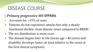
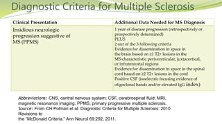
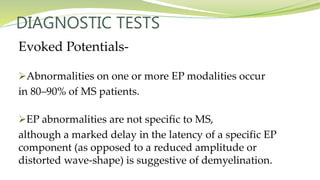
Dr. Farhana Faruque
IMO, Medicine Unit -2
SSMC & MH.
WHAT IS MULTIPLE SCLEROSIS ?
Multiple sclerosis (MS) is an autoimmune disease of the CNS
characterized by chronic inflammation, demyelination, gliosis
(scarring) and neuronal loss.
WHAT IS MULTIPLE SCLEROSIS ?
Lesions of MS typically develop at different times and in
different CNS locations (i.e., MS is said to be disseminated in
time and space).
Approximately 350,000 individuals in the United States and
2.5 million individuals worldwide are affected.
The clinical course can be extremely variable, ranging from a
benign condition to a rapidly evolving disease.
PATHOGENESIS
 New MS lesions begin with perivenular cuffing by
inflammatory mononuclear cells, predominantly T cells and
macrophages, which also infiltrate the surrounding white
matter.
 At sites of inflammation, the blood-brain barrier (BBB) is
disrupted, but unlike vasculitis, the vessel wall is preserved.
PATHOGENESIS
 Involvement of the humoral immune system is also evident;
small numbers of B lymphocytes also infiltrate the nervous
system, myelin-specific autoantibodies are present on
degenerating myelin sheaths, and complement is activated.
 A remarkable feature of MS plaques is that oligodendrocyte
precursor cells survive and in many lesions are present in
even greater numbers than in normal tissue but these cells
fail to differentiate into mature myelin-producing cells.
PATHOGENESIS
 In some lesions, surviving oligodendrocytes or those that
differentiate from precursor cells partially remyelinate the
surviving naked axons, producing so-called shadow plaques.
 As lesions evolve, there is prominent astrocytic proliferation
(gliosis).
 Over time, ectopic lymphocyte follicle-like structures, consisting
of aggregates of T and B cells resembling secondary lymphoid
tissue, appear in the meninges and especially overlying deep
cortical sulci and spaces.
PATHOGENESIS
 Partial or total axonal destruction can also occur, especially
within highly inflammatory lesions.
 Thus MS is not solely a disease of myelin, and neuronal
pathology is increasingly recognized as a major contributor
to irreversible neurologic disability.
Epidemiology
 MS is approximately threefold more common in women than men.
 The age of onset is typically between 20 and 40 years.
(slightly later in men than in women), but the disease can present
across the lifespan.
 Approximately 10% of cases begin before age 18 years of age, and a
small percentage of cases begin before the age of 10 years.
Epidemiology
 Highest known prevalence for MS (250 per 100,000)
present in the Orkney Islands, located north of Scotland.
 In other temperate zone areas (e.g. northern North
America, northern Europe, southern Australia, and
southern New Zealand), the prevalence of MS is 0.1–0.2%.
 By contrast, in the tropics (e.g., Asia, equatorial Africa,
and the Middle East), the prevalence is often 10- to 20-fold
less.
Well-established risk factors for MS include-
 Vitamin D deficiency.
 Exposure to Epstein-Barr virus (EBV).
 After early childhood.
 Cigarette smoking.
 Consumption of a high-salt diet.
RISK FACTORS
GENETIC CONSIDERATIONS
 Whites are inherently at higher risk for MS than Africans
or Asians.
 MS also aggregates within some families and adoption,
half-sibling, twin, and spousal studies indicate that
familial aggregation is due to genetic, and not
environmental.
GENETIC CONSIDERATIONS
 Susceptibility to MS is polygenic. The strongest susceptibility
signal in genome-wide studies maps to the HLA-DRB1 gene
in the class II region of the major histocompatibility complex
(MHC), and this association accounts for approximately 10%
of the disease risk.
CLINICAL MANIFESTATIONS
Weakness of the limbs may manifest as-
 Loss of strength, speed, or dexterity, as fatigue, or as a
disturbance of gait. Exercise-induced weakness is a
characteristic symptom of MS.
Weakness is upper motor neuron type (spasticity,
hyperreflexia, and Babinski signs.)
CLINICAL MANIFESTATIONS
Weakness of the limbs may manifest as-
Occasionally a tendon reflex may be lost (simulating a lower
motor neuron lesion) if an MS lesion disrupts the afferent
reflex fibers in the spinal cord .
CLINICAL MANIFESTATIONS
Spasticity is commonly associated with-
 Spontaneous and movement-induced muscle spasms.
 More than 30% of MS patients have moderate to severe
spasticity, especially in the legs. This Occasionally spasticity
provides support for the body weight during ambulation.
CLINICAL MANIFESTATIONS
Optic neuritis (ON) presents as-
 Diminished visual acuity,
 Dimness,
 Decreased color perception (desaturation).
CLINICAL MANIFESTATIONS
 Rarely, there is complete loss of light perception
 Visual symptoms are generally monocular but may be bilateral.
 An afferent pupillary defect is usually present.
Funduscopic examination –
May be normal or reveal optic disc swelling (papillitis).
Pallor of the optic disc (optic atrophy) commonly follows ON.
CLINICAL MANIFESTATIONS
Visual blurring may result from-
ON or diplopia (double vision)
If the symptom resolves when either eye is covered, the cause is
diplopia.
Diplopia may result from-
Internuclear ophthalmoplegia (INO) or Sixth cranial nerve palsy
(rarely the third or fourth).
An INO consists of impaired adduction of one eye due to a
lesion in the ipsilateral medial longitudinal fasciculus .
CLINICAL MANIFESTATIONS
 Prominent nystagmus is often observed in the abducting
eye.
 A bilateral INO is particularly suggestive of MS.
 Other common gaze disturbances in MS include
(1) a horizontal gaze palsy
(2) a “one and a half” syndrome (horizontal gaze palsy
plus an INO) and
(3) acquired pendular nystagmus.
CLINICAL MANIFESTATIONS
Sensory symptoms are varied and include both
 paresthesias (e.g., tingling, prickling sensations,
formications, “pins and needles,” or painful burning)
hypesthesia (e.g., reduced sensation, numbness, or a
“dead” feeling).
Unpleasant sensations (e.g., feelings that body parts are
swollen, wet, raw, or tightly wrapped) are also common.
CLINICAL MANIFESTATIONS
 Sensory impairment of the trunk and legs below a horizontal
line on the torso (a sensory level) indicates that the spinal cord
is the origin of the sensory disturbance.
It is often accompanied by-
 A bandlike sensation of tightness around the torso.
 Pain is common, experienced by >50% of patients.
 Pain can occur anywhere on the body and can change
locations over time.
CLINICAL MANIFESTATIONS
Bladder dysfunction
 Present in >90% of MS patients
 Detrusor hyperreflexia- impairment of suprasegmental inhibition,
causes urinary frequency, urgency, nocturia, and uncontrolled
bladder emptying.
 Detrusor sphincter dyssynergia- loss of synchronization between
detrusor and sphincter muscles, causes difficulty in initiating
and/or stopping the urinary stream, producing hesitancy, urinary
retention, overflow incontinence, and recurrent infection.
CLINICAL MANIFESTATIONS
Cognitive dysfunction include
-- Memory loss
-- Impaired attention
-- Difficulties in executive functioning, memory, and problem
solving
-- Slowed information processing and problems shifting
between cognitive tasks.
--Euphoria (elevated mood) ( occurring in <20% of patients.)
CLINICAL MANIFESTATIONS
OTHERS-
 Constipation( occurs in >30% of patients).
 Ataxia
 Depression
 Fatigue
 Sexual dysfunction
 Facial weakness ( not associated with ipsilateral loss of taste
sensation or retro-auricular pain)
 Vertigo
Initial Symptoms of MS
Symptom Percentage
of Cases
Symptom Percentage
of Cases
Sensory loss 37 Lhermitte 03
Optic neuritis 36 Pain 03
Weakness 35 Dementia 02
Paresthesias 24 Visual loss 02
Diplopia 15 Facial palsy 01
Ataxia 11 Impotence 01
Vertigo 06 Myokymia 01
Paroxysmal attacks 04 Epilepsy 01
Bladder 04 Falling 01
Ancillary Symptoms
 Heat sensitivity refers to neurologic symptoms produced
by an elevation of the body’s core temperature.
For example, unilateral visual blurring may occur
during a hot shower or with physical exercise
(Uhthoff ’s symptom). Such heat-related symptoms
probably result from transient conduction block .
Ancillary Symptoms
 Lhermitte’s symptom is an electric shock–like sensation
(typically induced by flexion or other movements of the
neck) that radiates down the back into the legs.
Rarely, it radiates into the arms. It is generally self-limited
but may persist for years.
Lhermitte’s symptom can also occur with other disorders of
the cervical spinal cord (e.g., cervical spondylosis).
Ancillary Symptoms
 Paroxysmal symptoms are result from spontaneous
discharges, arising at the edges of demyelinated plaques and
spreading to adjacent white matter tracts.
--brief duration (10 s to 2 min),
--high frequency (5–40 episodes per day)
-- lack of any alteration of consciousness or
--change in background electroencephalogram during episodes
--a self-limited course (generally lasting weeks to months)
--precipitated by hyperventilation or movement.
Ancillary Symptoms
These include –
--Lhermitte’s symptom
--Tonic contractions of a limb, face, or trunk (tonic seizures)
--Paroxysmal dysarthria and
--Ataxia
OTHERS:
 Trigeminal neuralgia, hemifacial spasm, and
glossopharyngeal neuralgia
 Facial myokymia
DISEASE COURSE
Four clinical types of MS exist :
1. Relapsing/remitting MS (RRMS) accounts for 85% of
MS cases at onset. Characterized by
-- Discrete attacks
-- Generally evolve over days to weeks (rarely over hours).
--With initial attacks, there is often substantial or complete
recovery over the ensuing weeks to months.
-- Between attacks, patients are neurologically stable.
DISEASE COURSE
2. Secondary progressive MS (SPMS)
 Always begins as RRMS
 Patient experiences a steady deterioration in function
unassociated with acute attacks (which may continue
or cease during the progressive phase)
 Produces a greater amount of fixed neurologic disability
than RRMS.
 A patient with RRMS, the risk of developing SPMS is
∼2% each year.
DISEASE COURSE
3. Primary progressive MS (PPMS)
 Accounts for ∼15% of cases
 Patients do not experience attacks but only a steady
functional decline from disease onset compared to RRMS
 The sex distribution is more even
 The disease begins later in life (mean age ∼40 years) and
disability develops faster (at least relative to the onset of
the first clinical symptom).
DISEASE COURSE
4. Progressive/relapsing MS (PRMS)
 Overlaps PPMS and SPMS and
 Accounts for ∼5% of MS patients.
 Like PPMS, these patients experience a steady deterioration in their
condition from disease onset.
 Like SPMS patients, they experience occasional attacks superimposed
upon their progressive course .
DIAGNOSIS
There is no definitive diagnostic test for MS. Diagnostic criteria
for clinically definite MS require documentation of two or more
episodes of symptoms and two or more signs that reflect
pathology in anatomically noncontiguous white matter tracts of
the CNS symptoms must last for >24 h and occur as distinct
episodes that are separated by a month or more.
DIAGNOSIS
 In patients who have only one of the two required signs on
neurologic examination, the second may be documented
by abnormal tests such as MRI or evoked potentials (EPs).
DIAGNOSIS
 In patients whose course is progressive from onset for ≥6
months without superimposed relapses, documentation of
intrathecal IgG synthesis may be used to support a
diagnosis of PPMS.
Diagnostic Criteria for Multiple Sclerosis
Clinical Presentation Additional Data Needed for MS Diagnosis
2 or more attacks; objective
clinical evidence of 2 or
more lesions or objective
clinical evidence of 1 lesion
with reasonable historical
evidence of a prior attack
None
2 or more attacks; objective
clinical evidence of
1 lesion
Dissemination in space, demonstrated by
• ≥1 T2 lesion on MRI in at least 2 out of 4
MS-typical regions of the CNS
(periventricular,
juxtacortical, infratentorial, or spinal cord)
OR
• Await a further clinical
Diagnostic Criteria for Multiple Sclerosis
Clinical Presentation Additional Data Needed for MS Diagnosis
1 attack; objective clinical
evidence of 2 or more
lesions
Dissemination in time, demonstrated by
• Simultaneous presence of asymptomatic
gadolinium-enhancing and nonenhancing
lesions at any time
OR
• A new T2 and/or gadolinium-enhancing
lesion(s) on follow-up MRI, irrespective of
its
timing with reference to a baseline scan
OR
• Await a second clinical attack
Diagnostic Criteria for Multiple Sclerosis
Clinical Presentation Additional Data Needed for MS Diagnosis
1 attack; objective clinical
evidence of 1 lesion (clinically
isolated syndrome)
Dissemination in space and time, demonstrated
by:
For dissemination in space
• ≥1 T2 lesion in at least 2 out of 4 MS-typical
regions of the CNS (periventricular, juxtacortical,
infratentorial, or spinal cord)
OR
• Await a second clinical attack implicating a
different CNS site
AND
For dissemination in time
• Simultaneous presence of asymptomatic
gadolinium-enhancing and nonenhancing
lesions at any time
OR
• A new T2 and/or gadolinium-enhancing
lesion(s) on follow-up MRI, irrespective of its
timing with reference to a baseline scan
OR
• Await a second clinical attack
Diagnostic Criteria for Multiple Sclerosis
Clinical Presentation Additional Data Needed for MS Diagnosis
Insidious neurologic
progression suggestive of
MS (PPMS)
1 year of disease progression (retrospectively or
prospectively determined)
PLUS
2 out of the 3 following criteria
Evidence for dissemination in space in
the brain based on ≥1 T2+ lesions in the
MS-characteristic periventricular, juxtacortical,
or infratentorial regions
Evidence for dissemination in space in the spinal
cord based on ≥2 T2+ lesions in the cord
Positive CSF (isoelectric focusing evidence of
oligoclonal bands and/or elevated IgG index)
Abbreviations: CNS, central nervous system; CSF, cerebrospinal fluid; MRI,
magnetic resonance imaging; PPMS, primary progressive multiple sclerosis.
Source: From CH Polman et al: Diagnostic Criteria for Multiple Sclerosis: 2010
Revisions to
the “McDonald Criteria.” Ann Neurol 69:292, 2011.
DIAGNOSTIC TESTS
Magnetic Resonance Imaging MRI
 Characteristic abnormalities are found in >95% of
patients, although more than 90% of the lesions visualized
by MRI are asymptomatic.
 An increase in vascular permeability from a breakdown
of the BBB is detected by leakage of intravenous
gadolinium (Gd) into the parenchyma & serves as a
useful marker of inflammation.
DIAGNOSTIC TESTS
Magnetic Resonance Imaging MRI
 Gd enhancement typically persists for approximately
1 month, and the residual MS plaque remains visible
indefinitely as a focal area of hyperintensity (a lesion) on
(T2-weighted) .
 Lesions are frequently oriented perpendicular
to the ventricular surface, corresponding to the pathologic
pattern of perivenous demyelination (Dawson’s fingers).
DIAGNOSTIC TESTS
Magnetic Resonance Imaging MRI
 Lesions are multifocal within the brain, brainstem, and
spinal cord. Lesions larger than 6 mm located in the
corpus callosum, periventricular white matter, brainstem,
cerebellum, or spinal cord are particularly diagnostic.
Approximately one-third of T2-weighted lesions appear
as hypointense lesions (black holes) on T1-weighted
imaging. Black holes may be a marker of irreversible
demyelination and axonal loss.
DIAGNOSTIC TESTS
Magnetic Resonance Imaging MRI
Newer MRI methods such as magnetization transfer ratio
(MTR) imaging and proton magnetic resonance spectroscopic
imaging (MRSI) may ultimately serve as surrogate markers of
clinical disability.
MRSI can quantitate molecules such as N-acetyl aspartate,
which is a marker of axonal integrity, and MTR may be able
to distinguish demyelination from edema.
DIAGNOSTIC TESTS
Evoked Potentials
 EP testing assesses function in afferent (visual, auditory,
and somatosensory) or efferent (motor) CNS pathways.
Eps use computer averaging to measure CNS electric
potentials evoked by repetitive stimulation of selected
peripheral nerves or of the brain.
DIAGNOSTIC TESTS
Evoked Potentials-
 These tests provide the most information when the
pathways studied are clinically uninvolved.
For example, in a patient with a remitting and relapsing
spinal cord syndrome with sensory deficits in the legs, an
abnormal somatosensory EP following posterior tibial nerve
stimulation provides little new information.
By contrast, an abnormal visual EP in this circumstance
would permit a diagnosis of clinically definite MS.
DIAGNOSTIC TESTS
Evoked Potentials-
Abnormalities on one or more EP modalities occur
in 80–90% of MS patients.
EP abnormalities are not specific to MS,
although a marked delay in the latency of a specific EP
component (as opposed to a reduced amplitude or
distorted wave-shape) is suggestive of demyelination.
DIAGNOSTIC TESTS
Cerebrospinal Fluid-
CSF abnormalities found in MS include a mononuclear
cell pleocytosis and an increased level of intrathecally
synthesized IgG. The total CSF protein is usually normal.
DIAGNOSTIC TESTS
Cerebrospinal Fluid-
IgG index, expresses the ratio of IgG to albumin in the CSF
divided by the same ratio in the serum. The IgG synthesis
rate uses serum and CSF IgG and albumin measurements to
calculate the rate of CNS IgG synthesis.
DIAGNOSTIC TESTS
Cerebrospinal Fluid-
 The measurement of oligoclonal bands (OCBs) by agarose
gel electrophoresis in the CSF also assesses intrathecal
production of IgG. Two or more discrete OCBs, not
present in a paired serum sample, are found in >75% of
patients with MS. OCBs may be absent at the onset of MS,
and in individual patients, the number of bands may
increase with time.
DIAGNOSTIC TESTS
Cerebrospinal Fluid-
 A mild CSF pleocytosis (>5 cells/μL) is present in ∼25% of
cases, usually in young patients with RRMS. A pleocytosis of
>75 cells/μL, the presence of polymorphonuclear leukocytes,
or a protein concentration >1 g/L (>100 mg/dL) in CSF should
raise concern that the patient may not have MS.
Disorders That Can Mimic Multiple Sclerosis (MS)
Acute disseminated
encephalomyelitis (ADEM)
Cerebral autosomal dominant
arteriopathy, subcortical infarcts,
and leukoencephalopathy
(CADASIL)
Congenital leukodystrophies (e.g.,
adrenoleukodystrophy,
metachromatic
leukodystrophy)
Antiphospholipid antibody
syndrome
Mitochondrial encephalopathy with
lactic acidosis and stroke (MELAS)
Systemic lupus erythematosus and
related collagen vascular disorders
Vascular malformations (especially
spinal dural AV fistulas)
Ischemic optic neuropathy (arteritic
and nonarteritic)
Neoplasms (e.g., lymphoma,
glioma, meningioma)
Human immunodeficiency virus
(HIV) infection
Tropical spastic paraparesis (HTLV-
1/2 infection)
Stroke and ischemic cerebrovascular
disease
Bechet's disease Lyme disease Sarcoidosis
Sjogren's syndrome Syphilis Vasculitis & Vitamin B12 deficiency
TREATMENT
Therapy for MS can be divided into several categories:
(1) Treatment of acute attacks
(2) Treatment with disease-modifying agents and
(3) Symptomatic therapy.
Treatments that promote remyelination or neural repair
do not currently exist, but several promising approaches
are being actively investigated.
TREATMENT
ACUTE ATTACKS OR INITIAL DEMYELINATING EPISODES
 When patients experience acute deterioration, it is important
to consider whether this is new disease activity or a
“pseudoexacerbation”.
TREATMENT
ACUTE ATTACKS OR INITIAL DEMYELINATING
EPISODES
 Glucocorticoids are used to manage either first attacks
or acute exacerbations.
 They provide short-term clinical benefit by reducing the
severity and shortening the duration of attacks.
TREATMENT
Glucocorticoid treatment is usually administered as
intravenous Methylprednisolone, 500–1000 mg/d for 3–5
days, either without a taper or followed by a course of oral
prednisone beginning at a dose of 60–80 mg/d and gradually
tapered over 2 weeks.
Orally administered methylprednisolone or dexamethasone
(in equivalent dosages) can be substituted for the
intravenous portion of the therapy.
TREATMENT
 Side effects of short-term glucocorticoid therapy include-
 Fluid retention
 Potassium loss
 Weight gain
 Gastric disturbances
 Acne and Emotional lability.
Concurrent use of a low-salt, potassium rich diet and
avoidance of potassium-wasting diuretics are advisable.
TREATMENT
 Lithium carbonate (300 mg orally bid) may help to manage
emotional lability and insomnia associated with
glucocorticoid therapy.
 Plasma exchange (five to seven exchanges: 40–60 mL/kg per
exchange, every other day for 14 days) may benefit patients
with fulminant attacks of demyelination that are
unresponsive to glucocorticoids. But cost is high, and
conclusive evidence of efficacy is lacking.
TREATMENT
DISEASE-MODIFYING THERAPIES FOR RELAPSING FORMS OF MS (RRMS,
SPMS WITH EXACERBATIONS)
Ten such agents are approved by the U.S. Food and Drug Administration (FDA):
All reduce relapse rate, number & size of lesions on MRI.
Some may reduce disability.
IFN-β-1a (Avonex) Fingolimod (Gilenya)
IFN-β-1a (Rebif) Dimethyl fumarate (Tecfidera)
IFN-β-1b (Betaseron or Extavia) Teriflunomide (Aubagio)
Glatiramer acetate (Copaxone) Mitoxantrone (Novantrone)
Natalizumab (Tysabri) Alemtuzumab (Lemtrada).
TREATMENT
Interferon-β-
IFN-β is a class I interferon. Efficacy in MS probably results
from immunomodulatory properties including-
(1) Downregulating expression of MHC molecules on
antigen-presenting cells.
(2) reducing proinflammatory and increasing regulatory
cytokine levels.
(3) inhibiting T cell proliferation and
(4) limiting the trafficking of inflammatory cells in the CNS.
TREATMENT
IFN-β-1a (Avonex), 30 μg, is administered by intramuscular
injection once every week.
IFN-β-1a (Rebif), 44 μg, is administered by subcutaneous
injection three times per week.
IFN-β-1b (Betaseron or Extavia), 250 μg, is administered by
subcutaneous injection every other day.
TREATMENT
Common side effects of IFN-β therapy-
 Flulike symptoms (e.g., fevers, chills, and myalgias) and
Mild abnormalities on routine laboratory evaluation (e.g.,
elevated liver function tests or lymphopenia).
Rarely, more severe hepatotoxicity may occur.
TREATMENT
Glatiramer Acetate-
Glatiramer acetate is a synthetic, random polypeptide composed of
four
amino acids (l-glutamic acid, l-lysine, l-alanine, and l-tyrosine).
Mechanism of action-
(1) Induction of antigen-specific suppressor T cells
(2) Binding to MHC molecules, thereby displacing bound MBP or
(3) Altering the balance between proinflammatory and regulatory
cytokines.
TREATMENT
Dose- Glatiramer acetate is administered by-
subcutaneous injection of either 20 mg everyday or
40 mg thrice weekly.
Side Effects-
One or more episodes of flushing, chest tightness, dyspnea,
palpitations, and anxiety after injection. This systemic
reaction is unpredictable, brief (duration <1 h), and tends
not to recur.
TREATMENT
 Natalizumab
Natalizumab is a humanized monoclonal antibody
directed against the α4 subunit of α4β1 integrin, a
cellular adhesion molecule expressed on the surface of
lymphocytes. It prevents lymphocytes from binding to
endothelial cells, thereby preventing lymphocytes from
penetrating the BBB and entering the CNS.
TREATMENT
Dose- Natalizumab, 300 mg, is administered by IV infusion each
month.
Side Effects-
 <10% of patients experience hypersensitivity reactions
(including anaphylaxis) and
 ∼6% develop neutralizing antibodies to the molecule (only half of
which persist).
 0.3% of patients treated with natalizumab develop PML resulting from
infection by John Cunningham (JC) virus.
TREATMENT
Fingolimod -
Fingolimod is a sphingosine-1-phosphate (S1P) inhibitor
that prevents the egress of lymphocytes from the
secondary lymphoid organs such as the lymph nodes and
spleen.
Mechanism of action is probably due, to the trapping of
lymphocytes in the periphery and inhibiting their
trafficking to the CNS.
TREATMENT
Dose- Fingolimod, 0.5 mg, is administered orally each day.
Side Effects-
 Elevated liver function tests
 Lymphopenia
 First- and second degree heart block
 Bradycardia
A 6-h period of observation (including electrocardiogram
monitoring) is recommended for all patients receiving their first
dose, and individuals with preexisting cardiac disease.
TREATMENT
Alemtuzumab
Alemtuzumab is a humanized monoclonal antibody directed
against the CD52 antigen, which is expressed on both
monocytes and lymphocytes.
It causes lymphocyte depletion (of both B and T cells) and a
change in the composition of lymphocyte composition of
lymphocyte subsets.
Side Effects-
(1) Autoimmune diseases including thyroiditis, Graves’ disease,
thrombocytopenia, hemolytic anemia, pancytopenia,
anti glomerular basement membrane disease, and membranous
glomerulonephritis.
(2) Malignancies including thyroid cancer, melanoma, breast cancer,
and human papillomavirus (HPV)–related cancers.
(3) Serious infections and
(4) Infusion reactions.
PROGNOSIS
Most patients with clinically evident MS ultimately experience
progressive neurologic disability.
15 years after onset, only 20% of patients had no functional
limitation, and between one-third and one-half progressed to
SPMS and required assistance with ambulation; 25 years after
onset, ∼80% of MS patients reached this levelof disability.
ON or sensory symptoms at onset fewer than two relapses in the
first year of illness; and minimal impairment after 5 years.
PROGNOSIS
 Some MS patients (<20%) have a benign variant of MS
and never develop neurologic disability.
With three or more typical T2-weighted lesions, the risk of
developing MS after 20 years is ∼80%. Conversely, with a
normal brain MRI, the likelihood of developing MS is
<20%.
PROGNOSIS
 Mortality as a direct consequence of MS is uncommon, although it
has been estimated that the 25-year survival is only 85% of expected.
Death can occur during an acute MS attack, although this is distinctly
rare. More commonly, death occurs as a complication of MS (e.g.,
pneumonia in a debilitated individual).
Death can also result from suicide. Early disease-modifying therapy
seems to reduce this excess mortality.
THANK YOU

MULTIPLE SCLEROSIS

  • 1.
    Dr. Farhana Faruque IMO,Medicine Unit -2 SSMC & MH.
  • 2.
    WHAT IS MULTIPLESCLEROSIS ? Multiple sclerosis (MS) is an autoimmune disease of the CNS characterized by chronic inflammation, demyelination, gliosis (scarring) and neuronal loss.
  • 3.
    WHAT IS MULTIPLESCLEROSIS ? Lesions of MS typically develop at different times and in different CNS locations (i.e., MS is said to be disseminated in time and space). Approximately 350,000 individuals in the United States and 2.5 million individuals worldwide are affected. The clinical course can be extremely variable, ranging from a benign condition to a rapidly evolving disease.
  • 4.
    PATHOGENESIS  New MSlesions begin with perivenular cuffing by inflammatory mononuclear cells, predominantly T cells and macrophages, which also infiltrate the surrounding white matter.  At sites of inflammation, the blood-brain barrier (BBB) is disrupted, but unlike vasculitis, the vessel wall is preserved.
  • 5.
    PATHOGENESIS  Involvement ofthe humoral immune system is also evident; small numbers of B lymphocytes also infiltrate the nervous system, myelin-specific autoantibodies are present on degenerating myelin sheaths, and complement is activated.  A remarkable feature of MS plaques is that oligodendrocyte precursor cells survive and in many lesions are present in even greater numbers than in normal tissue but these cells fail to differentiate into mature myelin-producing cells.
  • 6.
    PATHOGENESIS  In somelesions, surviving oligodendrocytes or those that differentiate from precursor cells partially remyelinate the surviving naked axons, producing so-called shadow plaques.  As lesions evolve, there is prominent astrocytic proliferation (gliosis).  Over time, ectopic lymphocyte follicle-like structures, consisting of aggregates of T and B cells resembling secondary lymphoid tissue, appear in the meninges and especially overlying deep cortical sulci and spaces.
  • 7.
    PATHOGENESIS  Partial ortotal axonal destruction can also occur, especially within highly inflammatory lesions.  Thus MS is not solely a disease of myelin, and neuronal pathology is increasingly recognized as a major contributor to irreversible neurologic disability.
  • 8.
    Epidemiology  MS isapproximately threefold more common in women than men.  The age of onset is typically between 20 and 40 years. (slightly later in men than in women), but the disease can present across the lifespan.  Approximately 10% of cases begin before age 18 years of age, and a small percentage of cases begin before the age of 10 years.
  • 9.
    Epidemiology  Highest knownprevalence for MS (250 per 100,000) present in the Orkney Islands, located north of Scotland.  In other temperate zone areas (e.g. northern North America, northern Europe, southern Australia, and southern New Zealand), the prevalence of MS is 0.1–0.2%.  By contrast, in the tropics (e.g., Asia, equatorial Africa, and the Middle East), the prevalence is often 10- to 20-fold less.
  • 10.
    Well-established risk factorsfor MS include-  Vitamin D deficiency.  Exposure to Epstein-Barr virus (EBV).  After early childhood.  Cigarette smoking.  Consumption of a high-salt diet. RISK FACTORS
  • 11.
    GENETIC CONSIDERATIONS  Whitesare inherently at higher risk for MS than Africans or Asians.  MS also aggregates within some families and adoption, half-sibling, twin, and spousal studies indicate that familial aggregation is due to genetic, and not environmental.
  • 12.
    GENETIC CONSIDERATIONS  Susceptibilityto MS is polygenic. The strongest susceptibility signal in genome-wide studies maps to the HLA-DRB1 gene in the class II region of the major histocompatibility complex (MHC), and this association accounts for approximately 10% of the disease risk.
  • 13.
    CLINICAL MANIFESTATIONS Weakness ofthe limbs may manifest as-  Loss of strength, speed, or dexterity, as fatigue, or as a disturbance of gait. Exercise-induced weakness is a characteristic symptom of MS. Weakness is upper motor neuron type (spasticity, hyperreflexia, and Babinski signs.)
  • 14.
    CLINICAL MANIFESTATIONS Weakness ofthe limbs may manifest as- Occasionally a tendon reflex may be lost (simulating a lower motor neuron lesion) if an MS lesion disrupts the afferent reflex fibers in the spinal cord .
  • 15.
    CLINICAL MANIFESTATIONS Spasticity iscommonly associated with-  Spontaneous and movement-induced muscle spasms.  More than 30% of MS patients have moderate to severe spasticity, especially in the legs. This Occasionally spasticity provides support for the body weight during ambulation.
  • 16.
    CLINICAL MANIFESTATIONS Optic neuritis(ON) presents as-  Diminished visual acuity,  Dimness,  Decreased color perception (desaturation).
  • 17.
    CLINICAL MANIFESTATIONS  Rarely,there is complete loss of light perception  Visual symptoms are generally monocular but may be bilateral.  An afferent pupillary defect is usually present. Funduscopic examination – May be normal or reveal optic disc swelling (papillitis). Pallor of the optic disc (optic atrophy) commonly follows ON.
  • 18.
    CLINICAL MANIFESTATIONS Visual blurringmay result from- ON or diplopia (double vision) If the symptom resolves when either eye is covered, the cause is diplopia. Diplopia may result from- Internuclear ophthalmoplegia (INO) or Sixth cranial nerve palsy (rarely the third or fourth). An INO consists of impaired adduction of one eye due to a lesion in the ipsilateral medial longitudinal fasciculus .
  • 19.
    CLINICAL MANIFESTATIONS  Prominentnystagmus is often observed in the abducting eye.  A bilateral INO is particularly suggestive of MS.  Other common gaze disturbances in MS include (1) a horizontal gaze palsy (2) a “one and a half” syndrome (horizontal gaze palsy plus an INO) and (3) acquired pendular nystagmus.
  • 20.
    CLINICAL MANIFESTATIONS Sensory symptomsare varied and include both  paresthesias (e.g., tingling, prickling sensations, formications, “pins and needles,” or painful burning) hypesthesia (e.g., reduced sensation, numbness, or a “dead” feeling). Unpleasant sensations (e.g., feelings that body parts are swollen, wet, raw, or tightly wrapped) are also common.
  • 21.
    CLINICAL MANIFESTATIONS  Sensoryimpairment of the trunk and legs below a horizontal line on the torso (a sensory level) indicates that the spinal cord is the origin of the sensory disturbance. It is often accompanied by-  A bandlike sensation of tightness around the torso.  Pain is common, experienced by >50% of patients.  Pain can occur anywhere on the body and can change locations over time.
  • 22.
    CLINICAL MANIFESTATIONS Bladder dysfunction Present in >90% of MS patients  Detrusor hyperreflexia- impairment of suprasegmental inhibition, causes urinary frequency, urgency, nocturia, and uncontrolled bladder emptying.  Detrusor sphincter dyssynergia- loss of synchronization between detrusor and sphincter muscles, causes difficulty in initiating and/or stopping the urinary stream, producing hesitancy, urinary retention, overflow incontinence, and recurrent infection.
  • 23.
    CLINICAL MANIFESTATIONS Cognitive dysfunctioninclude -- Memory loss -- Impaired attention -- Difficulties in executive functioning, memory, and problem solving -- Slowed information processing and problems shifting between cognitive tasks. --Euphoria (elevated mood) ( occurring in <20% of patients.)
  • 24.
    CLINICAL MANIFESTATIONS OTHERS-  Constipation(occurs in >30% of patients).  Ataxia  Depression  Fatigue  Sexual dysfunction  Facial weakness ( not associated with ipsilateral loss of taste sensation or retro-auricular pain)  Vertigo
  • 25.
    Initial Symptoms ofMS Symptom Percentage of Cases Symptom Percentage of Cases Sensory loss 37 Lhermitte 03 Optic neuritis 36 Pain 03 Weakness 35 Dementia 02 Paresthesias 24 Visual loss 02 Diplopia 15 Facial palsy 01 Ataxia 11 Impotence 01 Vertigo 06 Myokymia 01 Paroxysmal attacks 04 Epilepsy 01 Bladder 04 Falling 01
  • 26.
    Ancillary Symptoms  Heatsensitivity refers to neurologic symptoms produced by an elevation of the body’s core temperature. For example, unilateral visual blurring may occur during a hot shower or with physical exercise (Uhthoff ’s symptom). Such heat-related symptoms probably result from transient conduction block .
  • 27.
    Ancillary Symptoms  Lhermitte’ssymptom is an electric shock–like sensation (typically induced by flexion or other movements of the neck) that radiates down the back into the legs. Rarely, it radiates into the arms. It is generally self-limited but may persist for years. Lhermitte’s symptom can also occur with other disorders of the cervical spinal cord (e.g., cervical spondylosis).
  • 28.
    Ancillary Symptoms  Paroxysmalsymptoms are result from spontaneous discharges, arising at the edges of demyelinated plaques and spreading to adjacent white matter tracts. --brief duration (10 s to 2 min), --high frequency (5–40 episodes per day) -- lack of any alteration of consciousness or --change in background electroencephalogram during episodes --a self-limited course (generally lasting weeks to months) --precipitated by hyperventilation or movement.
  • 29.
    Ancillary Symptoms These include– --Lhermitte’s symptom --Tonic contractions of a limb, face, or trunk (tonic seizures) --Paroxysmal dysarthria and --Ataxia OTHERS:  Trigeminal neuralgia, hemifacial spasm, and glossopharyngeal neuralgia  Facial myokymia
  • 30.
    DISEASE COURSE Four clinicaltypes of MS exist : 1. Relapsing/remitting MS (RRMS) accounts for 85% of MS cases at onset. Characterized by -- Discrete attacks -- Generally evolve over days to weeks (rarely over hours). --With initial attacks, there is often substantial or complete recovery over the ensuing weeks to months. -- Between attacks, patients are neurologically stable.
  • 31.
    DISEASE COURSE 2. Secondaryprogressive MS (SPMS)  Always begins as RRMS  Patient experiences a steady deterioration in function unassociated with acute attacks (which may continue or cease during the progressive phase)  Produces a greater amount of fixed neurologic disability than RRMS.  A patient with RRMS, the risk of developing SPMS is ∼2% each year.
  • 32.
    DISEASE COURSE 3. Primaryprogressive MS (PPMS)  Accounts for ∼15% of cases  Patients do not experience attacks but only a steady functional decline from disease onset compared to RRMS  The sex distribution is more even  The disease begins later in life (mean age ∼40 years) and disability develops faster (at least relative to the onset of the first clinical symptom).
  • 33.
    DISEASE COURSE 4. Progressive/relapsingMS (PRMS)  Overlaps PPMS and SPMS and  Accounts for ∼5% of MS patients.  Like PPMS, these patients experience a steady deterioration in their condition from disease onset.  Like SPMS patients, they experience occasional attacks superimposed upon their progressive course .
  • 35.
    DIAGNOSIS There is nodefinitive diagnostic test for MS. Diagnostic criteria for clinically definite MS require documentation of two or more episodes of symptoms and two or more signs that reflect pathology in anatomically noncontiguous white matter tracts of the CNS symptoms must last for >24 h and occur as distinct episodes that are separated by a month or more.
  • 36.
    DIAGNOSIS  In patientswho have only one of the two required signs on neurologic examination, the second may be documented by abnormal tests such as MRI or evoked potentials (EPs).
  • 37.
    DIAGNOSIS  In patientswhose course is progressive from onset for ≥6 months without superimposed relapses, documentation of intrathecal IgG synthesis may be used to support a diagnosis of PPMS.
  • 38.
    Diagnostic Criteria forMultiple Sclerosis Clinical Presentation Additional Data Needed for MS Diagnosis 2 or more attacks; objective clinical evidence of 2 or more lesions or objective clinical evidence of 1 lesion with reasonable historical evidence of a prior attack None 2 or more attacks; objective clinical evidence of 1 lesion Dissemination in space, demonstrated by • ≥1 T2 lesion on MRI in at least 2 out of 4 MS-typical regions of the CNS (periventricular, juxtacortical, infratentorial, or spinal cord) OR • Await a further clinical
  • 39.
    Diagnostic Criteria forMultiple Sclerosis Clinical Presentation Additional Data Needed for MS Diagnosis 1 attack; objective clinical evidence of 2 or more lesions Dissemination in time, demonstrated by • Simultaneous presence of asymptomatic gadolinium-enhancing and nonenhancing lesions at any time OR • A new T2 and/or gadolinium-enhancing lesion(s) on follow-up MRI, irrespective of its timing with reference to a baseline scan OR • Await a second clinical attack
  • 40.
    Diagnostic Criteria forMultiple Sclerosis Clinical Presentation Additional Data Needed for MS Diagnosis 1 attack; objective clinical evidence of 1 lesion (clinically isolated syndrome) Dissemination in space and time, demonstrated by: For dissemination in space • ≥1 T2 lesion in at least 2 out of 4 MS-typical regions of the CNS (periventricular, juxtacortical, infratentorial, or spinal cord) OR • Await a second clinical attack implicating a different CNS site AND For dissemination in time • Simultaneous presence of asymptomatic gadolinium-enhancing and nonenhancing lesions at any time OR • A new T2 and/or gadolinium-enhancing lesion(s) on follow-up MRI, irrespective of its timing with reference to a baseline scan OR • Await a second clinical attack
  • 41.
    Diagnostic Criteria forMultiple Sclerosis Clinical Presentation Additional Data Needed for MS Diagnosis Insidious neurologic progression suggestive of MS (PPMS) 1 year of disease progression (retrospectively or prospectively determined) PLUS 2 out of the 3 following criteria Evidence for dissemination in space in the brain based on ≥1 T2+ lesions in the MS-characteristic periventricular, juxtacortical, or infratentorial regions Evidence for dissemination in space in the spinal cord based on ≥2 T2+ lesions in the cord Positive CSF (isoelectric focusing evidence of oligoclonal bands and/or elevated IgG index) Abbreviations: CNS, central nervous system; CSF, cerebrospinal fluid; MRI, magnetic resonance imaging; PPMS, primary progressive multiple sclerosis. Source: From CH Polman et al: Diagnostic Criteria for Multiple Sclerosis: 2010 Revisions to the “McDonald Criteria.” Ann Neurol 69:292, 2011.
  • 42.
    DIAGNOSTIC TESTS Magnetic ResonanceImaging MRI  Characteristic abnormalities are found in >95% of patients, although more than 90% of the lesions visualized by MRI are asymptomatic.  An increase in vascular permeability from a breakdown of the BBB is detected by leakage of intravenous gadolinium (Gd) into the parenchyma & serves as a useful marker of inflammation.
  • 43.
    DIAGNOSTIC TESTS Magnetic ResonanceImaging MRI  Gd enhancement typically persists for approximately 1 month, and the residual MS plaque remains visible indefinitely as a focal area of hyperintensity (a lesion) on (T2-weighted) .  Lesions are frequently oriented perpendicular to the ventricular surface, corresponding to the pathologic pattern of perivenous demyelination (Dawson’s fingers).
  • 44.
    DIAGNOSTIC TESTS Magnetic ResonanceImaging MRI  Lesions are multifocal within the brain, brainstem, and spinal cord. Lesions larger than 6 mm located in the corpus callosum, periventricular white matter, brainstem, cerebellum, or spinal cord are particularly diagnostic. Approximately one-third of T2-weighted lesions appear as hypointense lesions (black holes) on T1-weighted imaging. Black holes may be a marker of irreversible demyelination and axonal loss.
  • 45.
    DIAGNOSTIC TESTS Magnetic ResonanceImaging MRI Newer MRI methods such as magnetization transfer ratio (MTR) imaging and proton magnetic resonance spectroscopic imaging (MRSI) may ultimately serve as surrogate markers of clinical disability. MRSI can quantitate molecules such as N-acetyl aspartate, which is a marker of axonal integrity, and MTR may be able to distinguish demyelination from edema.
  • 47.
    DIAGNOSTIC TESTS Evoked Potentials EP testing assesses function in afferent (visual, auditory, and somatosensory) or efferent (motor) CNS pathways. Eps use computer averaging to measure CNS electric potentials evoked by repetitive stimulation of selected peripheral nerves or of the brain.
  • 48.
    DIAGNOSTIC TESTS Evoked Potentials- These tests provide the most information when the pathways studied are clinically uninvolved. For example, in a patient with a remitting and relapsing spinal cord syndrome with sensory deficits in the legs, an abnormal somatosensory EP following posterior tibial nerve stimulation provides little new information. By contrast, an abnormal visual EP in this circumstance would permit a diagnosis of clinically definite MS.
  • 49.
    DIAGNOSTIC TESTS Evoked Potentials- Abnormalitieson one or more EP modalities occur in 80–90% of MS patients. EP abnormalities are not specific to MS, although a marked delay in the latency of a specific EP component (as opposed to a reduced amplitude or distorted wave-shape) is suggestive of demyelination.
  • 50.
    DIAGNOSTIC TESTS Cerebrospinal Fluid- CSFabnormalities found in MS include a mononuclear cell pleocytosis and an increased level of intrathecally synthesized IgG. The total CSF protein is usually normal.
  • 51.
    DIAGNOSTIC TESTS Cerebrospinal Fluid- IgGindex, expresses the ratio of IgG to albumin in the CSF divided by the same ratio in the serum. The IgG synthesis rate uses serum and CSF IgG and albumin measurements to calculate the rate of CNS IgG synthesis.
  • 52.
    DIAGNOSTIC TESTS Cerebrospinal Fluid- The measurement of oligoclonal bands (OCBs) by agarose gel electrophoresis in the CSF also assesses intrathecal production of IgG. Two or more discrete OCBs, not present in a paired serum sample, are found in >75% of patients with MS. OCBs may be absent at the onset of MS, and in individual patients, the number of bands may increase with time.
  • 53.
    DIAGNOSTIC TESTS Cerebrospinal Fluid- A mild CSF pleocytosis (>5 cells/μL) is present in ∼25% of cases, usually in young patients with RRMS. A pleocytosis of >75 cells/μL, the presence of polymorphonuclear leukocytes, or a protein concentration >1 g/L (>100 mg/dL) in CSF should raise concern that the patient may not have MS.
  • 54.
    Disorders That CanMimic Multiple Sclerosis (MS) Acute disseminated encephalomyelitis (ADEM) Cerebral autosomal dominant arteriopathy, subcortical infarcts, and leukoencephalopathy (CADASIL) Congenital leukodystrophies (e.g., adrenoleukodystrophy, metachromatic leukodystrophy) Antiphospholipid antibody syndrome Mitochondrial encephalopathy with lactic acidosis and stroke (MELAS) Systemic lupus erythematosus and related collagen vascular disorders Vascular malformations (especially spinal dural AV fistulas) Ischemic optic neuropathy (arteritic and nonarteritic) Neoplasms (e.g., lymphoma, glioma, meningioma) Human immunodeficiency virus (HIV) infection Tropical spastic paraparesis (HTLV- 1/2 infection) Stroke and ischemic cerebrovascular disease Bechet's disease Lyme disease Sarcoidosis Sjogren's syndrome Syphilis Vasculitis & Vitamin B12 deficiency
  • 55.
    TREATMENT Therapy for MScan be divided into several categories: (1) Treatment of acute attacks (2) Treatment with disease-modifying agents and (3) Symptomatic therapy. Treatments that promote remyelination or neural repair do not currently exist, but several promising approaches are being actively investigated.
  • 56.
    TREATMENT ACUTE ATTACKS ORINITIAL DEMYELINATING EPISODES  When patients experience acute deterioration, it is important to consider whether this is new disease activity or a “pseudoexacerbation”.
  • 57.
    TREATMENT ACUTE ATTACKS ORINITIAL DEMYELINATING EPISODES  Glucocorticoids are used to manage either first attacks or acute exacerbations.  They provide short-term clinical benefit by reducing the severity and shortening the duration of attacks.
  • 58.
    TREATMENT Glucocorticoid treatment isusually administered as intravenous Methylprednisolone, 500–1000 mg/d for 3–5 days, either without a taper or followed by a course of oral prednisone beginning at a dose of 60–80 mg/d and gradually tapered over 2 weeks. Orally administered methylprednisolone or dexamethasone (in equivalent dosages) can be substituted for the intravenous portion of the therapy.
  • 59.
    TREATMENT  Side effectsof short-term glucocorticoid therapy include-  Fluid retention  Potassium loss  Weight gain  Gastric disturbances  Acne and Emotional lability. Concurrent use of a low-salt, potassium rich diet and avoidance of potassium-wasting diuretics are advisable.
  • 60.
    TREATMENT  Lithium carbonate(300 mg orally bid) may help to manage emotional lability and insomnia associated with glucocorticoid therapy.  Plasma exchange (five to seven exchanges: 40–60 mL/kg per exchange, every other day for 14 days) may benefit patients with fulminant attacks of demyelination that are unresponsive to glucocorticoids. But cost is high, and conclusive evidence of efficacy is lacking.
  • 61.
    TREATMENT DISEASE-MODIFYING THERAPIES FORRELAPSING FORMS OF MS (RRMS, SPMS WITH EXACERBATIONS) Ten such agents are approved by the U.S. Food and Drug Administration (FDA): All reduce relapse rate, number & size of lesions on MRI. Some may reduce disability. IFN-β-1a (Avonex) Fingolimod (Gilenya) IFN-β-1a (Rebif) Dimethyl fumarate (Tecfidera) IFN-β-1b (Betaseron or Extavia) Teriflunomide (Aubagio) Glatiramer acetate (Copaxone) Mitoxantrone (Novantrone) Natalizumab (Tysabri) Alemtuzumab (Lemtrada).
  • 62.
    TREATMENT Interferon-β- IFN-β is aclass I interferon. Efficacy in MS probably results from immunomodulatory properties including- (1) Downregulating expression of MHC molecules on antigen-presenting cells. (2) reducing proinflammatory and increasing regulatory cytokine levels. (3) inhibiting T cell proliferation and (4) limiting the trafficking of inflammatory cells in the CNS.
  • 63.
    TREATMENT IFN-β-1a (Avonex), 30μg, is administered by intramuscular injection once every week. IFN-β-1a (Rebif), 44 μg, is administered by subcutaneous injection three times per week. IFN-β-1b (Betaseron or Extavia), 250 μg, is administered by subcutaneous injection every other day.
  • 64.
    TREATMENT Common side effectsof IFN-β therapy-  Flulike symptoms (e.g., fevers, chills, and myalgias) and Mild abnormalities on routine laboratory evaluation (e.g., elevated liver function tests or lymphopenia). Rarely, more severe hepatotoxicity may occur.
  • 65.
    TREATMENT Glatiramer Acetate- Glatiramer acetateis a synthetic, random polypeptide composed of four amino acids (l-glutamic acid, l-lysine, l-alanine, and l-tyrosine). Mechanism of action- (1) Induction of antigen-specific suppressor T cells (2) Binding to MHC molecules, thereby displacing bound MBP or (3) Altering the balance between proinflammatory and regulatory cytokines.
  • 66.
    TREATMENT Dose- Glatiramer acetateis administered by- subcutaneous injection of either 20 mg everyday or 40 mg thrice weekly. Side Effects- One or more episodes of flushing, chest tightness, dyspnea, palpitations, and anxiety after injection. This systemic reaction is unpredictable, brief (duration <1 h), and tends not to recur.
  • 67.
    TREATMENT  Natalizumab Natalizumab isa humanized monoclonal antibody directed against the α4 subunit of α4β1 integrin, a cellular adhesion molecule expressed on the surface of lymphocytes. It prevents lymphocytes from binding to endothelial cells, thereby preventing lymphocytes from penetrating the BBB and entering the CNS.
  • 68.
    TREATMENT Dose- Natalizumab, 300mg, is administered by IV infusion each month. Side Effects-  <10% of patients experience hypersensitivity reactions (including anaphylaxis) and  ∼6% develop neutralizing antibodies to the molecule (only half of which persist).  0.3% of patients treated with natalizumab develop PML resulting from infection by John Cunningham (JC) virus.
  • 69.
    TREATMENT Fingolimod - Fingolimod isa sphingosine-1-phosphate (S1P) inhibitor that prevents the egress of lymphocytes from the secondary lymphoid organs such as the lymph nodes and spleen. Mechanism of action is probably due, to the trapping of lymphocytes in the periphery and inhibiting their trafficking to the CNS.
  • 70.
    TREATMENT Dose- Fingolimod, 0.5mg, is administered orally each day. Side Effects-  Elevated liver function tests  Lymphopenia  First- and second degree heart block  Bradycardia A 6-h period of observation (including electrocardiogram monitoring) is recommended for all patients receiving their first dose, and individuals with preexisting cardiac disease.
  • 71.
    TREATMENT Alemtuzumab Alemtuzumab is ahumanized monoclonal antibody directed against the CD52 antigen, which is expressed on both monocytes and lymphocytes. It causes lymphocyte depletion (of both B and T cells) and a change in the composition of lymphocyte composition of lymphocyte subsets.
  • 72.
    Side Effects- (1) Autoimmunediseases including thyroiditis, Graves’ disease, thrombocytopenia, hemolytic anemia, pancytopenia, anti glomerular basement membrane disease, and membranous glomerulonephritis. (2) Malignancies including thyroid cancer, melanoma, breast cancer, and human papillomavirus (HPV)–related cancers. (3) Serious infections and (4) Infusion reactions.
  • 75.
    PROGNOSIS Most patients withclinically evident MS ultimately experience progressive neurologic disability. 15 years after onset, only 20% of patients had no functional limitation, and between one-third and one-half progressed to SPMS and required assistance with ambulation; 25 years after onset, ∼80% of MS patients reached this levelof disability. ON or sensory symptoms at onset fewer than two relapses in the first year of illness; and minimal impairment after 5 years.
  • 76.
    PROGNOSIS  Some MSpatients (<20%) have a benign variant of MS and never develop neurologic disability. With three or more typical T2-weighted lesions, the risk of developing MS after 20 years is ∼80%. Conversely, with a normal brain MRI, the likelihood of developing MS is <20%.
  • 77.
    PROGNOSIS  Mortality asa direct consequence of MS is uncommon, although it has been estimated that the 25-year survival is only 85% of expected. Death can occur during an acute MS attack, although this is distinctly rare. More commonly, death occurs as a complication of MS (e.g., pneumonia in a debilitated individual). Death can also result from suicide. Early disease-modifying therapy seems to reduce this excess mortality.
  • 78.