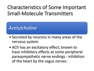
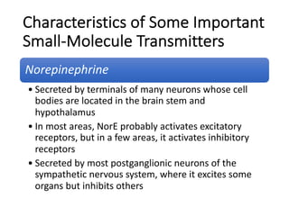
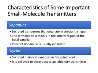
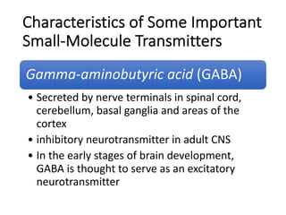
Download to read offline

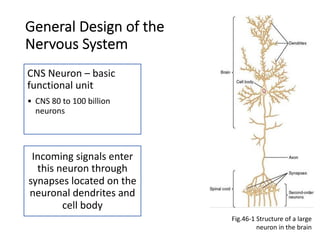
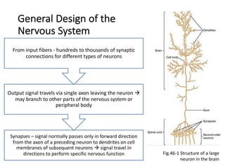
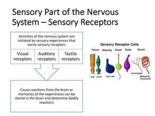
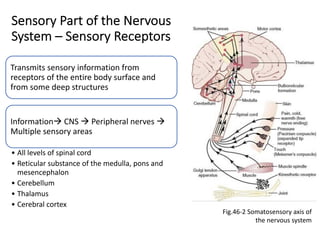
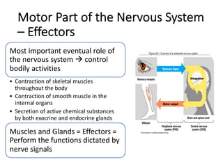
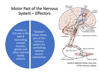
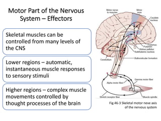
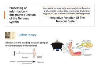
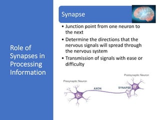
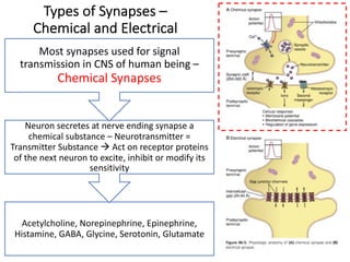
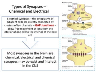
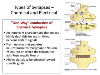
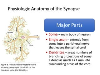


The document provides an overview of the organization and functions of the nervous system. It discusses the following key points: - Neurons are the basic functional units of the central nervous system, with dendrites that receive signals and a single axon that transmits output signals. Signals normally pass from axons to dendrites at synaptic connections. - Sensory receptors detect stimuli and transmit sensory information to the central nervous system. The motor system controls effectors like muscles and glands. - Information processing in the nervous system involves integration of sensory input and output responses. Synapses determine the pathways of signal transmission and storage of memories. - The central nervous system is organized into different levels like the spinal cord