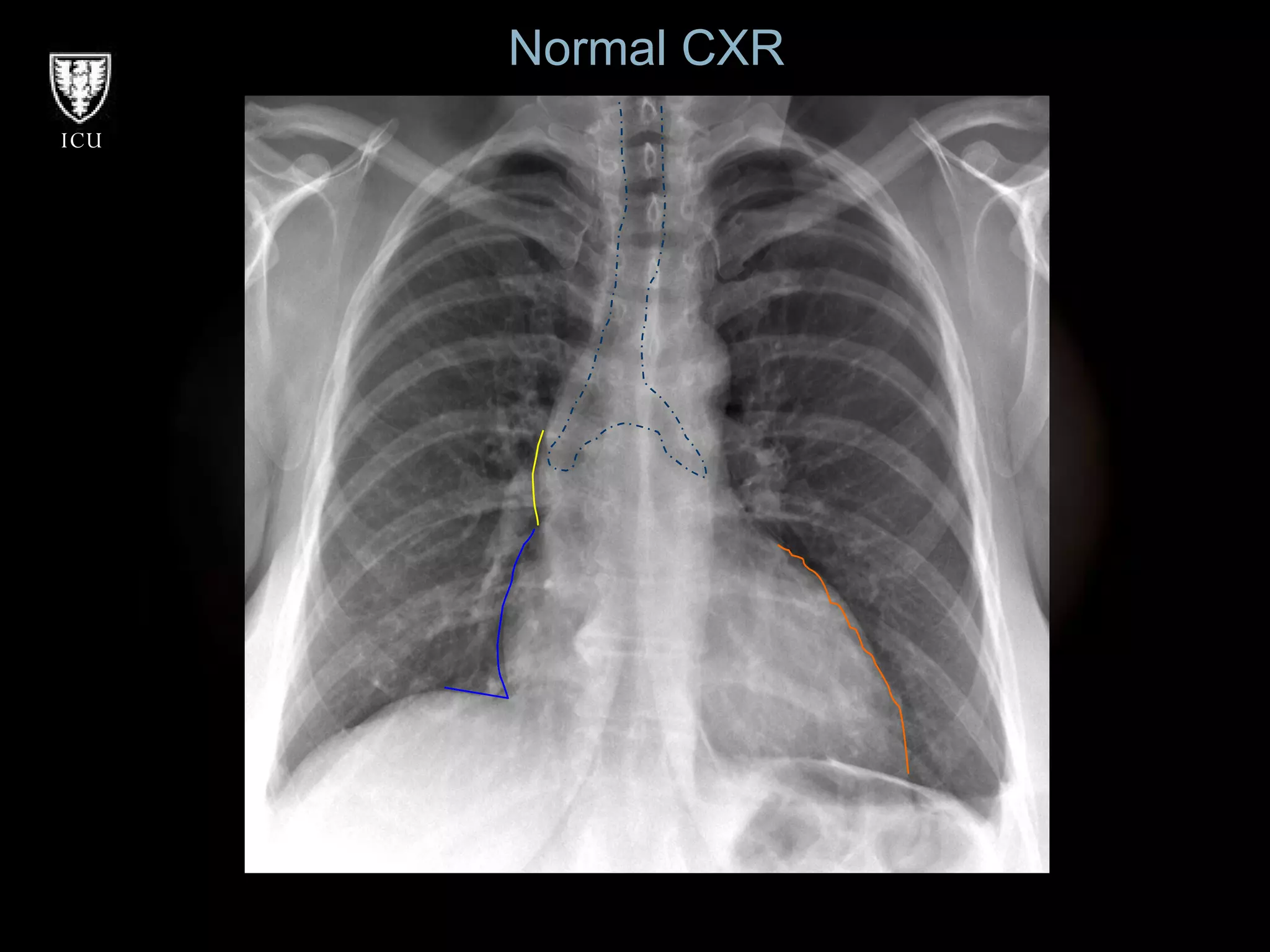
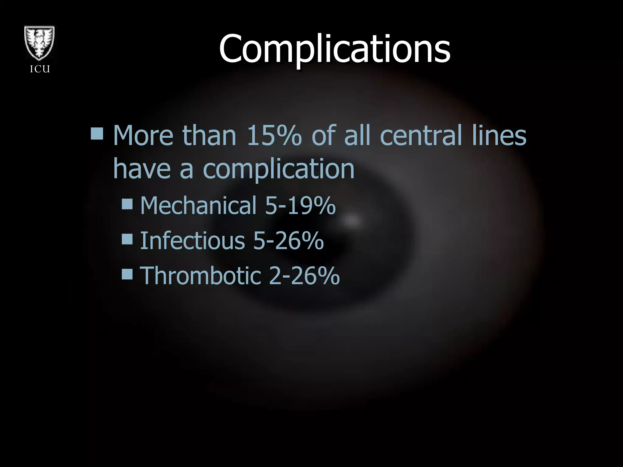
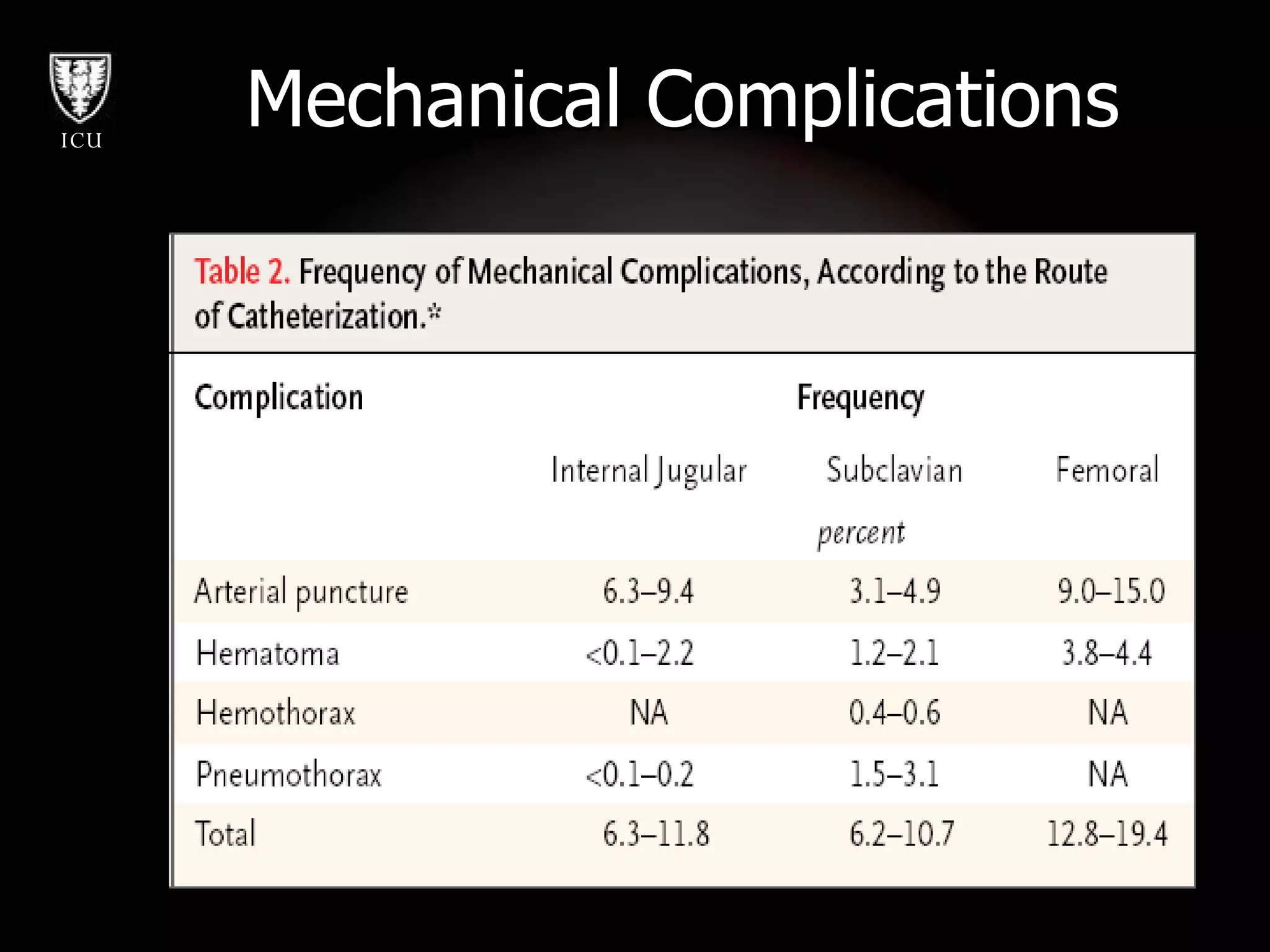
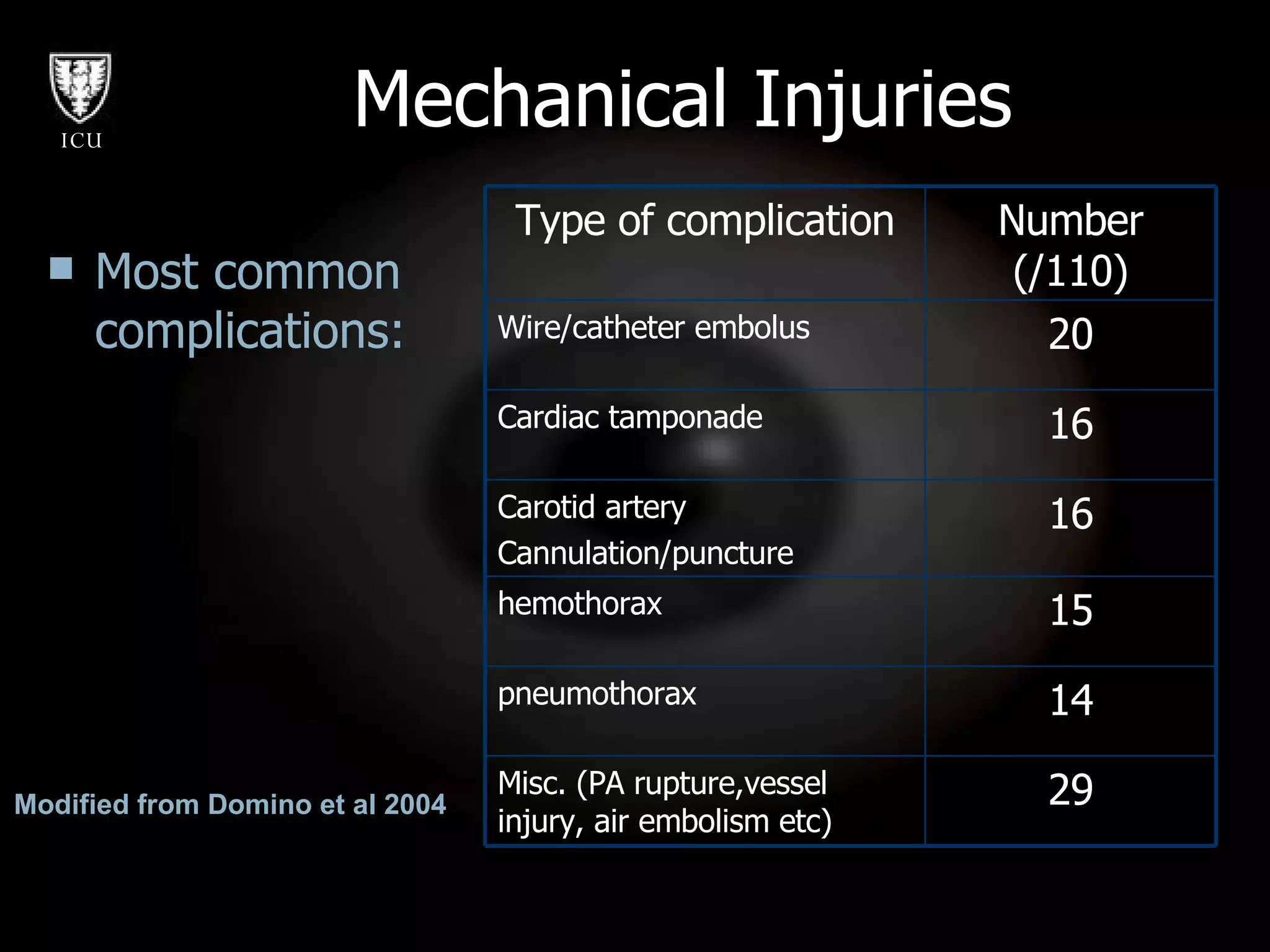
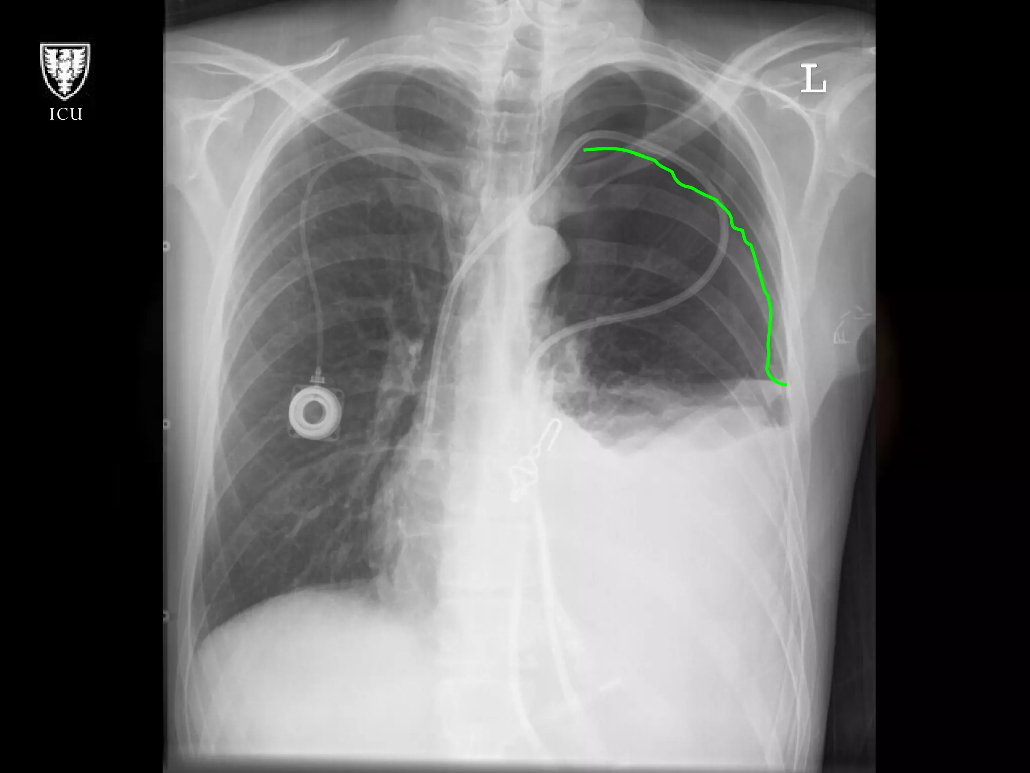
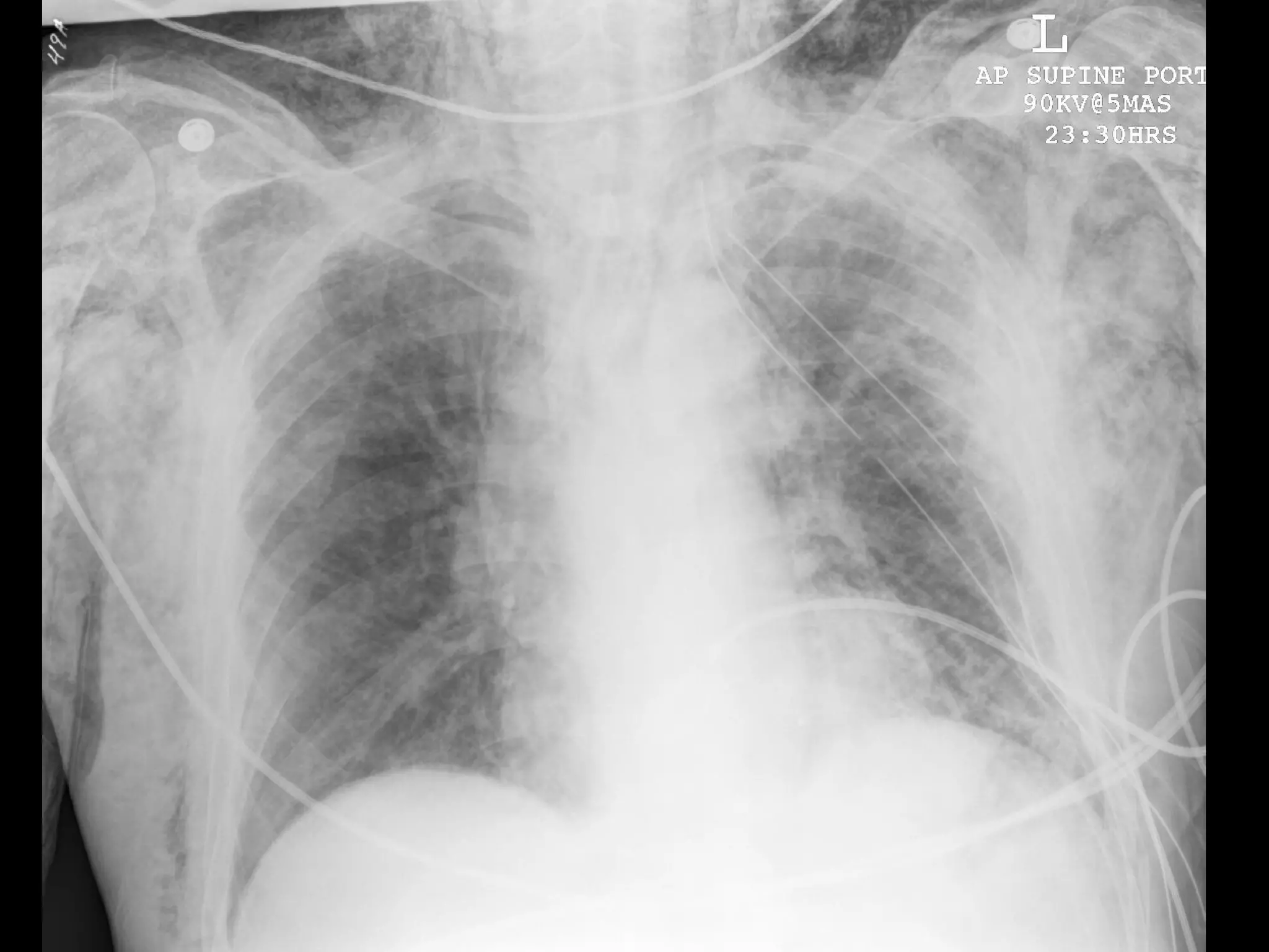
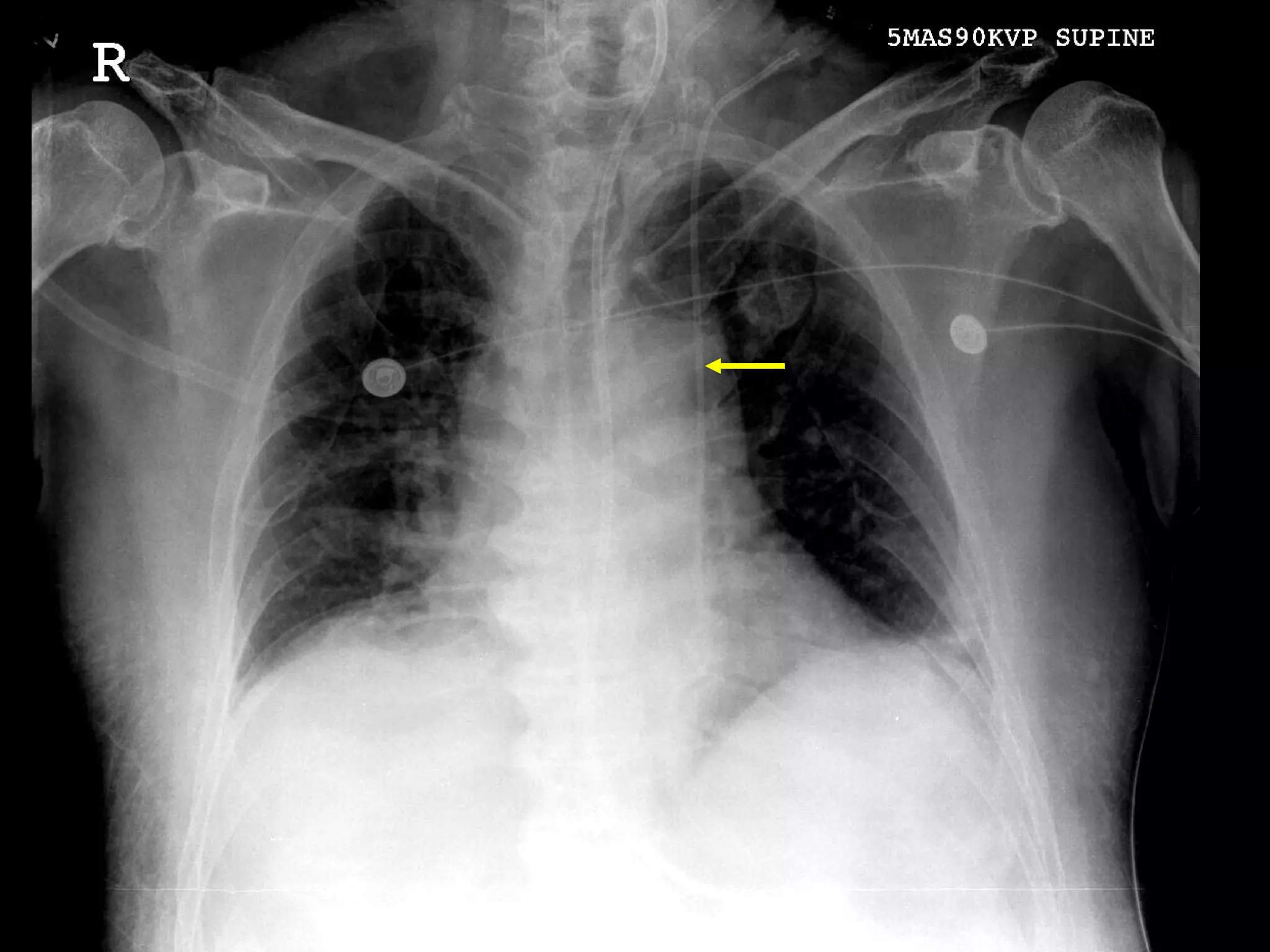
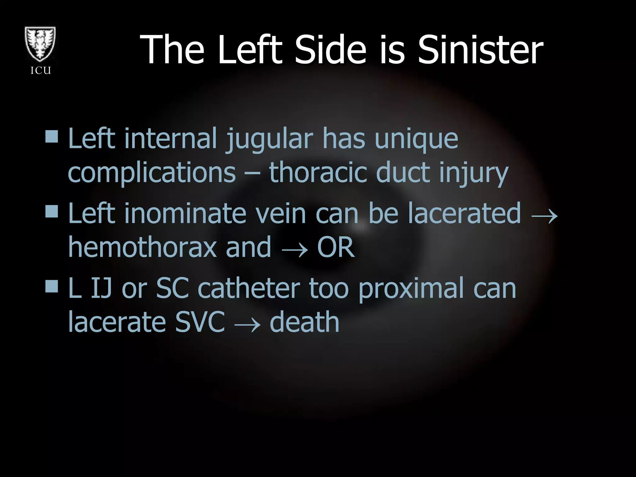
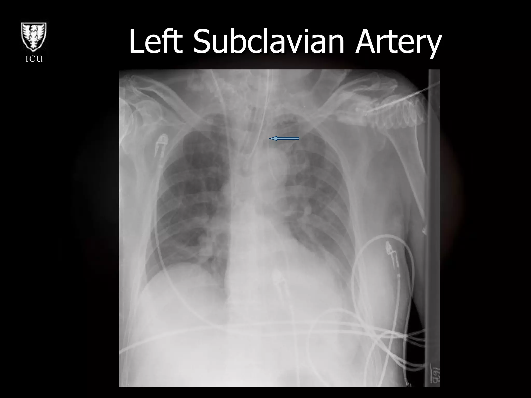
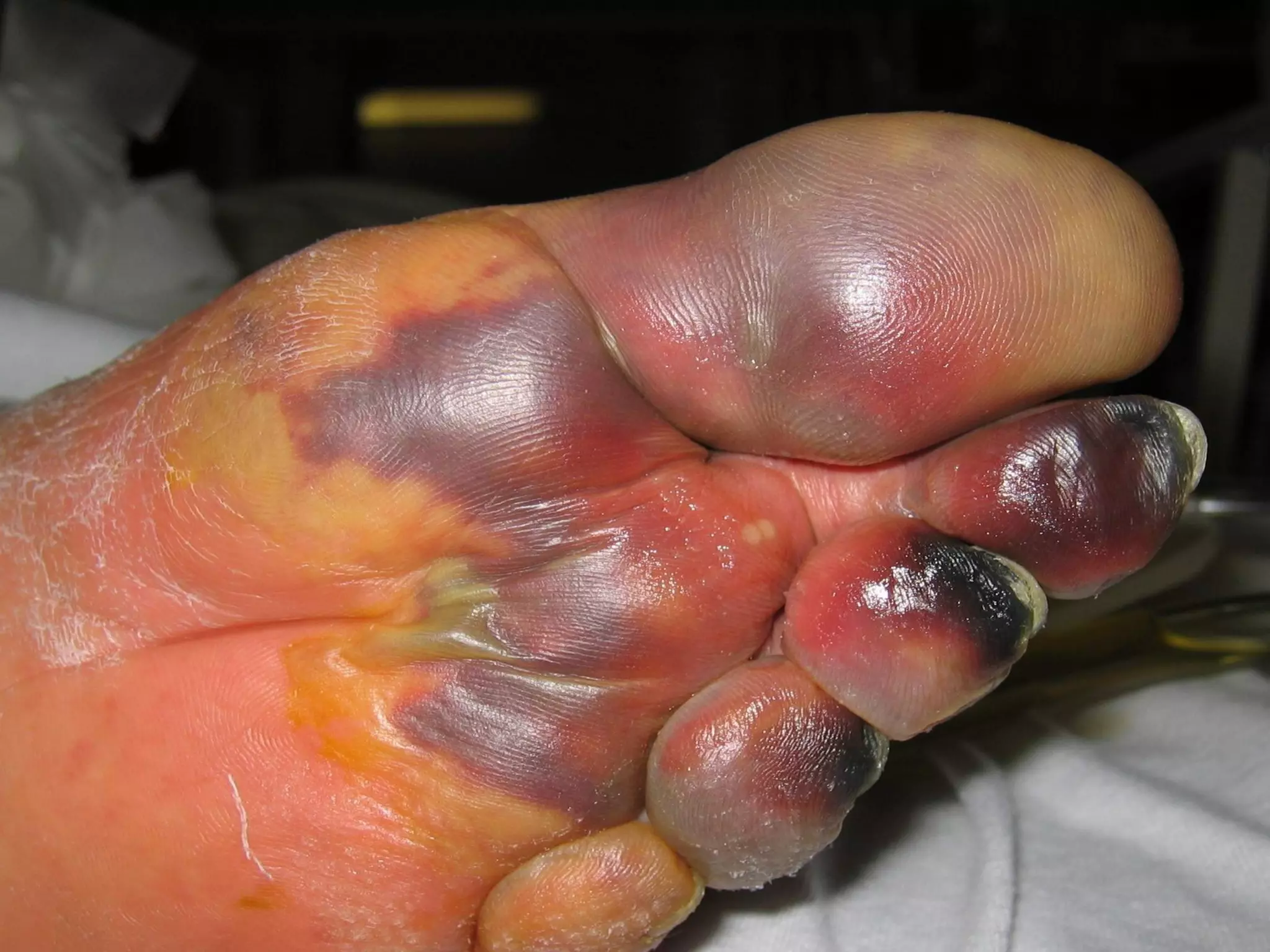
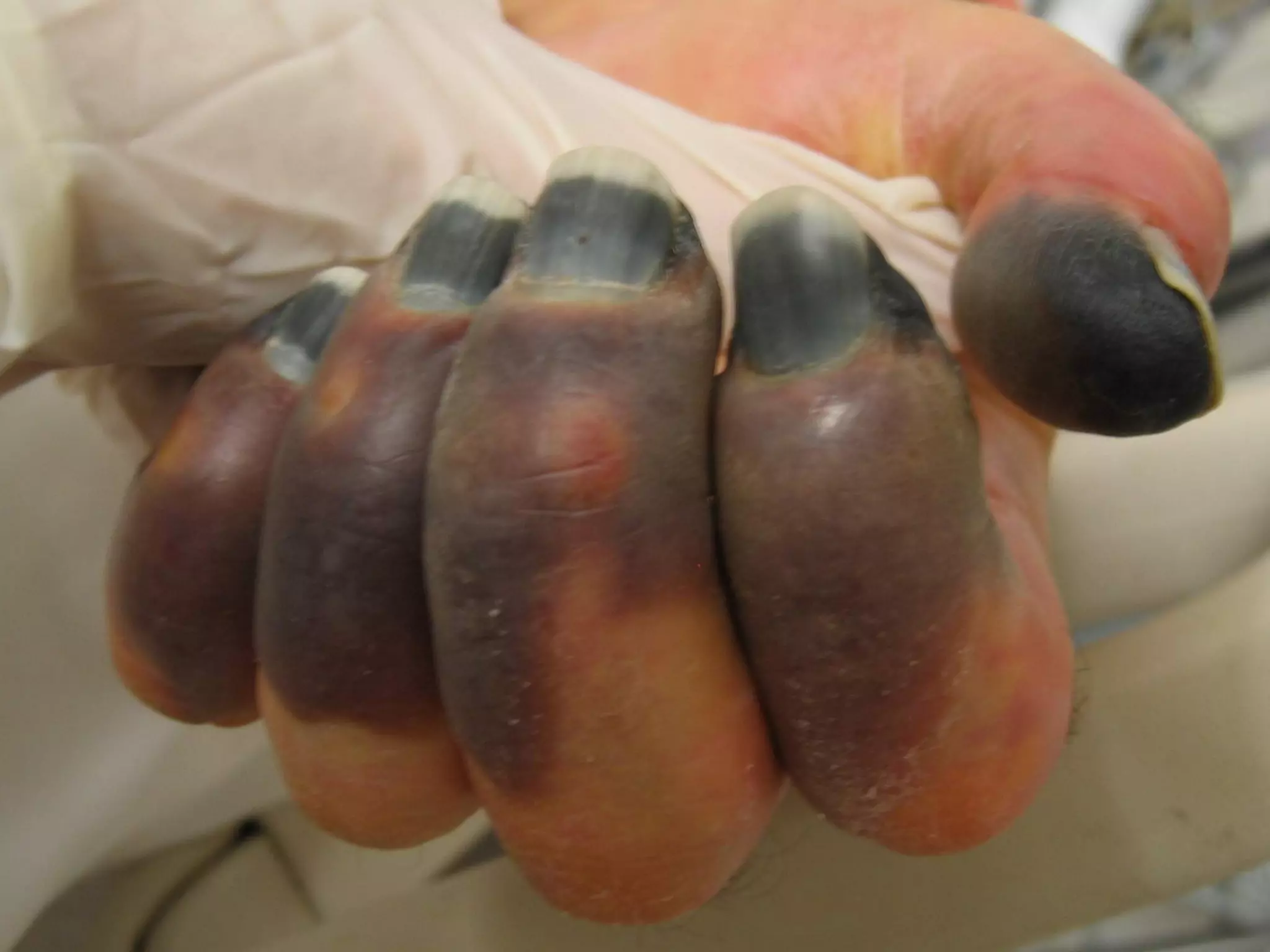
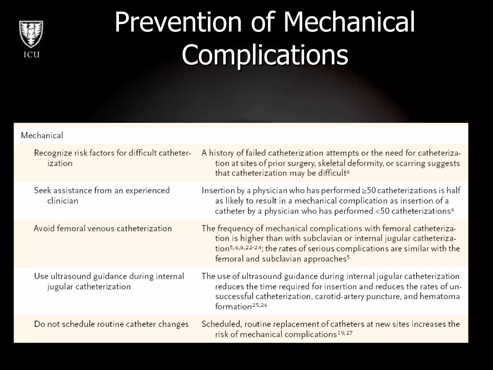
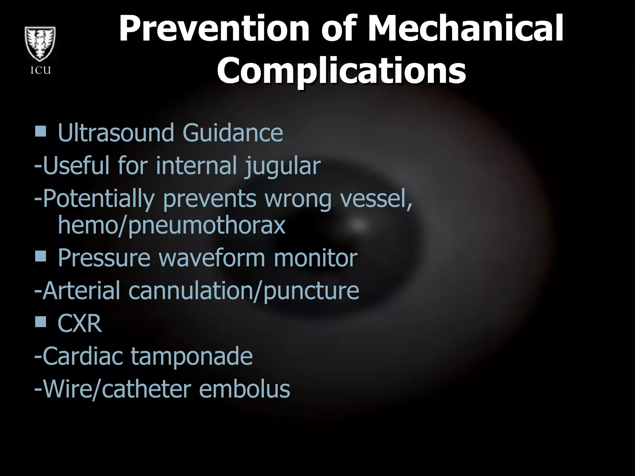
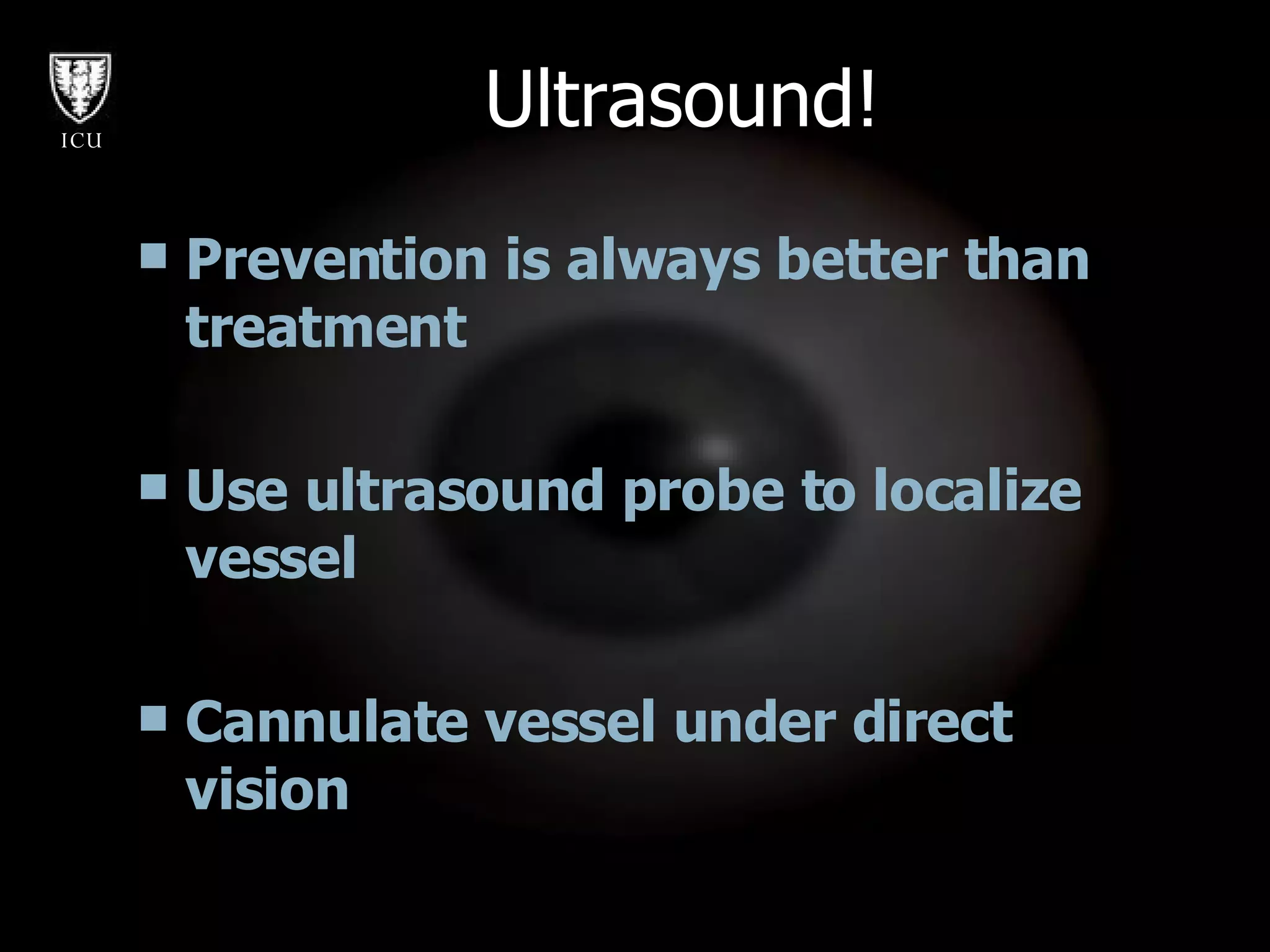
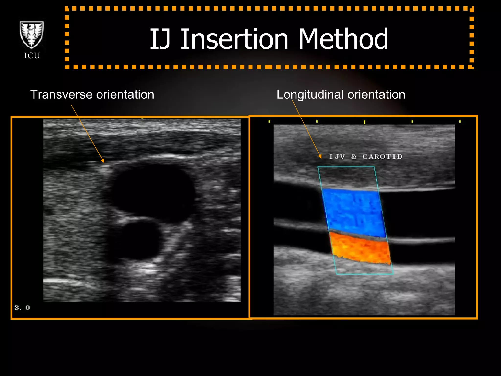
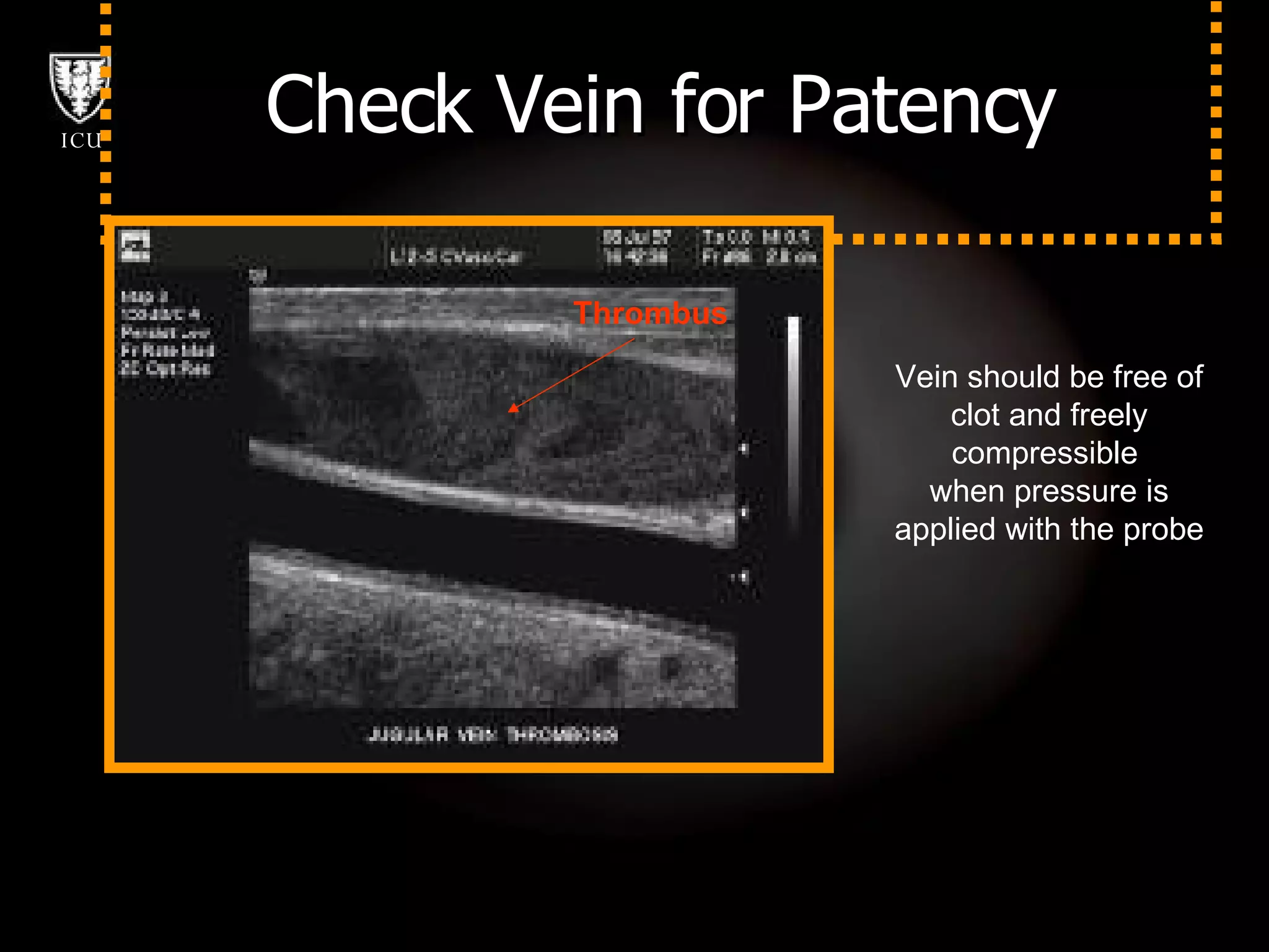
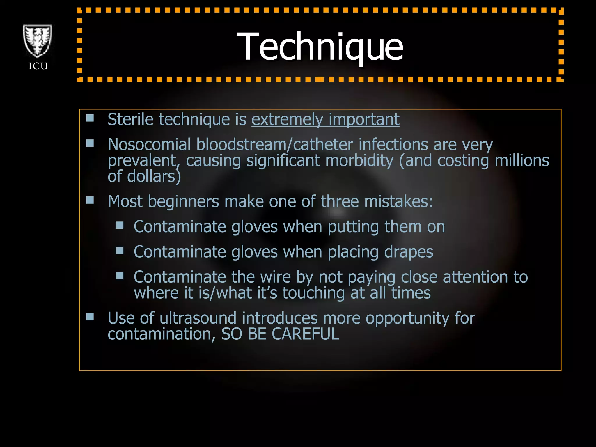
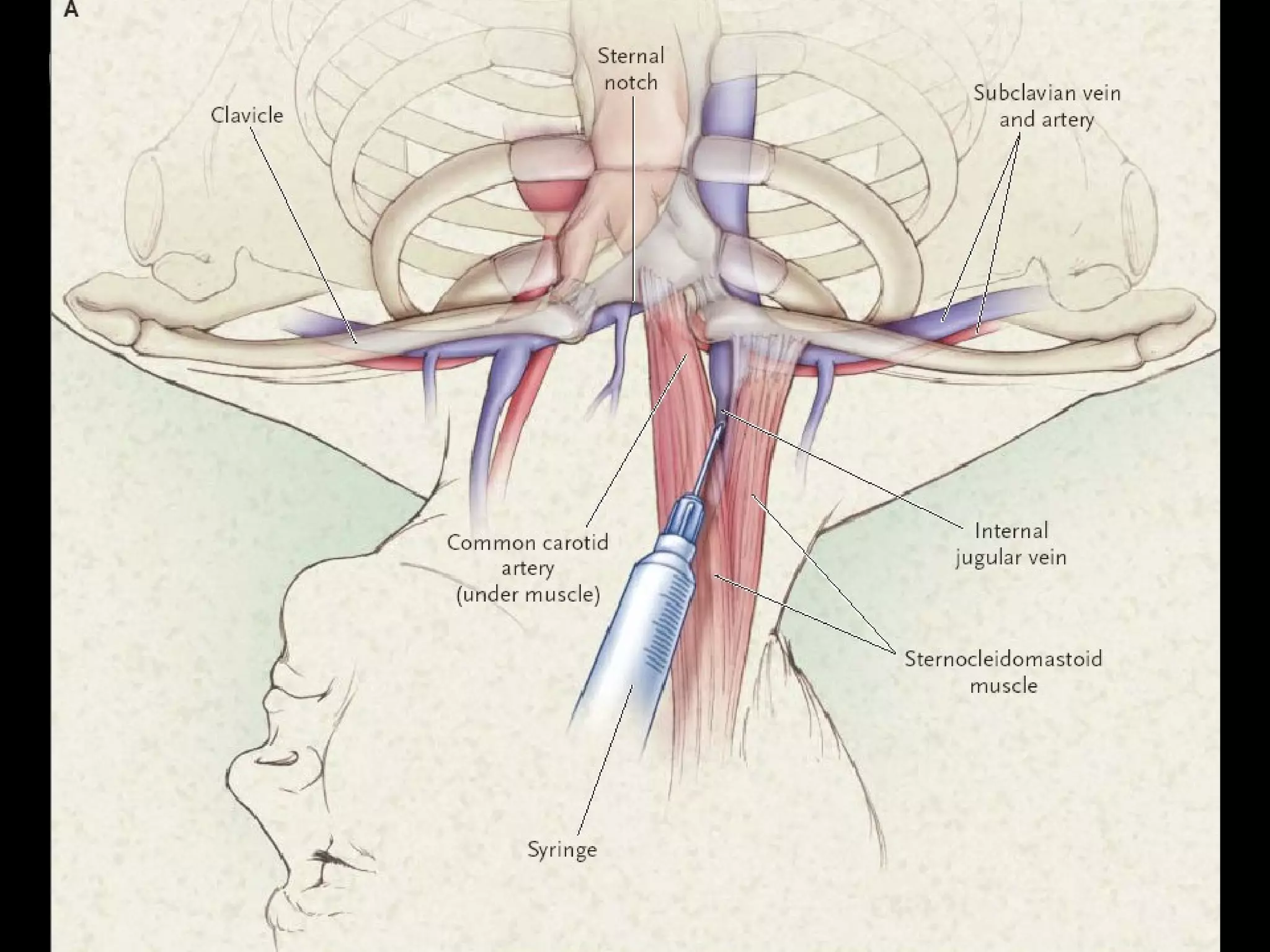
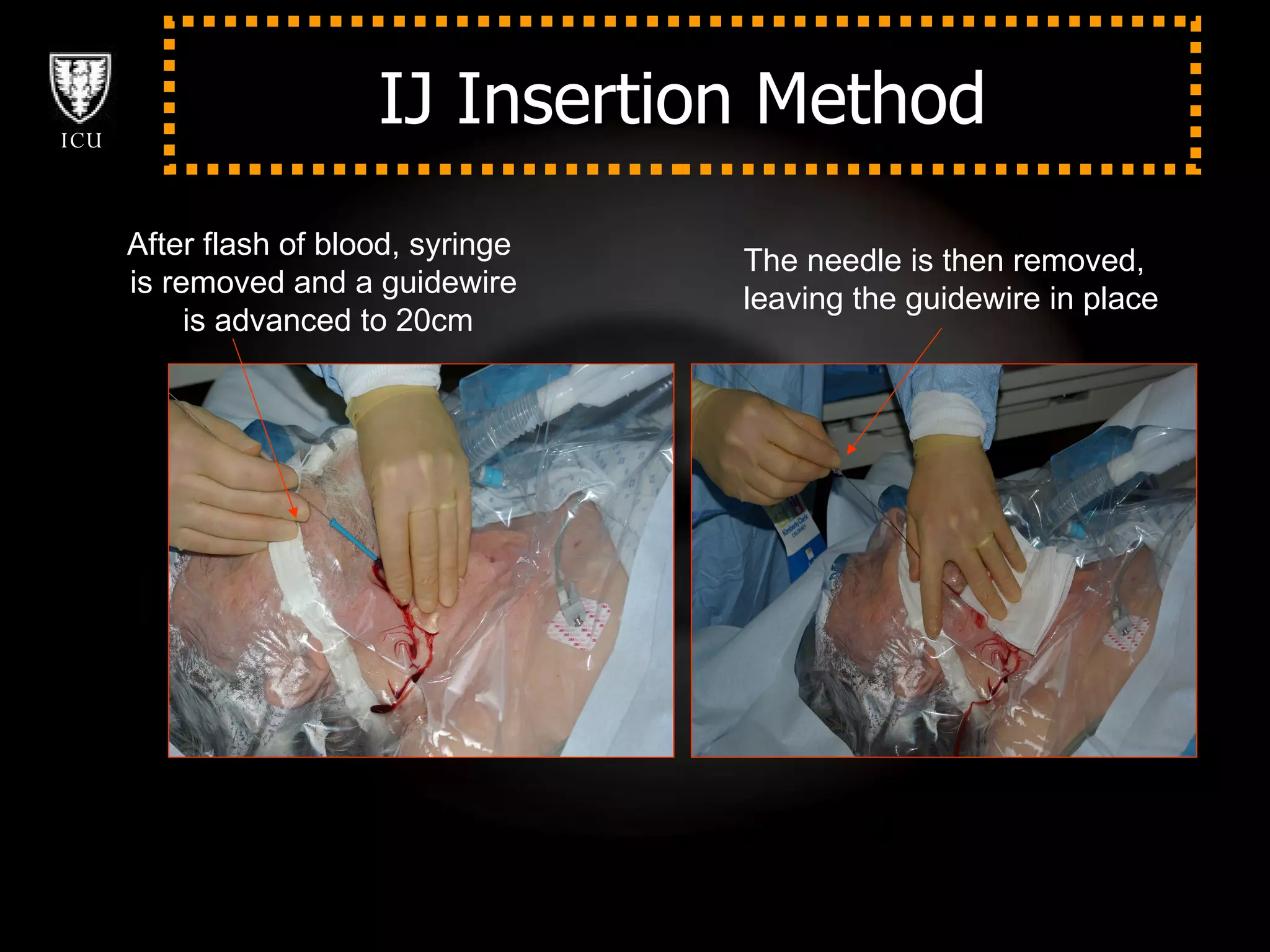
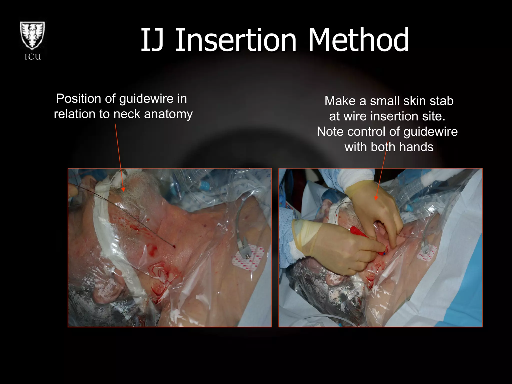
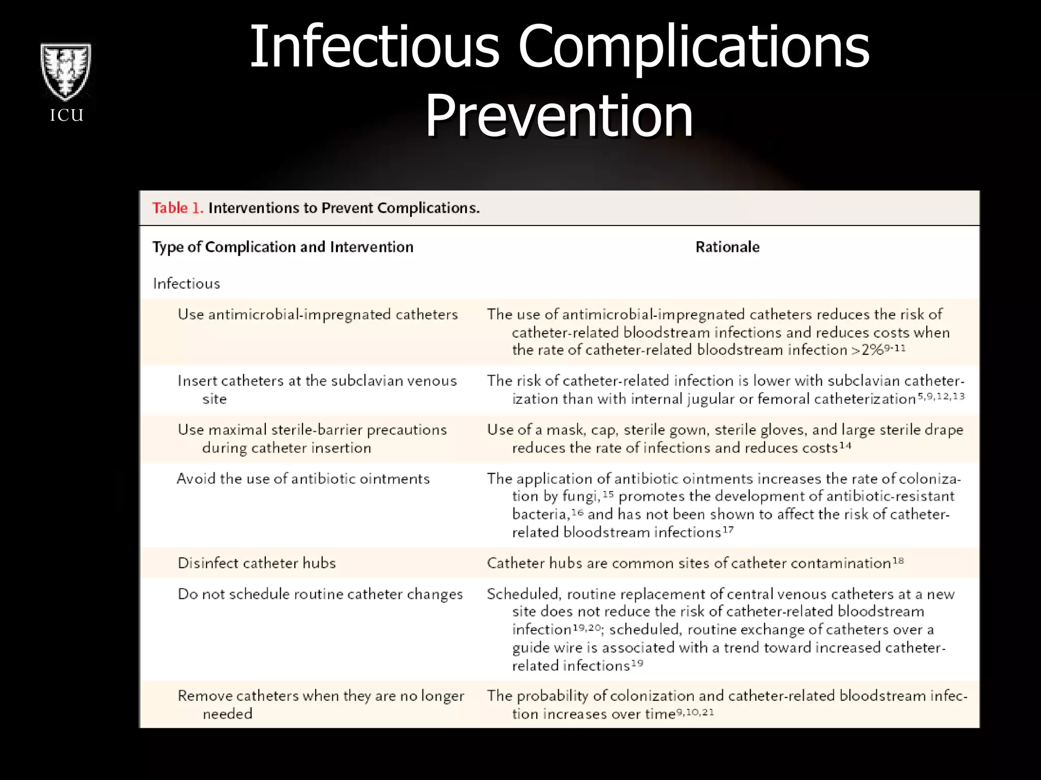
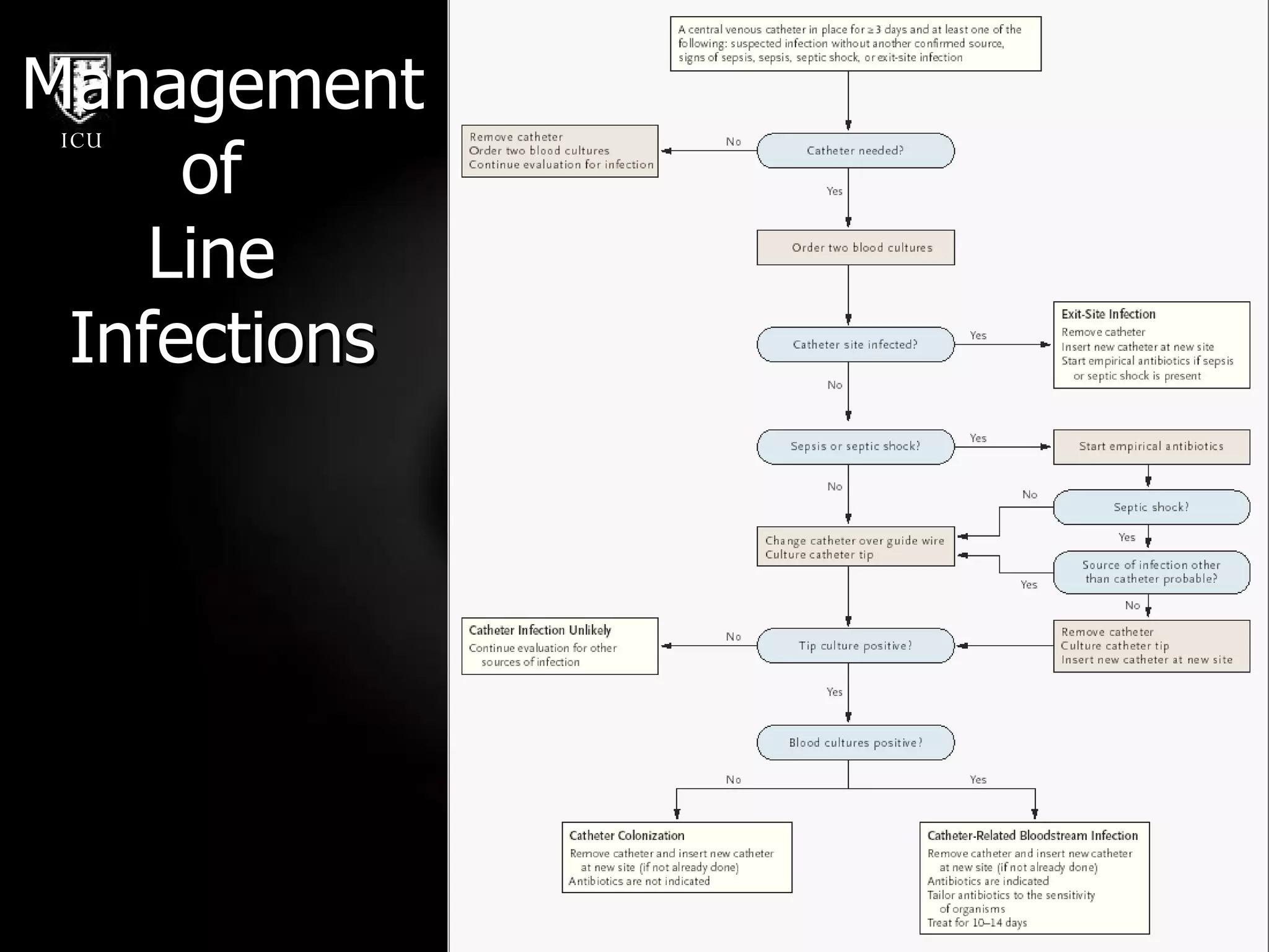
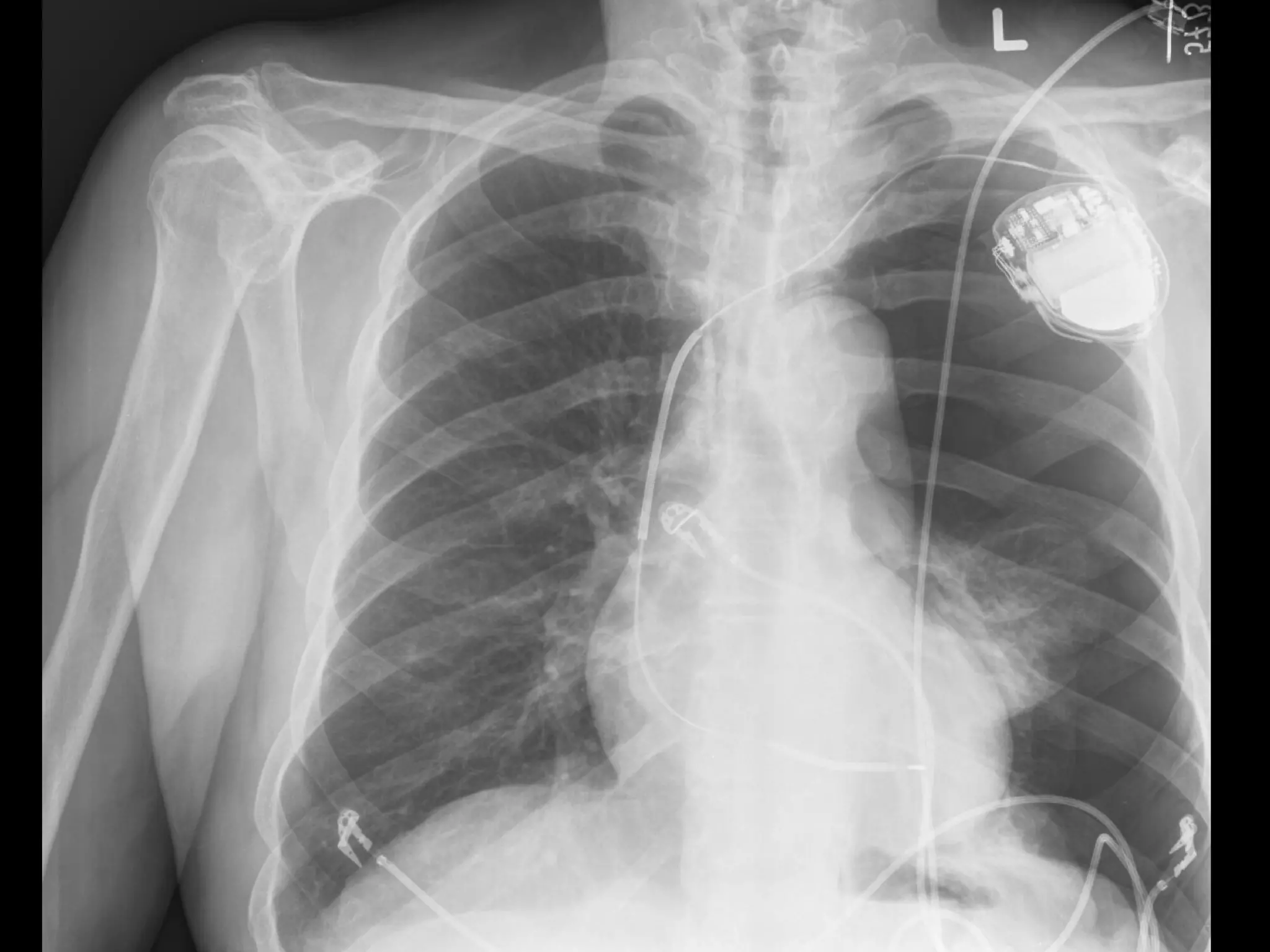
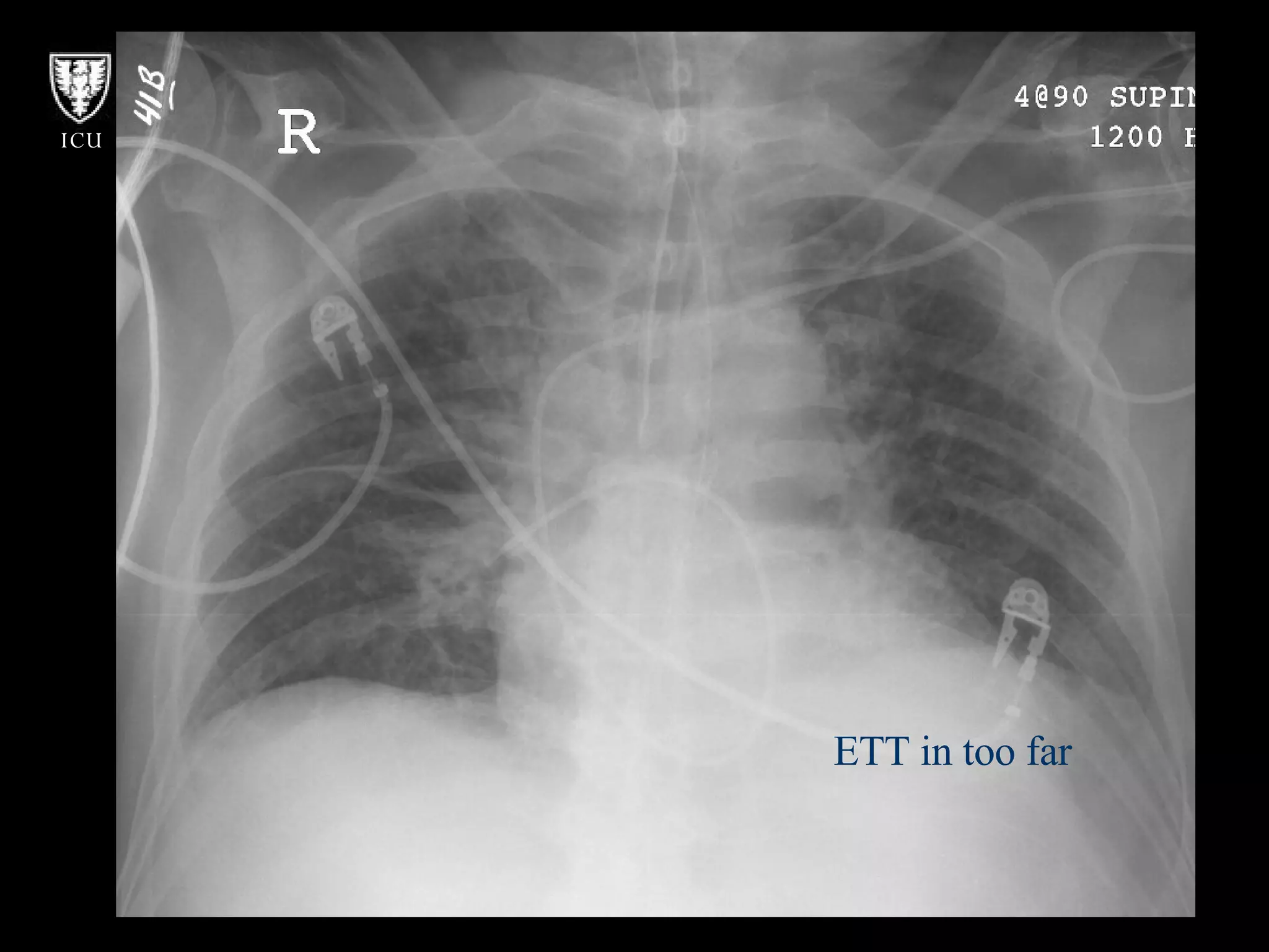
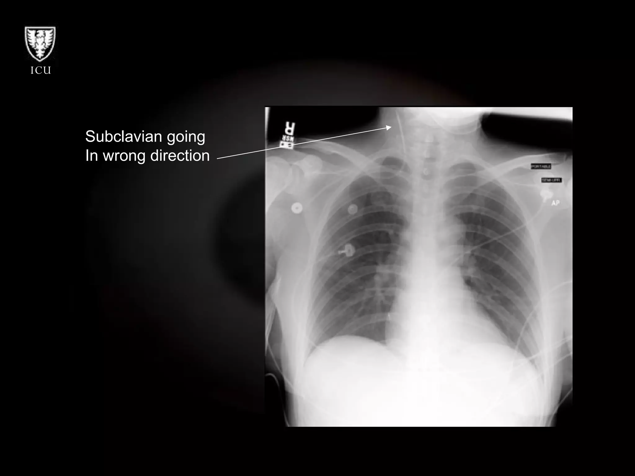
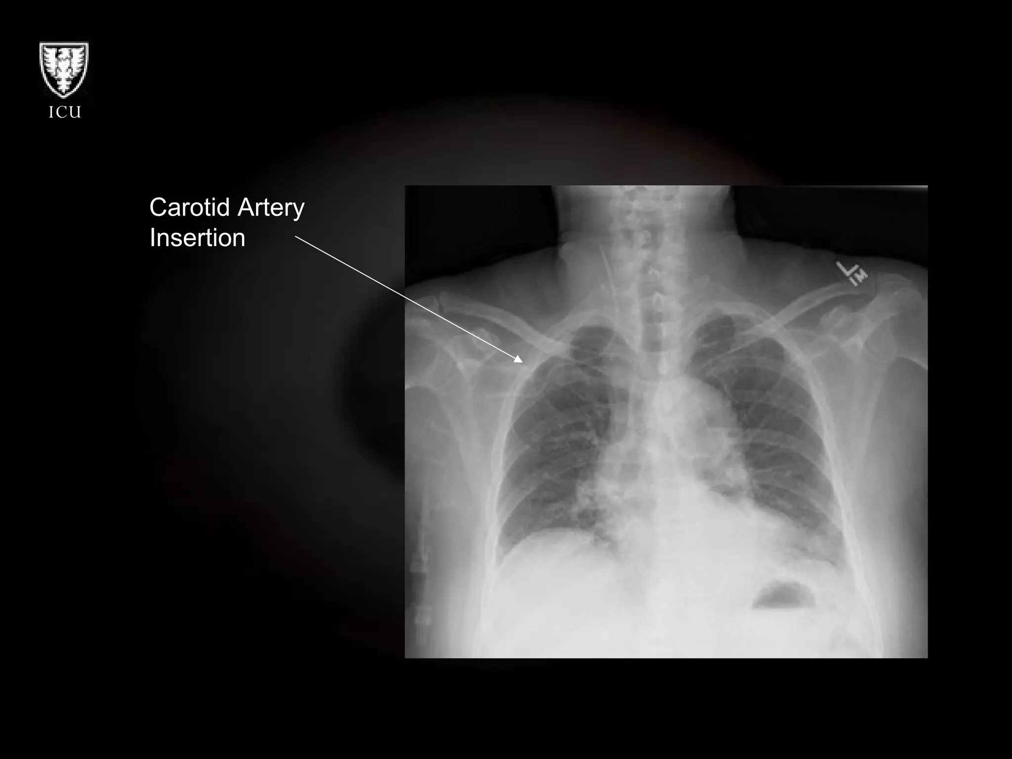
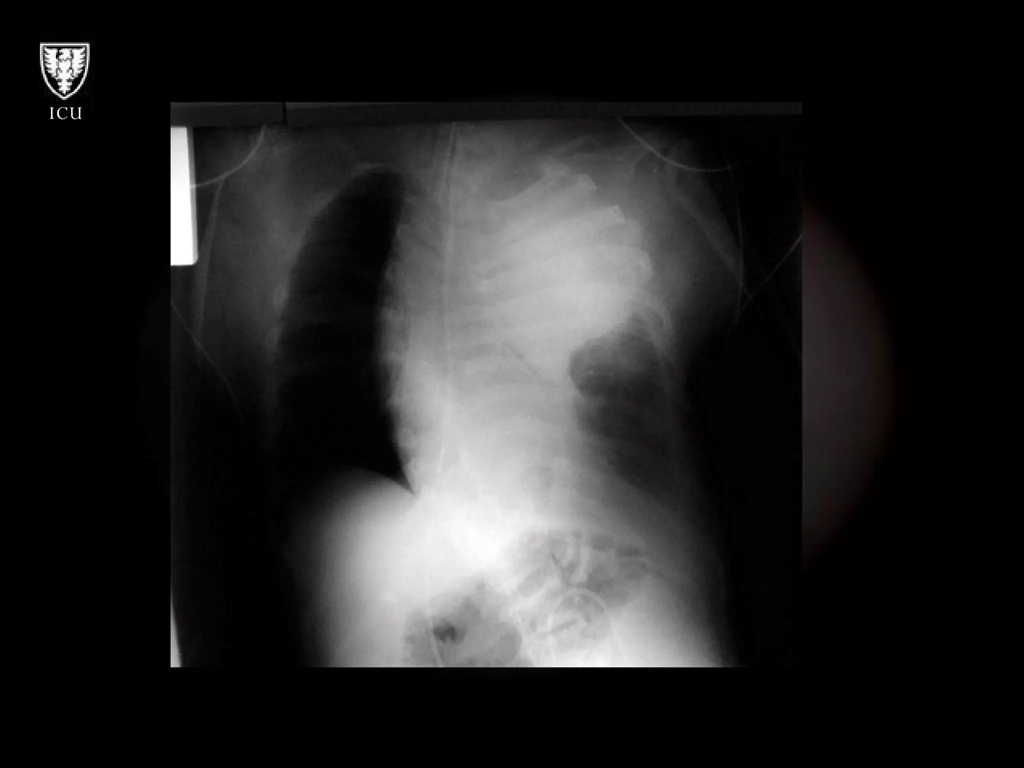
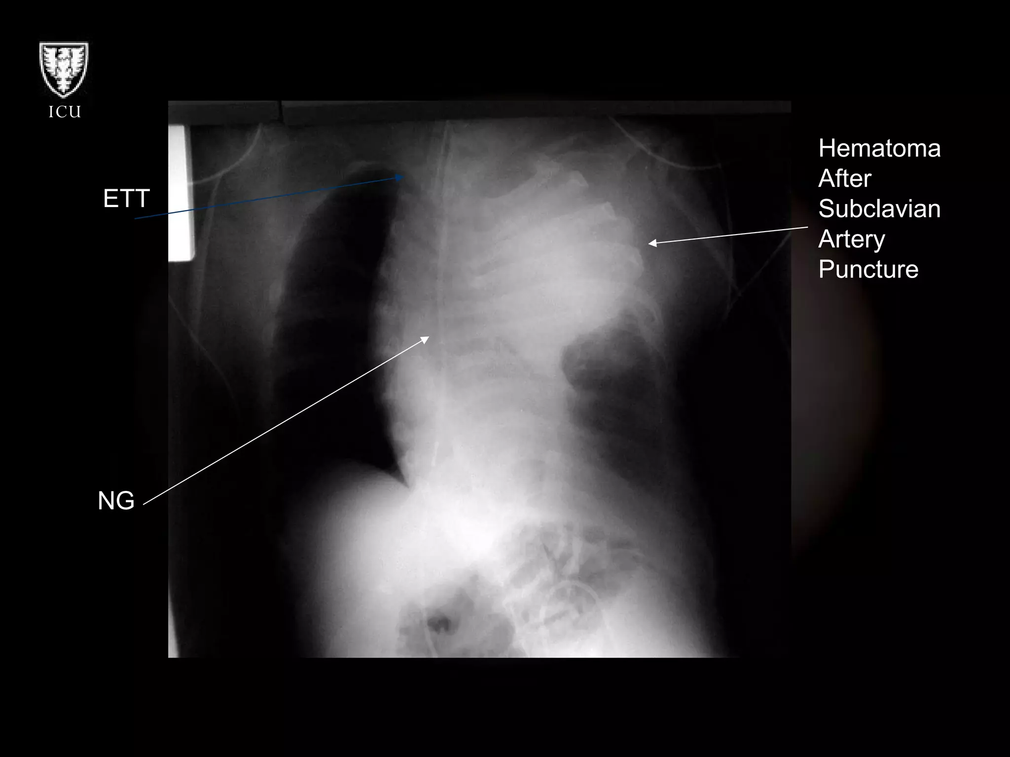
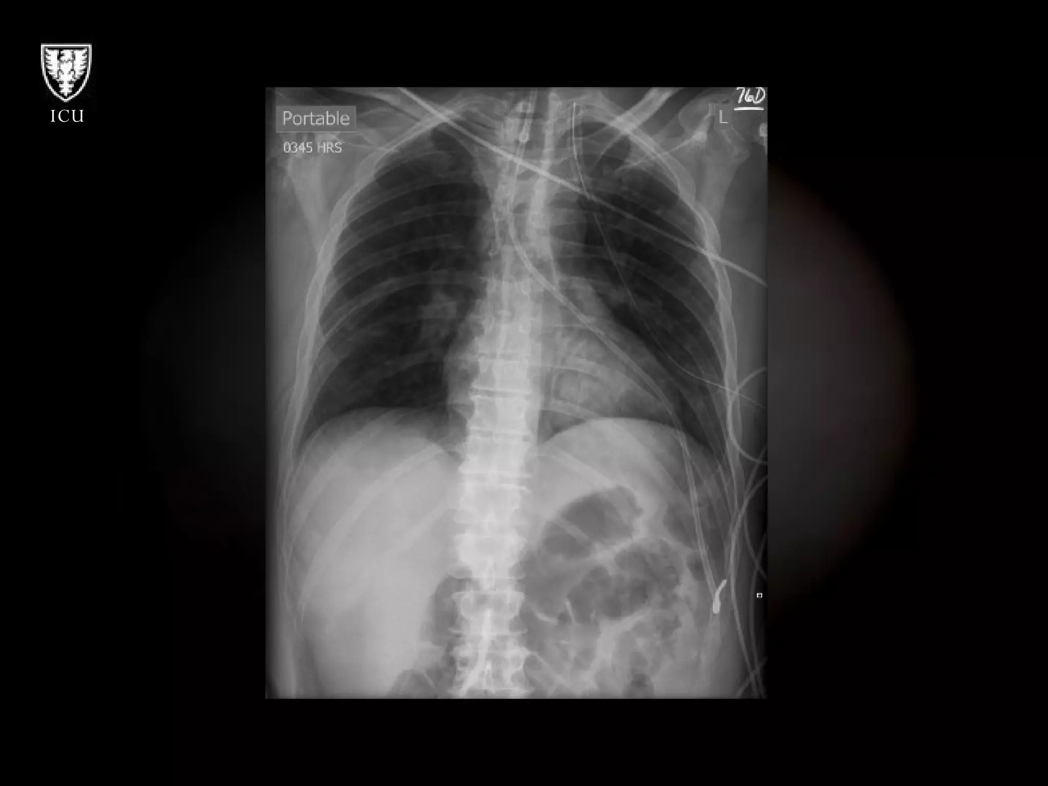
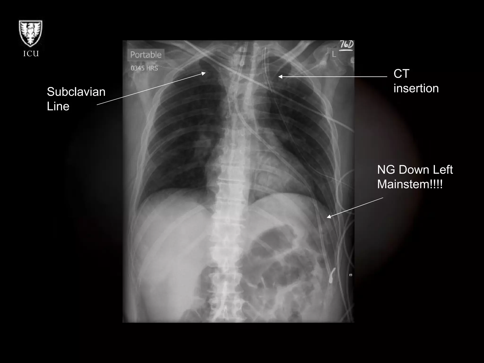
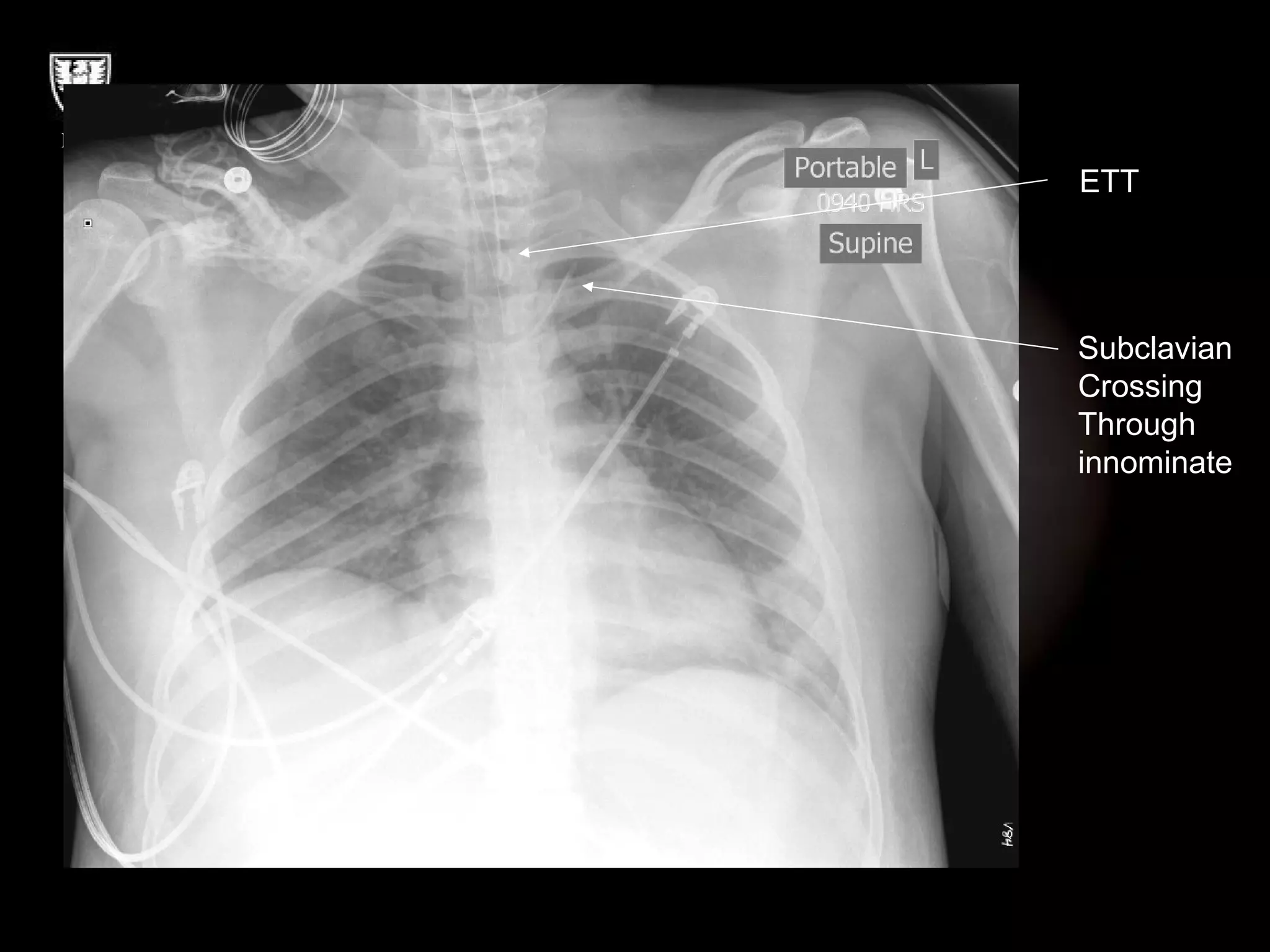
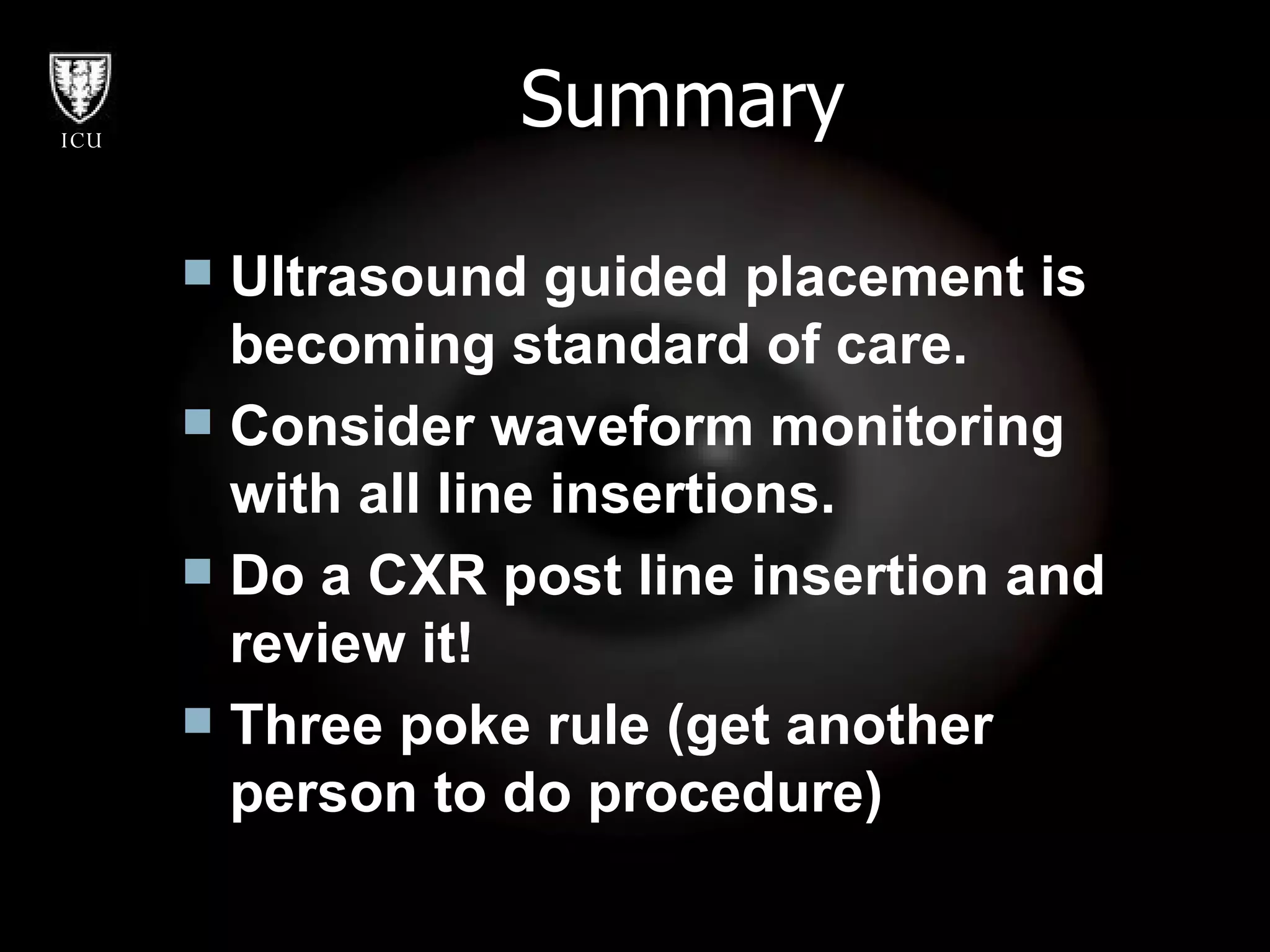
Line Complications Dalhousie University Critical Care Lecture Series
Objectives Know the indications and contraindications for central line insertion Review the technique of central line insertion Know the most common complications of central lines and arterial lines Know how to prevent line complications Know how to recognize and manage line complications Examples
CVP Catheter Indications Pressure monitoring Mixed venous oxygen saturation Fluid administration during volume resuscitation (may not be as good as large-bore peripheral lines!!) Administration of corrosive or hypertonic fluids Administration of vasoactive agents Lack of other sites
CVP Contraindications Severe coagulopathy Obstruction or congenital abnormality  Patient refusal (if competent) Restless and uncooperative patient, unless sedation can be utilized Lack of experience and no expert supervision,  OR  previous failed attempt by experienced physician Injury or infection at the site
Normal CXR
Complications More than 15% of all central lines have a complication Mechanical 5-19% Infectious 5-26% Thrombotic 2-26%
Mechanical Complications
Mechanical Injuries Most common complications: Modified from Domino et al 2004 29 Misc. (PA rupture,vessel injury, air embolism etc) 14 pneumothorax 15 hemothorax 16 Carotid artery Cannulation/puncture 16 Cardiac tamponade 20 Wire/catheter embolus Number (/110) Type of complication
Pneumothorax More common in subclavian Incidence ranges from 0.3 to 3% depending on experience Classic presentation = hypoxemia/hypotension/pleuritic CP Can be treated conservatively in non-ventilated patients
 
Treatment Chest tube to expand lung  Insertion of chest tube may create hole in lung parenchyma leading to another complication:
 
Wrong Vessel If finder needle or 18 g insertion needle in artery may remove and apply pressure If CVC or PAC introducer is in artery  remove catheter and apply pressure OR: Consult CV surgeon/thoracic surgeon ? OR for thoracotomy, removal of catheter and repair of vessel
 
The Left Side is Sinister Left internal jugular has unique complications – thoracic duct injury Left inominate vein can be lacerated    hemothorax and    OR L IJ or SC catheter too proximal can lacerate SVC    death
Left Subclavian Artery
 
 
Prevention of Mechanical Complications
Prevention of Mechanical Complications Ultrasound Guidance  -Useful for internal jugular -Potentially prevents wrong vessel, hemo/pneumothorax Pressure waveform monitor -Arterial cannulation/puncture CXR -Cardiac tamponade -Wire/catheter embolus
Ultrasound! Prevention is always better than treatment Use ultrasound probe to localize vessel Cannulate vessel under direct vision
Ultrasonographic Guidance: Dynamic vs. Static Dynamic Consists of ultrasonic localization, and image-guided cannulation More precise and “real time” Difficult to keep sterility of transducer and site. More hand to screen coordination, unless two persons involved  Static Consists of ultrasonic localization and marking of landmarks only Cannulation is not image-guided, but is separate Time delay between marking and cannulation Easy   to keep sterility of transducer and site Less technically demanding
Pressure Waveforms Several options: Connect sterile tubing to pressure tubing and flush Attach to needle in vessel and confirm venous trace  Remove syringe and confirm non-pulsatile blood Compare arterial blood sample with your sample
IJ Insertion Method Transverse orientation Longitudinal orientation
Check Vein for Patency Thrombus Vein should be free of clot and freely compressible  when pressure is applied with the probe
Transverse Orientation- “Finger Wiggle” Finger on one side of probe  Acoustic shadow of finger on same side of image
Orientation- “Mock Poke”
Orientation- “Mock Poke” Acoustic “shadow” of the  needle over the vein
Technique Steps: Obtain informed consent (unless emergency) Check radiograph for any existing pathology (put line on the same side as pathology) Check coagulation studies, if indicated Position patient appropriately (see individual slides for specifics) Prep/drape using sterile technique Inject local, making certain not to inject intravascularly Using ultrasound, assess for proper orientation, localize the vessel, and ensure patency Cannulate vessel and place catheter (see steps) Check radiograph to ensure correct position and to evaluate for complications (can also use ultrasound to evaluate for pneumothorax and to look for incorrect placement, i.e., subclavian to IJ positioning)
Technique Sterile technique is  extremely important Nosocomial bloodstream/catheter infections are very prevalent, causing significant morbidity (and costing millions of dollars) Most beginners make one of three mistakes: Contaminate gloves when putting them on Contaminate gloves when placing drapes  Contaminate the wire by not paying close attention to where it is/what it’s touching at all times Use of ultrasound introduces more opportunity for contamination, SO BE CAREFUL
 
IJ Insertion Method After flash of blood, syringe  is removed and a guidewire is advanced to 20cm The needle is then removed,  leaving the guidewire in place
IJ Insertion Method Position of guidewire in  relation to neck anatomy Make a small skin stab at wire insertion site.  Note control of guidewire  with both hands
IJ Insertion Method Dilate.  If awake, tell the patient “you’re going to feel some pressure.” Advance the catheter over the wire.  NEVER let go of the wire .  Grab it when it comes out the brown port
Most use infraclavicular approach (insert at “fossa” of deltopectoral groove, about 1-2 cm inferior to clavicle) Poor choice in coagulopathy (difficult to compress)  Higher PTX risk than internal jugular (1-5%) Less infection risk than IJ Trendelenburg’s position with towel roll under scapulae Direct needle toward sternal notch Keep needle parallel to floor; DO NOT AIM UNDER CLAVICLE OR YOU WILL CAUSE PNEUMOTHORAX Constant suction in and out Ultrasound not as useful Again, think after inserting needle 5 cm deep Insertion Method-Subclavian
 
Insertion Site-Femoral Vein is medial to femoral artery In anatomic position  (legs apart), axis of  vein is as pictured:  toward umbilicus Note the inguinal  ligament!
Reverse Trendelenburg position (legs DOWN) to reduce chance of air embolus Use for emergency access Try to remove after 72 hours Do not use if PA catheter needs to be placed Insertion Method-Femoral At 45 o  angle to vessel,  just medial to artery
Image and Orientation
Infectious Complications
Infectious Complications Prevention
Management of  Line  Infections
Pulmonary Artery Catheters PAC have unique set of complications: Arrhythmias inc. complete heart block Knotting of the PAC Pulmonary Artery infarction or rupture
Arterial Lines Most common site = radial artery Rare complications 1983 Slogoff and Keats prospective study 1699 radial artery cannulations no ischemia or disability of hand Pseudoaneurysm of the radial artery can occur  Bowdle Anesthesiology Clinics of NA 2002: 20
What’s Wrong With These Pictures?
 
ETT in too far
Chest tube in poor position
Left mainstem intubation
Feeding tube in lung
Feeding  Tube in  RLL
Subclavian going  In wrong direction
Carotid Artery  Insertion
 
Hematoma After Subclavian Artery Puncture NG ETT
 
CT insertion Subclavian  Line NG Down Left Mainstem!!!!
ETT Subclavian Crossing Through innominate
Summary Ultrasound guided placement is becoming standard of care. Consider waveform monitoring with all line insertions. Do a CXR post line insertion and review it! Three poke rule (get another person to do procedure)

Line Insertion Technique & Follies

  • 1.
    Line Complications DalhousieUniversity Critical Care Lecture Series
  • 2.
    Objectives Know theindications and contraindications for central line insertion Review the technique of central line insertion Know the most common complications of central lines and arterial lines Know how to prevent line complications Know how to recognize and manage line complications Examples
  • 3.
    CVP Catheter IndicationsPressure monitoring Mixed venous oxygen saturation Fluid administration during volume resuscitation (may not be as good as large-bore peripheral lines!!) Administration of corrosive or hypertonic fluids Administration of vasoactive agents Lack of other sites
  • 4.
    CVP Contraindications Severecoagulopathy Obstruction or congenital abnormality Patient refusal (if competent) Restless and uncooperative patient, unless sedation can be utilized Lack of experience and no expert supervision, OR previous failed attempt by experienced physician Injury or infection at the site
  • 5.
  • 6.
    Complications More than15% of all central lines have a complication Mechanical 5-19% Infectious 5-26% Thrombotic 2-26%
  • 7.
  • 8.
    Mechanical Injuries Mostcommon complications: Modified from Domino et al 2004 29 Misc. (PA rupture,vessel injury, air embolism etc) 14 pneumothorax 15 hemothorax 16 Carotid artery Cannulation/puncture 16 Cardiac tamponade 20 Wire/catheter embolus Number (/110) Type of complication
  • 9.
    Pneumothorax More commonin subclavian Incidence ranges from 0.3 to 3% depending on experience Classic presentation = hypoxemia/hypotension/pleuritic CP Can be treated conservatively in non-ventilated patients
  • 10.
  • 11.
    Treatment Chest tubeto expand lung Insertion of chest tube may create hole in lung parenchyma leading to another complication:
  • 12.
  • 13.
    Wrong Vessel Iffinder needle or 18 g insertion needle in artery may remove and apply pressure If CVC or PAC introducer is in artery remove catheter and apply pressure OR: Consult CV surgeon/thoracic surgeon ? OR for thoracotomy, removal of catheter and repair of vessel
  • 14.
  • 15.
    The Left Sideis Sinister Left internal jugular has unique complications – thoracic duct injury Left inominate vein can be lacerated  hemothorax and  OR L IJ or SC catheter too proximal can lacerate SVC  death
  • 16.
  • 17.
  • 18.
  • 19.
  • 20.
    Prevention of MechanicalComplications Ultrasound Guidance -Useful for internal jugular -Potentially prevents wrong vessel, hemo/pneumothorax Pressure waveform monitor -Arterial cannulation/puncture CXR -Cardiac tamponade -Wire/catheter embolus
  • 21.
    Ultrasound! Prevention isalways better than treatment Use ultrasound probe to localize vessel Cannulate vessel under direct vision
  • 22.
    Ultrasonographic Guidance: Dynamicvs. Static Dynamic Consists of ultrasonic localization, and image-guided cannulation More precise and “real time” Difficult to keep sterility of transducer and site. More hand to screen coordination, unless two persons involved Static Consists of ultrasonic localization and marking of landmarks only Cannulation is not image-guided, but is separate Time delay between marking and cannulation Easy to keep sterility of transducer and site Less technically demanding
  • 23.
    Pressure Waveforms Severaloptions: Connect sterile tubing to pressure tubing and flush Attach to needle in vessel and confirm venous trace Remove syringe and confirm non-pulsatile blood Compare arterial blood sample with your sample
  • 24.
    IJ Insertion MethodTransverse orientation Longitudinal orientation
  • 25.
    Check Vein forPatency Thrombus Vein should be free of clot and freely compressible when pressure is applied with the probe
  • 26.
    Transverse Orientation- “FingerWiggle” Finger on one side of probe Acoustic shadow of finger on same side of image
  • 27.
  • 28.
    Orientation- “Mock Poke”Acoustic “shadow” of the needle over the vein
  • 29.
    Technique Steps: Obtaininformed consent (unless emergency) Check radiograph for any existing pathology (put line on the same side as pathology) Check coagulation studies, if indicated Position patient appropriately (see individual slides for specifics) Prep/drape using sterile technique Inject local, making certain not to inject intravascularly Using ultrasound, assess for proper orientation, localize the vessel, and ensure patency Cannulate vessel and place catheter (see steps) Check radiograph to ensure correct position and to evaluate for complications (can also use ultrasound to evaluate for pneumothorax and to look for incorrect placement, i.e., subclavian to IJ positioning)
  • 30.
    Technique Sterile techniqueis extremely important Nosocomial bloodstream/catheter infections are very prevalent, causing significant morbidity (and costing millions of dollars) Most beginners make one of three mistakes: Contaminate gloves when putting them on Contaminate gloves when placing drapes Contaminate the wire by not paying close attention to where it is/what it’s touching at all times Use of ultrasound introduces more opportunity for contamination, SO BE CAREFUL
  • 31.
  • 32.
    IJ Insertion MethodAfter flash of blood, syringe is removed and a guidewire is advanced to 20cm The needle is then removed, leaving the guidewire in place
  • 33.
    IJ Insertion MethodPosition of guidewire in relation to neck anatomy Make a small skin stab at wire insertion site. Note control of guidewire with both hands
  • 34.
    IJ Insertion MethodDilate. If awake, tell the patient “you’re going to feel some pressure.” Advance the catheter over the wire. NEVER let go of the wire . Grab it when it comes out the brown port
  • 35.
    Most use infraclavicularapproach (insert at “fossa” of deltopectoral groove, about 1-2 cm inferior to clavicle) Poor choice in coagulopathy (difficult to compress) Higher PTX risk than internal jugular (1-5%) Less infection risk than IJ Trendelenburg’s position with towel roll under scapulae Direct needle toward sternal notch Keep needle parallel to floor; DO NOT AIM UNDER CLAVICLE OR YOU WILL CAUSE PNEUMOTHORAX Constant suction in and out Ultrasound not as useful Again, think after inserting needle 5 cm deep Insertion Method-Subclavian
  • 36.
  • 37.
    Insertion Site-Femoral Veinis medial to femoral artery In anatomic position (legs apart), axis of vein is as pictured: toward umbilicus Note the inguinal ligament!
  • 38.
    Reverse Trendelenburg position(legs DOWN) to reduce chance of air embolus Use for emergency access Try to remove after 72 hours Do not use if PA catheter needs to be placed Insertion Method-Femoral At 45 o angle to vessel, just medial to artery
  • 39.
  • 40.
  • 41.
  • 42.
    Management of Line Infections
  • 43.
    Pulmonary Artery CathetersPAC have unique set of complications: Arrhythmias inc. complete heart block Knotting of the PAC Pulmonary Artery infarction or rupture
  • 44.
    Arterial Lines Mostcommon site = radial artery Rare complications 1983 Slogoff and Keats prospective study 1699 radial artery cannulations no ischemia or disability of hand Pseudoaneurysm of the radial artery can occur Bowdle Anesthesiology Clinics of NA 2002: 20
  • 45.
    What’s Wrong WithThese Pictures?
  • 46.
  • 47.
  • 48.
    Chest tube inpoor position
  • 49.
  • 50.
  • 51.
  • 52.
    Subclavian going In wrong direction
  • 53.
    Carotid Artery Insertion
  • 54.
  • 55.
    Hematoma After SubclavianArtery Puncture NG ETT
  • 56.
  • 57.
    CT insertion Subclavian Line NG Down Left Mainstem!!!!
  • 58.
    ETT Subclavian CrossingThrough innominate
  • 59.
    Summary Ultrasound guidedplacement is becoming standard of care. Consider waveform monitoring with all line insertions. Do a CXR post line insertion and review it! Three poke rule (get another person to do procedure)