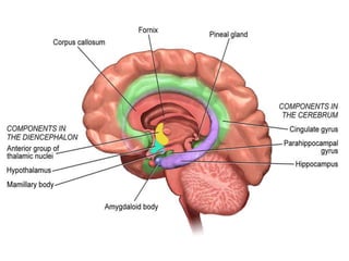
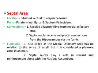
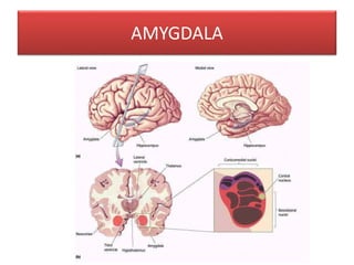
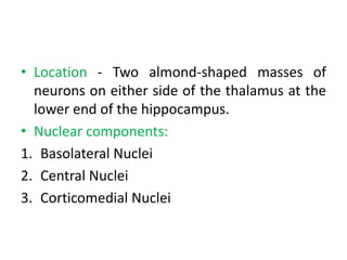
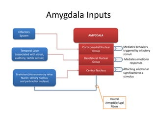
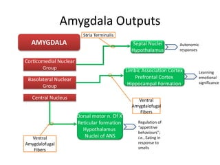
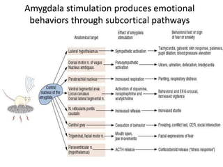
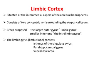
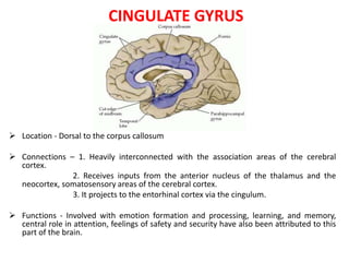
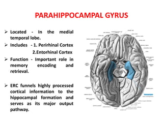
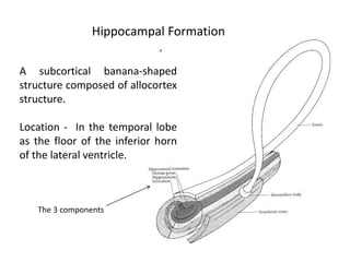
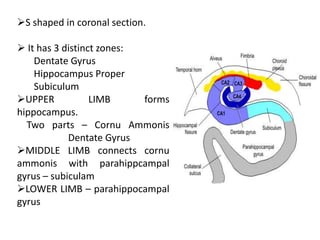
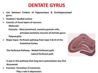
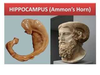
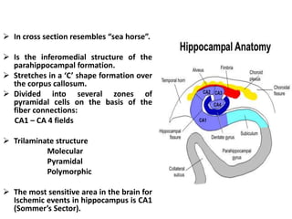
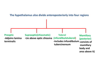
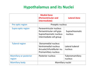
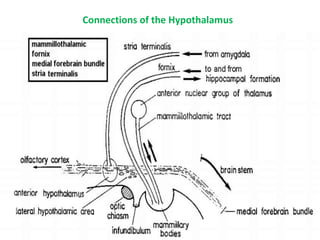
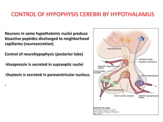
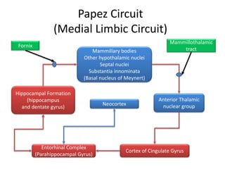
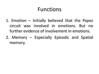
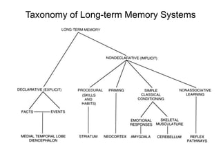
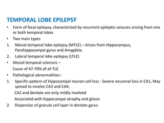
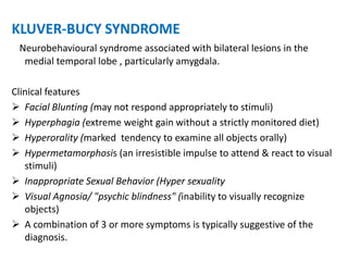
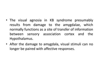
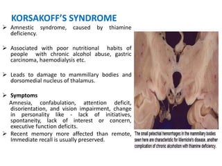
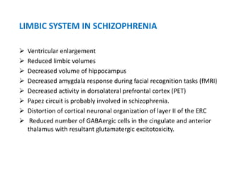
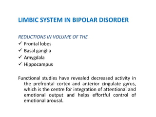
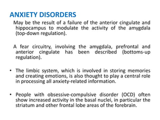
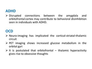
DR. BHARAT BHUSHAN
(DM-NEUROLOGY)
ASSOCIATE PROFESSOR
GOVERNMENT MEDICAL COLLEGE,KOTA
Contents
• Historical Aspects
• Introduction
• Components of Limbic System
• Papez Circuit
• Clinical Implications
Historical Aspects
Thomas Willis 1664 Cerebri Limbus
Paul Broca 1878 le grand lobe limbique
James Papez 1937 Papez Circuit
Paul Kluver & Heinrich Bucy First evidence of cortical
1939 representation
of emotions in limbic
system
Paul Maclean 1952 Limbic System
 Functional anatomic system of interconnected cortical and
sub cortical structures.
 Area of intimate processing between the hypothalamus and
cortical information.
 Plays a role in emotions, learning and autonomic regulation.
INTRODUCTION
THE BROAD FUNCTIONAL DIVISIONS
• Rostral limbic system: Amygdala, septum, orbitofrontal cortex,
anterior insula, and anterior cingulate.
– Important for emotion
• Caudal limbic system: Hippocampus, posterior parahippocampal
cortex and posterior cingulate.
– Important for memory and visual-spatial functions
COMPONENTS OF LIMBIC SYSTEM
Olfactory pathways
Anterior perforated substance
Pyriform lobe
Septal area
Amygdala
Limbic cortex
Hippocampal formation
Elements of diencephalon
Olfactory pathway
Olfactory epithelium
Olfactory nerve filaments
Olfactory bulb
Olfactory tract
Lateral Intermediate Medial
Blend with cortexOlfactory tubercalUncus
Anterior perforated substance
• Mass of gray matter on either side of optic chiasma
• Perforated by central branch of MCA
Pyriform lobe
• Sometimes also called as the Olfactory Cortex.
• Uncus – Curved portion of anterior extremity of hippocampal
gyrus
• Principal region of awareness of olfaction
• Fibers from lateral olfactory stria terminate in it
• Limen insulae – Cortex of most medial part of insula
• Entorhinal cortex- Anterior part of parahippcampal gyrus BA28
• "Area Tempestas“ - Epileptogenic trigger zone, From this site
chemical and electrically evoked seizures can be triggered and is the
site of action for the proconvulsant action of chemoconvulsants.
Septal Area
• Location - Situated ventral to corpus callosum.
• Parts - Paraterminal Gyrus & Septum Pellucidam.
• Connections – 1. Receive olfactory fibre from medial olfactory
stria.
2. Septal nuclei receive reciprocal connections
from the Hippocampus via the fornix.
• Functions – 1. Also called as the Medial Olfactory Area has no
relation to the sense of smell, but it is considered a pleasure
zone in animals.
2. Septal nuclei play a role in reward and
reinforcement along with the Nucleus Accumbens.
AMYGDALA
• Location - Two almond-shaped masses of
neurons on either side of the thalamus at the
lower end of the hippocampus.
• Nuclear components:
1. Basolateral Nuclei
2. Central Nuclei
3. Corticomedial Nuclei
Amygdala Inputs
AMYGDALA
Corticomedial Nuclear
Group
Basolateral Nuclear
Group
Central Nucleus
Olfactory
System
Temporal Lobe
(associated with visual,
auditory, tactile senses)
Brainstem (viscerosensory relay
Nuclei: solitary nucleus
and parbrachial nucleus)
Ventral
Amygdalofugal
Fibers
Mediates behaviors
triggered by olfactory
stimuli
Mediates emotional
responses
Attaching emotional
significance to a
stimulus
Amygdala Outputs
AMYGDALA
Corticomedial Nuclear
Group
Basolateral Nuclear
Group
Central Nucleus
Ventral
Amygdalofugal
Fibers
Septal Nuclei
Hypothalamus
Limbic Association Cortex
Prefrontal Cortex
Hippocampal Formation
Dorsal motor n. Of X
Reticular formation
Hypothalamus
Nuclei of ANS
Ventral
Amygdalofugal
Fibers
Stria Terminalis
Learning
emotional
significance
Autonomic
responses
Regulation of
“appetitive
behaviours”;
i.e., Eating in
response to
smells
Amygdala stimulation produces emotional
behaviors through subcortical pathways
•Emotional Learning - Formation and storage of memories
associated with emotional events.
•Modulation of Memory Consolidation - Following any learning
event, the long-term memory for the event is not formed
instantaneously. Rather, information regarding the event is slowly
assimilated into long-term storage over time, possibly via Long-
term Potentiation.
•Aggression - Animal studies have shown that stimulating the
amygdala appears to increase both sexual and aggressive behavior.
•Alcoholism and binge drinking - Alcoholism is associated with
dampened activation in brain networks responsible for emotional
processing, including the amygdala.
Functions of the Amygdala
• Project into the limbic system one’s current status in
relation to both surroundings and thoughts.
• Make the person behavioral response appropriate for
each occasion.
• Relate environmental stimuli to coordinated behavioral
autonomic and endocrine responses seen in species-
preservation.
• Responses include:
Feeding and Drinking
Fighting behavior
Mating and maternal care
Responses to physical or emotional stresses
Limbic Cortex
 Situated at the inferomedial aspect of the cerebral hemispheres.
 Consists of two concentric gyri surrounding the corpus callosum.
 Broca proposed - the larger outer gyrus-" limbic gyrus"
smaller inner one "the intralimbic gyrus".
 The limbic gyrus (limbic lobe) consists
Isthmus of the cingulate gyrus,
Parahippocampal gyrus
Subcallosal area.
CINGULATE GYRUS
 Location - Dorsal to the corpus callosum
 Connections – 1. Heavily interconnected with the association areas of the cerebral
cortex.
2. Receives inputs from the anterior nucleus of the thalamus and the
neocortex, somatosensory areas of the cerebral cortex.
3. It projects to the entorhinal cortex via the cingulum.
 Functions - Involved with emotion formation and processing, learning, and memory,
central role in attention, feelings of safety and security have also been attributed to this
part of the brain.
PARAHIPPOCAMPAL GYRUS
 Located - In the medial
temporal lobe.
 Includes - 1. Perirhinal Cortex
2.Entorhinal Cortex
 Function - Important role in
memory encoding and
retrieval.
 ERC funnels highly processed
cortical information to the
hippocampal formation and
serves as its major output
pathway.
A subcortical banana-shaped
structure composed of allocortex
structure.
Location - In the temporal lobe
as the floor of the inferior horn
of the lateral ventricle.
The 3 components
Hippocampal Formation
S shaped in coronal section.
 It has 3 distinct zones:
Dentate Gyrus
Hippocampus Proper
Subiculum
UPPER LIMB forms
hippocampus.
Two parts – Cornu Ammonis
Dentate Gyrus
MIDDLE LIMB connects cornu
ammonis with parahippcampal
gyrus – subiculam
LOWER LIMB – parahippocampal
gyrus
DENTATE GYRUS
 Lies between Fimbria of hippocampus & Parahippocampal
gyrus.
 Toothed / beaded surface
 Consists of three layers of neurons:
Molecular
Granular - Most prominent, contains granule cells,
principal excitatory neurons of dentate gyrus.
Polymorphic
 Major Input- Perforant pathway from layer II & III of the
Entorhinal Cortex
The Perforant Pathway - Medial Perforant path
Lateral Perforant path
It was in this pathway that long term potentiation was first
discovered.
 Function- Formation of memories
Play a role in depression.
HIPPOCAMPUS (Ammon’s Horn)
 In cross section resembles “sea horse”.
 Is the inferomedial structure of the
parahippocampal formation.
 Stretches in a ‘C’ shape formation over
the corpus callosum.
 Divided into several zones of
pyramidal cells on the basis of the
fiber connections:
CA1 – CA 4 fields
 Trilaminate structure
Molecular
Pyramidal
Polymorphic
 The most sensitive area in the brain for
Ischemic events in hippocampus is CA1
(Sommer’s Sector).
SUBICULAR COMPLEX
• Subiculum is the transitional zone between the six-layered
entorhinal cortex and the three-layered hippocampus.
• Components: Pre subiculum
Para subiculum
Subiculum
Hippocampal Circuits
FornixFornix
(septal-hippocampal pathway) (Hippocampal commissure)
Afferents
Efferents
HIPPOCAMPUS
(Cortico-entorhinal projections)
ERC/Sub
(PHG)
Cortex
Septal nuclei
Mammillary Body
Contralateral
Hippocampus
1. & 2.
The Hippocampus Dentate Complex
(HC-DG)
Afferent Pathways
Pyramidal cell
(CA1,2)
PHG (ERC, Sub)Dentate gyrus
(granule cells)
mossy fibers
Pyramidal cell
(CA3)
schaffer collaterals
perforant path
alvear path
Septal nuclei
septo-hippocampal
path - thru fornix
Functions of the Hippocampus
1. Declarative Memory – Facts and events.
2. Spatial Memory - The hippocampus contains
place cells that encode spatial memory
(where have I been?)
Recalling of place, and of the routes required
to navigate them requires hippocampal
activation.
Elements of Diencephalon
Habenular nucleus- Lies in habenular triangle at the root of
pineal body.
Mammilary Bodies
Anterior thalamic nucleus
Afferent from – Mamillary body
Efferent to – Cingulate gyrus
Thought to play a role in the modulation of alertness and
are involved in learning and episodic memory.
Hypothalamus
The hypothalamus consists of only 4 cm3 of neural tissue, or
0.3% of the total brain.
Mammilary Bodies
• Located at the ends of the
anterior arches of the fornix.
• Acts as a relay for impulses
coming from
the amygdalae and hippocampi,
via the mamillo-thalamic
tract to the thalamus.
• This circuit, from amygdalae to
mammillary bodies, and then on
to the thalamus, is part of the
larger 'Papez circuit'.
• Fuctions – 1. Episodic Memory
2. Adding the element
of smell to the memories.
HYPOTHALAMUS
Hypothalamus
Lamina terminalis
Mamillary body
Hypothalamic sulcusSeptum pellucidum
Pellucidum
Choroid plexus
Aqueduct
Interventricular foramen
Relations of the Hypothalamus
The hypothalamus also divide anteroposteriorly into four regions
Preoptic
-Adjoins lamina
terminalis
Supraoptic(chiasmatic)
-Lie above optic chiasma
Tuberal
(infundibulotuberal)
-includes infundibulum
tubercinereum
Mamillary
(posterior)
-consists of
mamillary
body and
area above it)
Medial Zone
(Periventricular and
intermediate)
Lateral Zone
Pre optic region Preoptic nucleus
Supra optic region Paraventricular nucleus
Periventricular cell grps
Suprachiasmatic nucleus
Intermediate cell group
Suprachiasmatic
nucleus
Tuberal region Dorsomedial nucleus
Ventromedial nucleus
Arcuate/infundibular nu.
Premamillary nucleus
Lateral tuberal
nucleus
Mamillary or posterior
region
Posterior nucleus Tuberomamillary
nucleus
Mamillary body Mamillary nuclei
Hypothalamus and its Nuclei
Connections of the Hypothalamus
CONTROL OF HYPOPHYSIS CEREBRI BY HYPOTHALAMUS
Neurons in some hypothalamic nuclei produce
bioactive peptides discharged to neighborhood
capillaries (neurosecretion)
Control of neurohypophysis (posterior lobe)
-Vasopressin is secreted in supraoptic nuclei
-Oxytocin is secreted in paraventricular nucleus.
-
Axons of paraventriculo-hypophyseal tract
join axons arising from supra-optic nucleus
to form supraoptico-hypophyseal tract.
The axons of supraoptico-hypophyseal tract pass
down into neurohypophysis where they branch
and end in relation to capillaries and release
their secretion.
Together known as
Hypoyhalamo-hypophyseal tract
Paraventicular nucleus
Hypophysis cerebri
Supraoptic
nucleusParaventriculo-
Hypophyseal tract
Supraoptico-hypophyseal
tract
Control of adenohypophysis by hypothalamus
Hypothalamus control adenohypophysis by producing number of releasing
factors.
Releasing factors travel through tubero-hypophyseal tract which receives
fibers
from various nuclei.
Release the factors into the capillaries
The capillaries carry the factors into the pars anterior of hypophysis cerebri
through hypothalamo-hypophseal portal system.
Functions
• Hormonal Release – Through its control of the anterior
and posterior pituitary.
• Hormonal and Behavioural Circadian Rhythms
• Control of food intake - Extreme lateral part of
the ventromedial nucleus of the hypothalamus is
responsible for the control of food intake. Stimulation
of this area causes increased food intake.
Bilateral lesion of this area causes complete cessation
of food intake.
• Fear processing - The medial zone of hypothalamus is
part of a circuitry that controls motivated behaviors,
like defensive behaviors.
Papez Circuit
(Medial Limbic Circuit)
Mammillary bodies
Other hypothalamic nuclei
Septal nuclei
Substantia innominata
(Basal nucleus of Meynert)
Hippocampal Formation
(hippocampus
and dentate gyrus)
Anterior Thalamic
nuclear group
Cortex of Cingulate Gyrus
Entorhinal Complex
(Parahippocampal Gyrus)
Neocortex
Fornix
Mammillothalamic
tract
Functions
1. Emotion – Initially believed that the Papez
circuit was involved in emotions. But no
further evidence of involvement in emotions.
2. Memory – Especially Episodic and Spatial
memory.
Taxonomy of Long-term Memory Systems
LIMBIC SYSTEM - CLINICAL IMPLICATIONS
TEMPORAL LOBE EPILEPSY
• Form of focal epilepsy, characterized by recurrent epileptic seizures arising from one
or both temporal lobes
• Two main types
1. Mesial temporal lobe epilepsy (MTLE) – Arises from Hippocampus,
Parahippocampal gyrus and Amygdala.
2. Lateral temporal lobe epilepsy (LTLE)
• Mesial temporal sclerosis –
Cause of 47-70% of all TLE
• Pathological abnormalities:-
1. Specific pattern of hippocampal neuron cell loss - Severe neuronal loss in CA1, May
spread to involve CA3 and CA4,
CA2 and dentate are only mildly involved
Associated with hippocampal atrophy and gliosis
2. Dispersion of granule cell layer in dentate gyrus
Signs and Symptoms
• Pre-ictal symptoms –
1. Autonomic sensations – Fullness of stomach
Blushing
Changes in respiration
2. Cognitive sensations – Deja vu, Jamais vu, forced thinking, dreamy states
3. Affective States – Fear, Depression, Elation
4. Automatisms – Lip smacking, rubbing, chewing
• Ictal Symptoms – Recuurent seizures usually simple partial but can be
complex partial also.
 Duration – 1-2 minutes
 Sensory hallucinations – visual auditory, olfactory (m.c.) and gustatory.
 Absence seizures
 Spiritual religious experience
• Interictal Symptoms –
1. Personaltiy changes –
 Hyposexuality
 Emotional intensity
 Perseverative approach to interactions (viscosity)
 References to personal destiny and philosophical themes (left TLE)
 Excessive emotionality (right TLE)
2. Psychotic symptoms – Interictal are more common than intraictal psychosis.
 10% of all complex partial epilepsy patients develop psychotic symptoms.
 Classically they appear in those who have had epilepsy for a long time and
developed personality changes.
 Most characteristic symptoms – Halluciantions and paranoid delusions.
 Patient is usually warm and has appropriate affect in contrast to schizophrenia
3. Violence
4. Affective symptoms – Seen less than psychotic symptoms.
 Episodic
 High incidence of suicide in patients with epilepsy.
LIMBIC ENCEPHALITIS
 Limbic encephalitis is a form of encephalitis.
 An inflammatory process involving the hippocampi, amygdala and less
frequently frontobasal and insular regions of the limbic system and other
parts of the brain.
 Clinical features:-
1. Severe impairment of short-term memory (cardinal sign)
2. Confusion
3. Psychiatric symptoms (changes in behavior & mood –
irritability, depressive , sleep disturbances),
4. Seizures
 60% of the time, limbic encephalitis is paraneoplastic in origin.
 Paraneoplastic limbic encephalitis (PLE) is a particularly severe form of
limbic encephalitis caused by neoplasms most commonly associated with
small cell lung carcinoma. Whereas the majority of encephalities are viral in
nature, PLE is often associated with cancer
ALZHEIMERS’ DISEASE
 Neurodegenerative changes in
limbic system.
 Amyloid proteins build up and form
amyloid plaques (outside cells).
 Neurofibrilllary tangles (inside cells),
leads to neuronal death.
 Hippocampus is one of first areas to
degenerate, leads to anterograde
amnesia.
 Cortex also degenerates early, leads
to retrograde amnesia and
dementia.
KLUVER-BUCY SYNDROME
Neurobehavioural syndrome associated with bilateral lesions in the
medial temporal lobe , particularly amygdala.
Clinical features
 Facial Blunting (may not respond appropriately to stimuli)
 Hyperphagia (extreme weight gain without a strictly monitored diet)
 Hyperorality (marked tendency to examine all objects orally)
 Hypermetamorphosis (an irresistible impulse to attend & react to visual
stimuli)
 Inappropriate Sexual Behavior (Hyper sexuality
 Visual Agnosia/ "psychic blindness" (inability to visually recognize
objects)
 A combination of 3 or more symptoms is typically suggestive of the
diagnosis.
• The visual agnosia in KB syndrome presumably
results from damage to the amygdalae, which
normally functions as a site of transfer of information
between sensory association cortex and the
Hypothalamus.
• After the damage to amygdala, visual stimuli can no
longer be paired with affective responses.
KORSAKOFF’S SYNDROME
 Amnestic syndrome, caused by thiamine
deficiency.
 Associated with poor nutritional habits of
people with chronic alcohol abuse, gastric
carcinoma, haemodialysis etc.
 Leads to damage to mammillary bodies and
dorsomedial nucleus of thalamus.
 Symptoms
Amnesia, confabulation, attention deficit,
disorientation, and vision impairment, change
in personality like - lack of initiatives,
spontaneity, lack of interest or concern,
executive function deficits.
 Recent memory more affected than remote,
Immediate recall is usually preserved.
Semantic Dementia
•Rare degenerative disorder that exhibits
defects in all semantic memory functions,
including naming, single word comprehension
and impoverished general knowledge, with
relative preservation of other components of
speech, perceptual and nonverbal problem-
solving skills, and episodic memory.
•Damage to mammillary bodies, ventral
anterior nucleus, and ventral lateral nucleus.
LIMBIC SYSTEM IN SCHIZOPHRENIA
 Ventricular enlargement
 Reduced limbic volumes
 Decreased volume of hippocampus
 Decreased amygdala response during facial recognition tasks (fMRI)
 Decreased activity in dorsolateral prefrontal cortex (PET)
 Papez circuit is probably involved in schizophrenia.
 Distortion of cortical neuronal organization of layer II of the ERC
 Reduced number of GABAergic cells in the cingulate and anterior
thalamus with resultant glutamatergic excitotoxicity.
LIMBIC SYSTEM IN BIPOLAR DISORDER
REDUCTIONS IN VOLUME OF THE
 Frontal lobes
 Basal ganglia
 Amygdala
 Hippocampus
Functional studies have revealed decreased activity in
the prefrontal cortex and anterior cingulate gyrus,
which is the centre for integration of attentional and
emotional output and helps effortful control of
emotional arousal.
ANXIETY DISORDERS
May be the result of a failure of the anterior cingulate and
hippocampus to modulate the activity of the amygdala
(top-down regulation).
A fear circuitry, involving the amygdala, prefrontal and
anterior cingulate has been described (bottoms-up
regulation).
• The limbic system, which is involved in storing memories
and creating emotions, is also thought to play a central role
in processing all anxiety-related information.
• People with obsessive-compulsive disorder (OCD) often
show increased activity in the basal nuclei, in particular the
striatum and other frontal lobe areas of the forebrain.
ADHD
 Disrupted connections between the amygdala and
orbitofrontalcortex may contribute to behavioral disinhibition
seen in individuals with ADHD.
OCD
 Neuro-imaging has implicated the cortical-striatal-thalamic
circuit
 PET imaging shows increased glucose metabolism in the
orbital gyri
 It is postulated that orbitofrontal – thalamic hyperactivity
gives rise to obsessive thoughts
AUTISM
 Limbic structures involved include the cingulate gyrus and
amygdala, which mediate cognitive and affective processing.
 The basolateral circuit integral for social cognition is disrupted
in autism spectrum disorders.
Psychosurgery
• All forms of psychosurgeries in use today target the limbic system –
1. Anterior cingulotomy – Most commonly used psychosurgery in U.S.
Target Site – Anterior Cingulate cortex
Disconnects the thalamic and posterior frontal regions and damages the
anterior cingulate region.
2. Anterior capsulotomy – Disconnects the orbitofrontal cortex and thalamic
nuclei.
3. Subcaudate tractotomy -
Target Site- Lower medial quadrant of the frontal lobes. Disconnects limbic
system and supra-orbital part of the frontal lobe.
4. Limbic leucotomy - Combination of subcaudate tractotomy and anterior
cingulotomy.
5. Amygdalotomy –
Target Site - Amygdala
It was developed as a treatment for aggression in 1961.
Henry Molaison
Popularly known as Patient H.M.
H.M. suffered from temporal lobe
epilepsy , which could not controlled
with drug alone.
So in 1953 – Surgery to control TLE
Bilateral removal of part of temporal
lobes including hippocampus.
Anterograde amnesia: inability to
form new memories.
Moderate retrograde amnesia which
inhibited his ability to remember any
events one to three years before his
surgery.
Retained his short-term memory and
procedural memory as well.

Limbic system

  • 1.
    DR. BHARAT BHUSHAN (DM-NEUROLOGY) ASSOCIATEPROFESSOR GOVERNMENT MEDICAL COLLEGE,KOTA
  • 2.
    Contents • Historical Aspects •Introduction • Components of Limbic System • Papez Circuit • Clinical Implications
  • 3.
    Historical Aspects Thomas Willis1664 Cerebri Limbus Paul Broca 1878 le grand lobe limbique James Papez 1937 Papez Circuit Paul Kluver & Heinrich Bucy First evidence of cortical 1939 representation of emotions in limbic system Paul Maclean 1952 Limbic System
  • 4.
     Functional anatomicsystem of interconnected cortical and sub cortical structures.  Area of intimate processing between the hypothalamus and cortical information.  Plays a role in emotions, learning and autonomic regulation. INTRODUCTION
  • 6.
    THE BROAD FUNCTIONALDIVISIONS • Rostral limbic system: Amygdala, septum, orbitofrontal cortex, anterior insula, and anterior cingulate. – Important for emotion • Caudal limbic system: Hippocampus, posterior parahippocampal cortex and posterior cingulate. – Important for memory and visual-spatial functions
  • 7.
    COMPONENTS OF LIMBICSYSTEM Olfactory pathways Anterior perforated substance Pyriform lobe Septal area Amygdala Limbic cortex Hippocampal formation Elements of diencephalon
  • 8.
    Olfactory pathway Olfactory epithelium Olfactorynerve filaments Olfactory bulb Olfactory tract Lateral Intermediate Medial Blend with cortexOlfactory tubercalUncus
  • 9.
    Anterior perforated substance •Mass of gray matter on either side of optic chiasma • Perforated by central branch of MCA Pyriform lobe • Sometimes also called as the Olfactory Cortex. • Uncus – Curved portion of anterior extremity of hippocampal gyrus • Principal region of awareness of olfaction • Fibers from lateral olfactory stria terminate in it • Limen insulae – Cortex of most medial part of insula • Entorhinal cortex- Anterior part of parahippcampal gyrus BA28 • "Area Tempestas“ - Epileptogenic trigger zone, From this site chemical and electrically evoked seizures can be triggered and is the site of action for the proconvulsant action of chemoconvulsants.
  • 10.
    Septal Area • Location- Situated ventral to corpus callosum. • Parts - Paraterminal Gyrus & Septum Pellucidam. • Connections – 1. Receive olfactory fibre from medial olfactory stria. 2. Septal nuclei receive reciprocal connections from the Hippocampus via the fornix. • Functions – 1. Also called as the Medial Olfactory Area has no relation to the sense of smell, but it is considered a pleasure zone in animals. 2. Septal nuclei play a role in reward and reinforcement along with the Nucleus Accumbens.
  • 11.
  • 12.
    • Location -Two almond-shaped masses of neurons on either side of the thalamus at the lower end of the hippocampus. • Nuclear components: 1. Basolateral Nuclei 2. Central Nuclei 3. Corticomedial Nuclei
  • 13.
    Amygdala Inputs AMYGDALA Corticomedial Nuclear Group BasolateralNuclear Group Central Nucleus Olfactory System Temporal Lobe (associated with visual, auditory, tactile senses) Brainstem (viscerosensory relay Nuclei: solitary nucleus and parbrachial nucleus) Ventral Amygdalofugal Fibers Mediates behaviors triggered by olfactory stimuli Mediates emotional responses Attaching emotional significance to a stimulus
  • 14.
    Amygdala Outputs AMYGDALA Corticomedial Nuclear Group BasolateralNuclear Group Central Nucleus Ventral Amygdalofugal Fibers Septal Nuclei Hypothalamus Limbic Association Cortex Prefrontal Cortex Hippocampal Formation Dorsal motor n. Of X Reticular formation Hypothalamus Nuclei of ANS Ventral Amygdalofugal Fibers Stria Terminalis Learning emotional significance Autonomic responses Regulation of “appetitive behaviours”; i.e., Eating in response to smells
  • 15.
    Amygdala stimulation producesemotional behaviors through subcortical pathways
  • 16.
    •Emotional Learning -Formation and storage of memories associated with emotional events. •Modulation of Memory Consolidation - Following any learning event, the long-term memory for the event is not formed instantaneously. Rather, information regarding the event is slowly assimilated into long-term storage over time, possibly via Long- term Potentiation. •Aggression - Animal studies have shown that stimulating the amygdala appears to increase both sexual and aggressive behavior. •Alcoholism and binge drinking - Alcoholism is associated with dampened activation in brain networks responsible for emotional processing, including the amygdala. Functions of the Amygdala
  • 17.
    • Project intothe limbic system one’s current status in relation to both surroundings and thoughts. • Make the person behavioral response appropriate for each occasion. • Relate environmental stimuli to coordinated behavioral autonomic and endocrine responses seen in species- preservation. • Responses include: Feeding and Drinking Fighting behavior Mating and maternal care Responses to physical or emotional stresses
  • 18.
    Limbic Cortex  Situatedat the inferomedial aspect of the cerebral hemispheres.  Consists of two concentric gyri surrounding the corpus callosum.  Broca proposed - the larger outer gyrus-" limbic gyrus" smaller inner one "the intralimbic gyrus".  The limbic gyrus (limbic lobe) consists Isthmus of the cingulate gyrus, Parahippocampal gyrus Subcallosal area.
  • 19.
    CINGULATE GYRUS  Location- Dorsal to the corpus callosum  Connections – 1. Heavily interconnected with the association areas of the cerebral cortex. 2. Receives inputs from the anterior nucleus of the thalamus and the neocortex, somatosensory areas of the cerebral cortex. 3. It projects to the entorhinal cortex via the cingulum.  Functions - Involved with emotion formation and processing, learning, and memory, central role in attention, feelings of safety and security have also been attributed to this part of the brain.
  • 20.
    PARAHIPPOCAMPAL GYRUS  Located- In the medial temporal lobe.  Includes - 1. Perirhinal Cortex 2.Entorhinal Cortex  Function - Important role in memory encoding and retrieval.  ERC funnels highly processed cortical information to the hippocampal formation and serves as its major output pathway.
  • 21.
    A subcortical banana-shaped structurecomposed of allocortex structure. Location - In the temporal lobe as the floor of the inferior horn of the lateral ventricle. The 3 components Hippocampal Formation
  • 22.
    S shaped incoronal section.  It has 3 distinct zones: Dentate Gyrus Hippocampus Proper Subiculum UPPER LIMB forms hippocampus. Two parts – Cornu Ammonis Dentate Gyrus MIDDLE LIMB connects cornu ammonis with parahippcampal gyrus – subiculam LOWER LIMB – parahippocampal gyrus
  • 23.
    DENTATE GYRUS  Liesbetween Fimbria of hippocampus & Parahippocampal gyrus.  Toothed / beaded surface  Consists of three layers of neurons: Molecular Granular - Most prominent, contains granule cells, principal excitatory neurons of dentate gyrus. Polymorphic  Major Input- Perforant pathway from layer II & III of the Entorhinal Cortex The Perforant Pathway - Medial Perforant path Lateral Perforant path It was in this pathway that long term potentiation was first discovered.  Function- Formation of memories Play a role in depression.
  • 24.
  • 25.
     In crosssection resembles “sea horse”.  Is the inferomedial structure of the parahippocampal formation.  Stretches in a ‘C’ shape formation over the corpus callosum.  Divided into several zones of pyramidal cells on the basis of the fiber connections: CA1 – CA 4 fields  Trilaminate structure Molecular Pyramidal Polymorphic  The most sensitive area in the brain for Ischemic events in hippocampus is CA1 (Sommer’s Sector).
  • 26.
    SUBICULAR COMPLEX • Subiculumis the transitional zone between the six-layered entorhinal cortex and the three-layered hippocampus. • Components: Pre subiculum Para subiculum Subiculum
  • 27.
    Hippocampal Circuits FornixFornix (septal-hippocampal pathway)(Hippocampal commissure) Afferents Efferents HIPPOCAMPUS (Cortico-entorhinal projections) ERC/Sub (PHG) Cortex Septal nuclei Mammillary Body Contralateral Hippocampus 1. & 2.
  • 28.
    The Hippocampus DentateComplex (HC-DG) Afferent Pathways Pyramidal cell (CA1,2) PHG (ERC, Sub)Dentate gyrus (granule cells) mossy fibers Pyramidal cell (CA3) schaffer collaterals perforant path alvear path Septal nuclei septo-hippocampal path - thru fornix
  • 29.
    Functions of theHippocampus 1. Declarative Memory – Facts and events. 2. Spatial Memory - The hippocampus contains place cells that encode spatial memory (where have I been?) Recalling of place, and of the routes required to navigate them requires hippocampal activation.
  • 30.
    Elements of Diencephalon Habenularnucleus- Lies in habenular triangle at the root of pineal body. Mammilary Bodies Anterior thalamic nucleus Afferent from – Mamillary body Efferent to – Cingulate gyrus Thought to play a role in the modulation of alertness and are involved in learning and episodic memory. Hypothalamus The hypothalamus consists of only 4 cm3 of neural tissue, or 0.3% of the total brain.
  • 31.
    Mammilary Bodies • Locatedat the ends of the anterior arches of the fornix. • Acts as a relay for impulses coming from the amygdalae and hippocampi, via the mamillo-thalamic tract to the thalamus. • This circuit, from amygdalae to mammillary bodies, and then on to the thalamus, is part of the larger 'Papez circuit'. • Fuctions – 1. Episodic Memory 2. Adding the element of smell to the memories.
  • 32.
  • 33.
    Hypothalamus Lamina terminalis Mamillary body HypothalamicsulcusSeptum pellucidum Pellucidum Choroid plexus Aqueduct Interventricular foramen Relations of the Hypothalamus
  • 34.
    The hypothalamus alsodivide anteroposteriorly into four regions Preoptic -Adjoins lamina terminalis Supraoptic(chiasmatic) -Lie above optic chiasma Tuberal (infundibulotuberal) -includes infundibulum tubercinereum Mamillary (posterior) -consists of mamillary body and area above it)
  • 35.
    Medial Zone (Periventricular and intermediate) LateralZone Pre optic region Preoptic nucleus Supra optic region Paraventricular nucleus Periventricular cell grps Suprachiasmatic nucleus Intermediate cell group Suprachiasmatic nucleus Tuberal region Dorsomedial nucleus Ventromedial nucleus Arcuate/infundibular nu. Premamillary nucleus Lateral tuberal nucleus Mamillary or posterior region Posterior nucleus Tuberomamillary nucleus Mamillary body Mamillary nuclei Hypothalamus and its Nuclei
  • 36.
    Connections of theHypothalamus
  • 37.
    CONTROL OF HYPOPHYSISCEREBRI BY HYPOTHALAMUS Neurons in some hypothalamic nuclei produce bioactive peptides discharged to neighborhood capillaries (neurosecretion) Control of neurohypophysis (posterior lobe) -Vasopressin is secreted in supraoptic nuclei -Oxytocin is secreted in paraventricular nucleus. -
  • 38.
    Axons of paraventriculo-hypophysealtract join axons arising from supra-optic nucleus to form supraoptico-hypophyseal tract. The axons of supraoptico-hypophyseal tract pass down into neurohypophysis where they branch and end in relation to capillaries and release their secretion. Together known as Hypoyhalamo-hypophyseal tract Paraventicular nucleus Hypophysis cerebri Supraoptic nucleusParaventriculo- Hypophyseal tract Supraoptico-hypophyseal tract
  • 39.
    Control of adenohypophysisby hypothalamus Hypothalamus control adenohypophysis by producing number of releasing factors. Releasing factors travel through tubero-hypophyseal tract which receives fibers from various nuclei. Release the factors into the capillaries The capillaries carry the factors into the pars anterior of hypophysis cerebri through hypothalamo-hypophseal portal system.
  • 40.
    Functions • Hormonal Release– Through its control of the anterior and posterior pituitary. • Hormonal and Behavioural Circadian Rhythms • Control of food intake - Extreme lateral part of the ventromedial nucleus of the hypothalamus is responsible for the control of food intake. Stimulation of this area causes increased food intake. Bilateral lesion of this area causes complete cessation of food intake. • Fear processing - The medial zone of hypothalamus is part of a circuitry that controls motivated behaviors, like defensive behaviors.
  • 41.
    Papez Circuit (Medial LimbicCircuit) Mammillary bodies Other hypothalamic nuclei Septal nuclei Substantia innominata (Basal nucleus of Meynert) Hippocampal Formation (hippocampus and dentate gyrus) Anterior Thalamic nuclear group Cortex of Cingulate Gyrus Entorhinal Complex (Parahippocampal Gyrus) Neocortex Fornix Mammillothalamic tract
  • 42.
    Functions 1. Emotion –Initially believed that the Papez circuit was involved in emotions. But no further evidence of involvement in emotions. 2. Memory – Especially Episodic and Spatial memory.
  • 43.
    Taxonomy of Long-termMemory Systems
  • 44.
    LIMBIC SYSTEM -CLINICAL IMPLICATIONS
  • 45.
    TEMPORAL LOBE EPILEPSY •Form of focal epilepsy, characterized by recurrent epileptic seizures arising from one or both temporal lobes • Two main types 1. Mesial temporal lobe epilepsy (MTLE) – Arises from Hippocampus, Parahippocampal gyrus and Amygdala. 2. Lateral temporal lobe epilepsy (LTLE) • Mesial temporal sclerosis – Cause of 47-70% of all TLE • Pathological abnormalities:- 1. Specific pattern of hippocampal neuron cell loss - Severe neuronal loss in CA1, May spread to involve CA3 and CA4, CA2 and dentate are only mildly involved Associated with hippocampal atrophy and gliosis 2. Dispersion of granule cell layer in dentate gyrus
  • 46.
    Signs and Symptoms •Pre-ictal symptoms – 1. Autonomic sensations – Fullness of stomach Blushing Changes in respiration 2. Cognitive sensations – Deja vu, Jamais vu, forced thinking, dreamy states 3. Affective States – Fear, Depression, Elation 4. Automatisms – Lip smacking, rubbing, chewing • Ictal Symptoms – Recuurent seizures usually simple partial but can be complex partial also.  Duration – 1-2 minutes  Sensory hallucinations – visual auditory, olfactory (m.c.) and gustatory.  Absence seizures  Spiritual religious experience
  • 47.
    • Interictal Symptoms– 1. Personaltiy changes –  Hyposexuality  Emotional intensity  Perseverative approach to interactions (viscosity)  References to personal destiny and philosophical themes (left TLE)  Excessive emotionality (right TLE) 2. Psychotic symptoms – Interictal are more common than intraictal psychosis.  10% of all complex partial epilepsy patients develop psychotic symptoms.  Classically they appear in those who have had epilepsy for a long time and developed personality changes.  Most characteristic symptoms – Halluciantions and paranoid delusions.  Patient is usually warm and has appropriate affect in contrast to schizophrenia 3. Violence 4. Affective symptoms – Seen less than psychotic symptoms.  Episodic  High incidence of suicide in patients with epilepsy.
  • 48.
    LIMBIC ENCEPHALITIS  Limbicencephalitis is a form of encephalitis.  An inflammatory process involving the hippocampi, amygdala and less frequently frontobasal and insular regions of the limbic system and other parts of the brain.  Clinical features:- 1. Severe impairment of short-term memory (cardinal sign) 2. Confusion 3. Psychiatric symptoms (changes in behavior & mood – irritability, depressive , sleep disturbances), 4. Seizures  60% of the time, limbic encephalitis is paraneoplastic in origin.  Paraneoplastic limbic encephalitis (PLE) is a particularly severe form of limbic encephalitis caused by neoplasms most commonly associated with small cell lung carcinoma. Whereas the majority of encephalities are viral in nature, PLE is often associated with cancer
  • 49.
    ALZHEIMERS’ DISEASE  Neurodegenerativechanges in limbic system.  Amyloid proteins build up and form amyloid plaques (outside cells).  Neurofibrilllary tangles (inside cells), leads to neuronal death.  Hippocampus is one of first areas to degenerate, leads to anterograde amnesia.  Cortex also degenerates early, leads to retrograde amnesia and dementia.
  • 50.
    KLUVER-BUCY SYNDROME Neurobehavioural syndromeassociated with bilateral lesions in the medial temporal lobe , particularly amygdala. Clinical features  Facial Blunting (may not respond appropriately to stimuli)  Hyperphagia (extreme weight gain without a strictly monitored diet)  Hyperorality (marked tendency to examine all objects orally)  Hypermetamorphosis (an irresistible impulse to attend & react to visual stimuli)  Inappropriate Sexual Behavior (Hyper sexuality  Visual Agnosia/ "psychic blindness" (inability to visually recognize objects)  A combination of 3 or more symptoms is typically suggestive of the diagnosis.
  • 51.
    • The visualagnosia in KB syndrome presumably results from damage to the amygdalae, which normally functions as a site of transfer of information between sensory association cortex and the Hypothalamus. • After the damage to amygdala, visual stimuli can no longer be paired with affective responses.
  • 52.
    KORSAKOFF’S SYNDROME  Amnesticsyndrome, caused by thiamine deficiency.  Associated with poor nutritional habits of people with chronic alcohol abuse, gastric carcinoma, haemodialysis etc.  Leads to damage to mammillary bodies and dorsomedial nucleus of thalamus.  Symptoms Amnesia, confabulation, attention deficit, disorientation, and vision impairment, change in personality like - lack of initiatives, spontaneity, lack of interest or concern, executive function deficits.  Recent memory more affected than remote, Immediate recall is usually preserved.
  • 53.
    Semantic Dementia •Rare degenerativedisorder that exhibits defects in all semantic memory functions, including naming, single word comprehension and impoverished general knowledge, with relative preservation of other components of speech, perceptual and nonverbal problem- solving skills, and episodic memory. •Damage to mammillary bodies, ventral anterior nucleus, and ventral lateral nucleus.
  • 54.
    LIMBIC SYSTEM INSCHIZOPHRENIA  Ventricular enlargement  Reduced limbic volumes  Decreased volume of hippocampus  Decreased amygdala response during facial recognition tasks (fMRI)  Decreased activity in dorsolateral prefrontal cortex (PET)  Papez circuit is probably involved in schizophrenia.  Distortion of cortical neuronal organization of layer II of the ERC  Reduced number of GABAergic cells in the cingulate and anterior thalamus with resultant glutamatergic excitotoxicity.
  • 55.
    LIMBIC SYSTEM INBIPOLAR DISORDER REDUCTIONS IN VOLUME OF THE  Frontal lobes  Basal ganglia  Amygdala  Hippocampus Functional studies have revealed decreased activity in the prefrontal cortex and anterior cingulate gyrus, which is the centre for integration of attentional and emotional output and helps effortful control of emotional arousal.
  • 56.
    ANXIETY DISORDERS May bethe result of a failure of the anterior cingulate and hippocampus to modulate the activity of the amygdala (top-down regulation). A fear circuitry, involving the amygdala, prefrontal and anterior cingulate has been described (bottoms-up regulation). • The limbic system, which is involved in storing memories and creating emotions, is also thought to play a central role in processing all anxiety-related information. • People with obsessive-compulsive disorder (OCD) often show increased activity in the basal nuclei, in particular the striatum and other frontal lobe areas of the forebrain.
  • 57.
    ADHD  Disrupted connectionsbetween the amygdala and orbitofrontalcortex may contribute to behavioral disinhibition seen in individuals with ADHD. OCD  Neuro-imaging has implicated the cortical-striatal-thalamic circuit  PET imaging shows increased glucose metabolism in the orbital gyri  It is postulated that orbitofrontal – thalamic hyperactivity gives rise to obsessive thoughts
  • 58.
    AUTISM  Limbic structuresinvolved include the cingulate gyrus and amygdala, which mediate cognitive and affective processing.  The basolateral circuit integral for social cognition is disrupted in autism spectrum disorders.
  • 59.
    Psychosurgery • All formsof psychosurgeries in use today target the limbic system – 1. Anterior cingulotomy – Most commonly used psychosurgery in U.S. Target Site – Anterior Cingulate cortex Disconnects the thalamic and posterior frontal regions and damages the anterior cingulate region. 2. Anterior capsulotomy – Disconnects the orbitofrontal cortex and thalamic nuclei. 3. Subcaudate tractotomy - Target Site- Lower medial quadrant of the frontal lobes. Disconnects limbic system and supra-orbital part of the frontal lobe. 4. Limbic leucotomy - Combination of subcaudate tractotomy and anterior cingulotomy. 5. Amygdalotomy – Target Site - Amygdala It was developed as a treatment for aggression in 1961.
  • 60.
    Henry Molaison Popularly knownas Patient H.M. H.M. suffered from temporal lobe epilepsy , which could not controlled with drug alone. So in 1953 – Surgery to control TLE Bilateral removal of part of temporal lobes including hippocampus. Anterograde amnesia: inability to form new memories. Moderate retrograde amnesia which inhibited his ability to remember any events one to three years before his surgery. Retained his short-term memory and procedural memory as well.