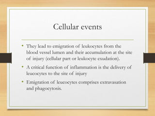
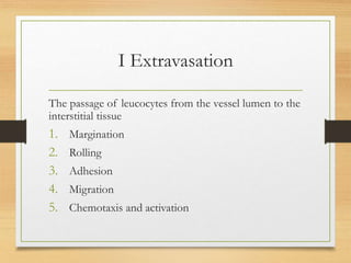
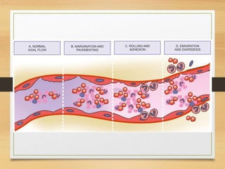
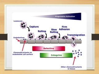
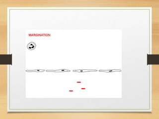
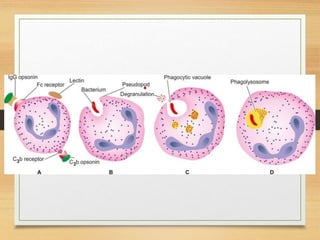
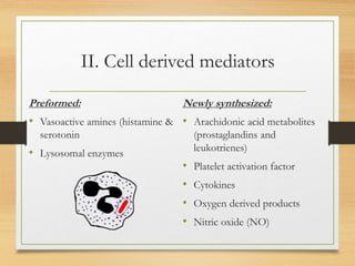
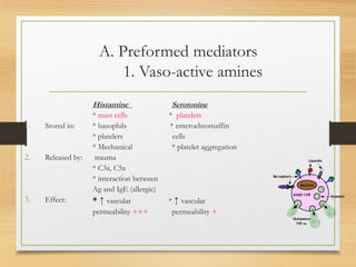
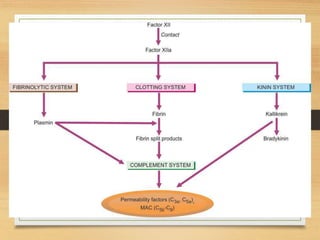
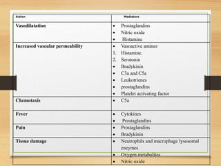
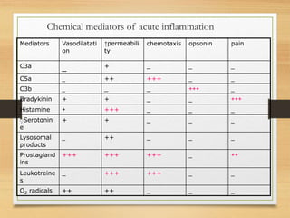
Inflammation
Learning objectives:
• Describe the leukocyte cellular events in acute
inflammation
• Illustrate the steps of leukocyte cellular events
• Define chemotaxis
• Identify chemotactic agents
• Define phagocytosis
• Recognize its steps and causes of its failure
• Discuss the role of cellular changes in the
pathogenesis of acute inflammation
• List the different classes of mediators
• Describe their role in inflammation
Cellular events
• They lead to emigration of leukocytes from the
blood vessel lumen and their accumulation at the site
of injury (cellular part or leukocyte exudation).
• A critical function of inflammation is the delivery of
leucocytes to the site of injury
• Emigration of leucocytes comprises extravasation
and phagocytosis.
I Extravasation
The passage of leucocytes from the vessel lumen to the
interstitial tissue
1. Margination
2. Rolling
3. Adhesion
4. Migration
5. Chemotaxis and activation
• Margination
• Leucocytes fallout of the central axis stream and
assume position in contact with the endothelium
• Rolling
• Transient and loose adhesion of leucocytes to the
endothelium
• Adhesion
• Firm adhesion of leucocytes to endothelium due to
interactions between adhesive molecules on
leucocytes and endothelium: ICAM-1 and PECAM-1
• Transmigration
• Adherent leucocytes emigrate through
interendothelial junctions and traverse the basement
membrane by collagenase enzyme
Chemotaxis
• It is defined as a unidirectional migration of cells
towards an injured tissue along a chemical gradient.
• All granulocytes, monocytes and to a lesser extent
lymphocytes respond to chemotactic stimuli at
varying rates of speed (neutrophils emigrate first and
monocytes follow).
• • Chemotactic agents (attractants) include: exogenous and
endogenous agents e.g.
• a) Soluble bacterial products
• b) Components of the complement system particularly
C5a.
• c) Products of the lipoxygenase pathway of
arachidonic acid metabolism, mainly leukotrienes B4
(LTB4).
• d) Cytokines
• Mechanism of Chemotaxis:
• Chemotactic agents bind to specific receptors on
the leukocyte surface, eventually resulting in
increased intracellular calcium which triggers
movement of leukocytes.
• The leukocytes extend pseudopods that anchor
them to the extracellular matrix and then pull the
remainder of the cell after.
• Leukocyte activation:-
• leukocyte activation occurs through out inflammation
and is initiated by binding of leukocytes to: the
endothelial cells or interleukins or chemotactic agents
to receptors on the surface of leukocytes. This leads
to the “leukocytes activation”
• Leukocyte activation is manifested by:-
• Changes in the leukocyte cytoskeleton leading to the
change in the leukocyte shape with formation of
pseudopodia and start to move towards the stimulus and
phagocytosis
• Degranulation and lysosomal enzymes e.g., collagenase
which allow leukocytes to penetrate the basement
membrane
• Activation of oxidative burst.
• Production of chemical mediators .
II Phagocytosis and degranulation
• Phagocytosis: is a process by which neutrophils and
macrophages engulf the offending pathogen such as
bacteria, dead cells and foreign bodies,-
• Phagocytosis involves (three steps):
• a) Recognition and attachment: The pathogens
are coated with serum proteins (opsonins as C3b,
IgG) that attach to C3b receptor and Fc receptor of
IgG on the surface of leukocytes
• b) Engulfment: Binding of opsonized
particles triggers engulfment by pseudopods of
leukocytes encircling the phagocytosed
particle, creating a phagosome. Fusion of
lysosomal granules with the phagosome with
the formation of phagolysosomes.
• c) Killing and degradation: It is the ultimate step in
phagocytosis.
• A-intracellular mechanisms
• There are two mechanisms in the process of killing:
• (i) Oxygen-dependent mechanisms: This is triggered by
activation of leukocyte-NADPH oxidase, which oxidizes
NADPH and liberates activated bactericidal oxygen
metabolites.
• (ii) Oxygen-independent mechanisms: This occurs by
substances in the leukocytic granules as; lysozyme and
lactoferrin. Killed organisms are then degraded by hydrolases
and other enzymes in lysosomes.
• B. EXTRACELLULAR MECHANISMS
• i) Granules : Degranulation of macrophages and neutrophils
continues to exert its effects of proteolysis outside the cells as
well.
• ii) Immune mechanisms : immune-mediated lysis of microbes
takes place outside the cells by mechanisms of cytolysis,
antibody mediated lysis and by cell-mediated cytotoxicity.
MEDIATORS OF
INFLAMMATION
• These are a large and increasing number of endogenous
chemical substances which mediate the process of acute
inflammation.
• These mediators are released either from the cells or are
derived from plasma proteins:
• ”Cell-derived mediators are released either from
their storage in the cell granules or are synthesized
in the cells.
• The most common site of synthesis of plasma-
derived mediators is the liver. After their release
from the liver, these mediators require activation.
• Once activated and released, most mediators, either
quickly decay, or are inactivated by enzymes and
inhibitors.
II. Cell derived mediators
Preformed:
• Vasoactive amines (histamine &
serotonin
• Lysosomal enzymes
Newly synthesized:
• Arachidonic acid metabolites
(prostaglandins and
leukotrienes)
• Platelet activation factor
• Cytokines
• Oxygen derived products
• Nitric oxide (NO)
A. Preformed mediators
1. Vaso-active amines
1. Stored in:
2. Released by:
3. Effect:
Histamine Serotonine
* mast cells * platelets
* basophils * enterochromaffin
* platelets cells
* Mechanical * platelet aggregation
trauma
* C3a, C5a
* interaction between
Ag and IgE (allergic)
* ↑ vascular * ↑ vascular
permeability +++ permeability +
2. Lysosomal products
• Sources:
PNL, monocytes (macrophages)
• Effects:
 proteolysis (collagenase & elastase)
 initiation of coagulation through activation of
factor XII
 ↑ vascular permeability
 chemotactic
2-Newly synthesized mediators
Arachidonic acid metabolites
• Arachidonic acid is a major constituent of the cell
membrane phospholipids. It is released from these
phospholipids by the action of phospholipase enzyme
which is activated by physical, chemical, or mechanical
agents or other mediators e.g. C5a.
• Arachidonic acid is metabolized through two major
pathways:
• i. Cyclo-oxygenase pathway; leading to the formation of prostaglandins.
• ii. Lipoxygenase pathway; with formation of leukotrienes.
• Prostaglandins
Vasodilatation+++
↑Vascular permeability
+++
Chemotaxis +++
Fever and pain
• Leukotreines
Vasoconstriction and
bronchospasm
↑Vascular permeability
+++
Chemotaxis +++
Platelet activating factor (PAF)
• increased vascular permeability
• vasodilatation in low concentration
• Bronchoconstriction
• adhesion of leucocytes to endothelium
• chemotaxis.
Cytokines
Source: Polypeptide products of many cells mainly
activated lymphocytes and macrophages.
Include:
Interleukins (IL1, IL2, IL3,…_
Interferon (α, β, γ)
Tumor necrosis factor (TNF)
Chemokines (chemotactic polypeptides)
• They can exert:
• Chemotaxins for polymorphs and macrophages.
• Systemic acute phase reaction e.g., fever, neutrophilia
and increased acute phase proteins.
Oxygen-derived products
A. Oxygen-derived free radicals
B. Nitric oxide (NO)
A. Oxygen-derived free radicals: O2
- , OH, H2O2
• ٍSources: neutrophils and macrophages
• Effects (cell injury)
Endothelial injury ↑vascular permeability
Injury to RBCs, parenchymal cells and tumor cells
Bactericidal effects
B. Nitric oxide (NO)
Potent vasodilator
Acts as microbicidal agent in activated macrophages.
I. Plasma derived chemical mediators
These include various products derived from activation
and interaction of 4 interlinked systems:
• kinin,
• clotting,
• fibrinolytic
• and complement.
The kinin system
• Kinin system is directly triggered by activation of
Hageman factor (XII). Activated Hageman factor (Factor
XII a) converts plasma prekallikrein into kallikrein. The
latter cleaves a plasma protein of high molecular weight,
kininogen, into bradykinin that induces:
• i) smooth muscle contraction;
• ii) vasodilatation;
• iii) increased vascular permeability; and
• iv) pain.
• The complement system:
• The complement system is a series of plasma
proteins, present as inactive forms in the plasma.
They are numbered from Cl to C9. The most critical
step in inflammation is activation of the third
component (C3) to form C3a and C3b. with
subsequent formation of C5a and assembly of C5-
C9 membrane attack complex.
C3a ↑vascular permeability
C5a ↑vascular permeability
and powerful chemotactic
agent
C3b opsonin
C5-9 membrane attack
(cytolysis)
Coagulation/ fibrinolytic system
•Stimulus:
Inflammation endothelial injury exposure
of blood to subendothelial collagen activation of
factor XII
Action Mediators
Vasodilatation  Prostaglandins
 Nitric oxide
 Histamine
Increased vascular permeability  Vasoactive amines
1. Histamine.
2. Serotonin
 Bradykinin
 C3a and C5a
 Leukotrienes
 prostaglandins
 Platelet activating factor
Chemotaxis  C5a
Fever  Cytokines
 Prostaglandins
Pain  Prostaglandins
 Bradykinin
Tissue damage  Neutrophils and macrophage lysosomal
enzymes
 Oxygen metabolites
 Nitric oxide
Chemical mediators of acute inflammation
painopsoninchemotaxis↑permeabili
ty
Vasodilatati
on
Mediators
___+_C3a
__+++++_C5a
_+++___C3b
+++__++Bradykinin
___++++Histamine
___++ٍSerotonin
e
___++_Lysosomal
products
++_+++++++++Prostagland
ins
__++++++_Leukotreine
s
___++++O2 radicals

Inflammation 2new

  • 1.
  • 2.
    Learning objectives: • Describethe leukocyte cellular events in acute inflammation • Illustrate the steps of leukocyte cellular events • Define chemotaxis • Identify chemotactic agents • Define phagocytosis • Recognize its steps and causes of its failure • Discuss the role of cellular changes in the pathogenesis of acute inflammation • List the different classes of mediators • Describe their role in inflammation
  • 3.
    Cellular events • Theylead to emigration of leukocytes from the blood vessel lumen and their accumulation at the site of injury (cellular part or leukocyte exudation). • A critical function of inflammation is the delivery of leucocytes to the site of injury • Emigration of leucocytes comprises extravasation and phagocytosis.
  • 4.
    I Extravasation The passageof leucocytes from the vessel lumen to the interstitial tissue 1. Margination 2. Rolling 3. Adhesion 4. Migration 5. Chemotaxis and activation
  • 8.
    • Margination • Leucocytesfallout of the central axis stream and assume position in contact with the endothelium • Rolling • Transient and loose adhesion of leucocytes to the endothelium
  • 9.
    • Adhesion • Firmadhesion of leucocytes to endothelium due to interactions between adhesive molecules on leucocytes and endothelium: ICAM-1 and PECAM-1 • Transmigration • Adherent leucocytes emigrate through interendothelial junctions and traverse the basement membrane by collagenase enzyme
  • 10.
    Chemotaxis • It isdefined as a unidirectional migration of cells towards an injured tissue along a chemical gradient. • All granulocytes, monocytes and to a lesser extent lymphocytes respond to chemotactic stimuli at varying rates of speed (neutrophils emigrate first and monocytes follow).
  • 11.
    • • Chemotacticagents (attractants) include: exogenous and endogenous agents e.g. • a) Soluble bacterial products • b) Components of the complement system particularly C5a. • c) Products of the lipoxygenase pathway of arachidonic acid metabolism, mainly leukotrienes B4 (LTB4). • d) Cytokines
  • 12.
    • Mechanism ofChemotaxis: • Chemotactic agents bind to specific receptors on the leukocyte surface, eventually resulting in increased intracellular calcium which triggers movement of leukocytes. • The leukocytes extend pseudopods that anchor them to the extracellular matrix and then pull the remainder of the cell after.
  • 13.
    • Leukocyte activation:- •leukocyte activation occurs through out inflammation and is initiated by binding of leukocytes to: the endothelial cells or interleukins or chemotactic agents to receptors on the surface of leukocytes. This leads to the “leukocytes activation”
  • 14.
    • Leukocyte activationis manifested by:- • Changes in the leukocyte cytoskeleton leading to the change in the leukocyte shape with formation of pseudopodia and start to move towards the stimulus and phagocytosis • Degranulation and lysosomal enzymes e.g., collagenase which allow leukocytes to penetrate the basement membrane • Activation of oxidative burst. • Production of chemical mediators .
  • 15.
    II Phagocytosis anddegranulation • Phagocytosis: is a process by which neutrophils and macrophages engulf the offending pathogen such as bacteria, dead cells and foreign bodies,- • Phagocytosis involves (three steps): • a) Recognition and attachment: The pathogens are coated with serum proteins (opsonins as C3b, IgG) that attach to C3b receptor and Fc receptor of IgG on the surface of leukocytes
  • 17.
    • b) Engulfment:Binding of opsonized particles triggers engulfment by pseudopods of leukocytes encircling the phagocytosed particle, creating a phagosome. Fusion of lysosomal granules with the phagosome with the formation of phagolysosomes.
  • 19.
    • c) Killingand degradation: It is the ultimate step in phagocytosis. • A-intracellular mechanisms • There are two mechanisms in the process of killing: • (i) Oxygen-dependent mechanisms: This is triggered by activation of leukocyte-NADPH oxidase, which oxidizes NADPH and liberates activated bactericidal oxygen metabolites. • (ii) Oxygen-independent mechanisms: This occurs by substances in the leukocytic granules as; lysozyme and lactoferrin. Killed organisms are then degraded by hydrolases and other enzymes in lysosomes.
  • 20.
    • B. EXTRACELLULARMECHANISMS • i) Granules : Degranulation of macrophages and neutrophils continues to exert its effects of proteolysis outside the cells as well. • ii) Immune mechanisms : immune-mediated lysis of microbes takes place outside the cells by mechanisms of cytolysis, antibody mediated lysis and by cell-mediated cytotoxicity.
  • 21.
    MEDIATORS OF INFLAMMATION • Theseare a large and increasing number of endogenous chemical substances which mediate the process of acute inflammation. • These mediators are released either from the cells or are derived from plasma proteins:
  • 22.
    • ”Cell-derived mediatorsare released either from their storage in the cell granules or are synthesized in the cells. • The most common site of synthesis of plasma- derived mediators is the liver. After their release from the liver, these mediators require activation. • Once activated and released, most mediators, either quickly decay, or are inactivated by enzymes and inhibitors.
  • 23.
    II. Cell derivedmediators Preformed: • Vasoactive amines (histamine & serotonin • Lysosomal enzymes Newly synthesized: • Arachidonic acid metabolites (prostaglandins and leukotrienes) • Platelet activation factor • Cytokines • Oxygen derived products • Nitric oxide (NO)
  • 24.
    A. Preformed mediators 1.Vaso-active amines 1. Stored in: 2. Released by: 3. Effect: Histamine Serotonine * mast cells * platelets * basophils * enterochromaffin * platelets cells * Mechanical * platelet aggregation trauma * C3a, C5a * interaction between Ag and IgE (allergic) * ↑ vascular * ↑ vascular permeability +++ permeability +
  • 25.
    2. Lysosomal products •Sources: PNL, monocytes (macrophages) • Effects:  proteolysis (collagenase & elastase)  initiation of coagulation through activation of factor XII  ↑ vascular permeability  chemotactic
  • 26.
    2-Newly synthesized mediators Arachidonicacid metabolites • Arachidonic acid is a major constituent of the cell membrane phospholipids. It is released from these phospholipids by the action of phospholipase enzyme which is activated by physical, chemical, or mechanical agents or other mediators e.g. C5a. • Arachidonic acid is metabolized through two major pathways: • i. Cyclo-oxygenase pathway; leading to the formation of prostaglandins. • ii. Lipoxygenase pathway; with formation of leukotrienes.
  • 27.
    • Prostaglandins Vasodilatation+++ ↑Vascular permeability +++ Chemotaxis+++ Fever and pain • Leukotreines Vasoconstriction and bronchospasm ↑Vascular permeability +++ Chemotaxis +++
  • 28.
    Platelet activating factor(PAF) • increased vascular permeability • vasodilatation in low concentration • Bronchoconstriction • adhesion of leucocytes to endothelium • chemotaxis.
  • 29.
    Cytokines Source: Polypeptide productsof many cells mainly activated lymphocytes and macrophages. Include: Interleukins (IL1, IL2, IL3,…_ Interferon (α, β, γ) Tumor necrosis factor (TNF) Chemokines (chemotactic polypeptides)
  • 30.
    • They canexert: • Chemotaxins for polymorphs and macrophages. • Systemic acute phase reaction e.g., fever, neutrophilia and increased acute phase proteins.
  • 31.
    Oxygen-derived products A. Oxygen-derivedfree radicals B. Nitric oxide (NO) A. Oxygen-derived free radicals: O2 - , OH, H2O2 • ٍSources: neutrophils and macrophages • Effects (cell injury) Endothelial injury ↑vascular permeability Injury to RBCs, parenchymal cells and tumor cells Bactericidal effects B. Nitric oxide (NO) Potent vasodilator Acts as microbicidal agent in activated macrophages.
  • 32.
    I. Plasma derivedchemical mediators These include various products derived from activation and interaction of 4 interlinked systems: • kinin, • clotting, • fibrinolytic • and complement.
  • 33.
    The kinin system •Kinin system is directly triggered by activation of Hageman factor (XII). Activated Hageman factor (Factor XII a) converts plasma prekallikrein into kallikrein. The latter cleaves a plasma protein of high molecular weight, kininogen, into bradykinin that induces: • i) smooth muscle contraction; • ii) vasodilatation; • iii) increased vascular permeability; and • iv) pain.
  • 34.
    • The complementsystem: • The complement system is a series of plasma proteins, present as inactive forms in the plasma. They are numbered from Cl to C9. The most critical step in inflammation is activation of the third component (C3) to form C3a and C3b. with subsequent formation of C5a and assembly of C5- C9 membrane attack complex.
  • 35.
    C3a ↑vascular permeability C5a↑vascular permeability and powerful chemotactic agent C3b opsonin C5-9 membrane attack (cytolysis)
  • 36.
    Coagulation/ fibrinolytic system •Stimulus: Inflammationendothelial injury exposure of blood to subendothelial collagen activation of factor XII
  • 38.
    Action Mediators Vasodilatation Prostaglandins  Nitric oxide  Histamine Increased vascular permeability  Vasoactive amines 1. Histamine. 2. Serotonin  Bradykinin  C3a and C5a  Leukotrienes  prostaglandins  Platelet activating factor Chemotaxis  C5a Fever  Cytokines  Prostaglandins Pain  Prostaglandins  Bradykinin Tissue damage  Neutrophils and macrophage lysosomal enzymes  Oxygen metabolites  Nitric oxide
  • 39.
    Chemical mediators ofacute inflammation painopsoninchemotaxis↑permeabili ty Vasodilatati on Mediators ___+_C3a __+++++_C5a _+++___C3b +++__++Bradykinin ___++++Histamine ___++ٍSerotonin e ___++_Lysosomal products ++_+++++++++Prostagland ins __++++++_Leukotreine s ___++++O2 radicals