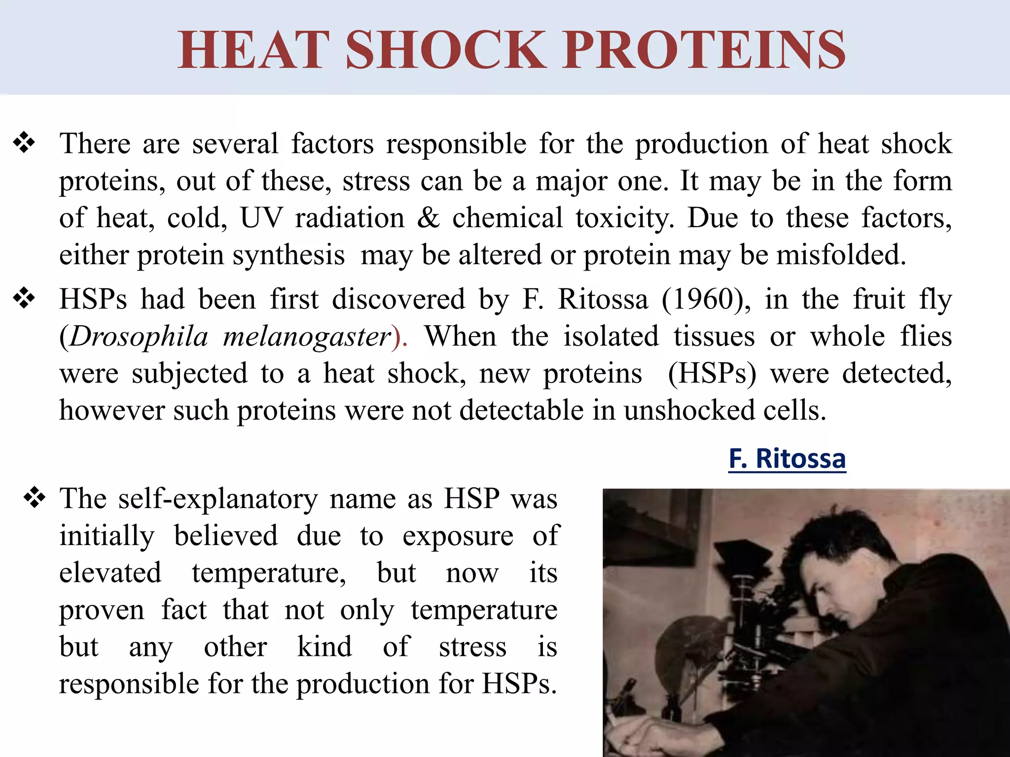
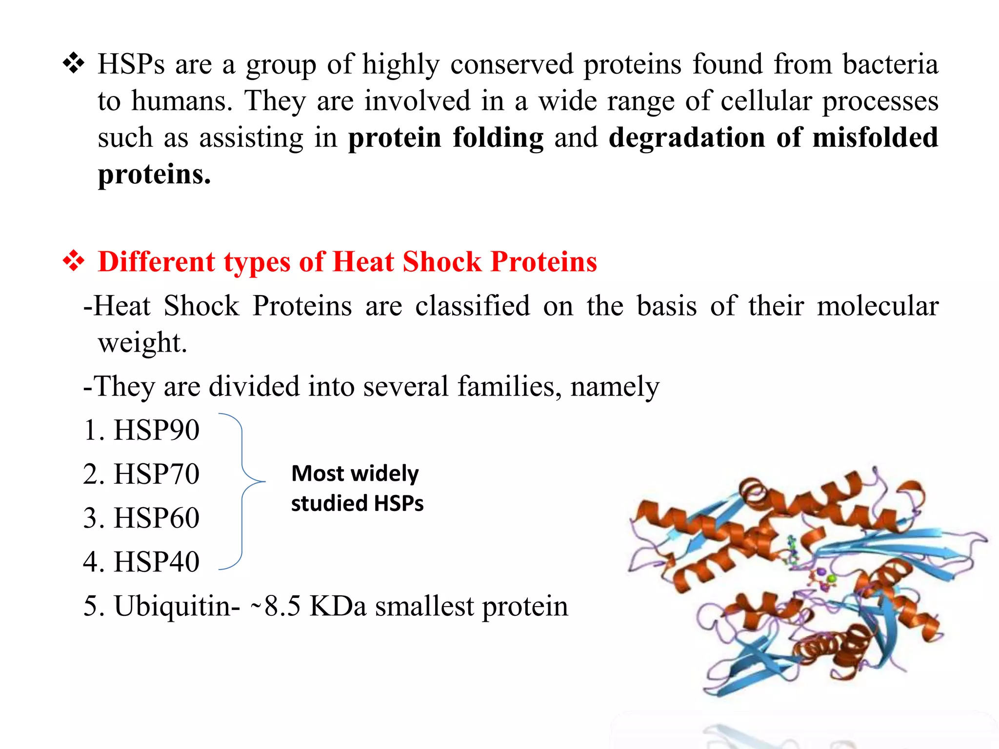
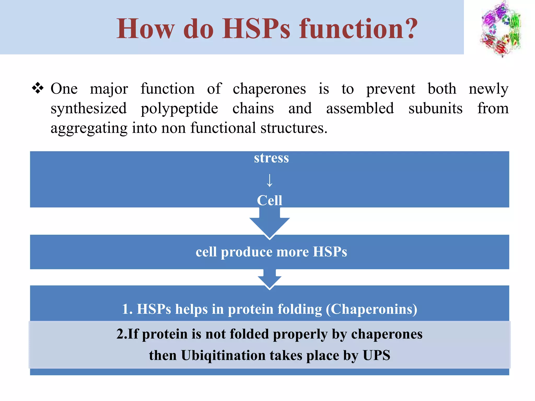
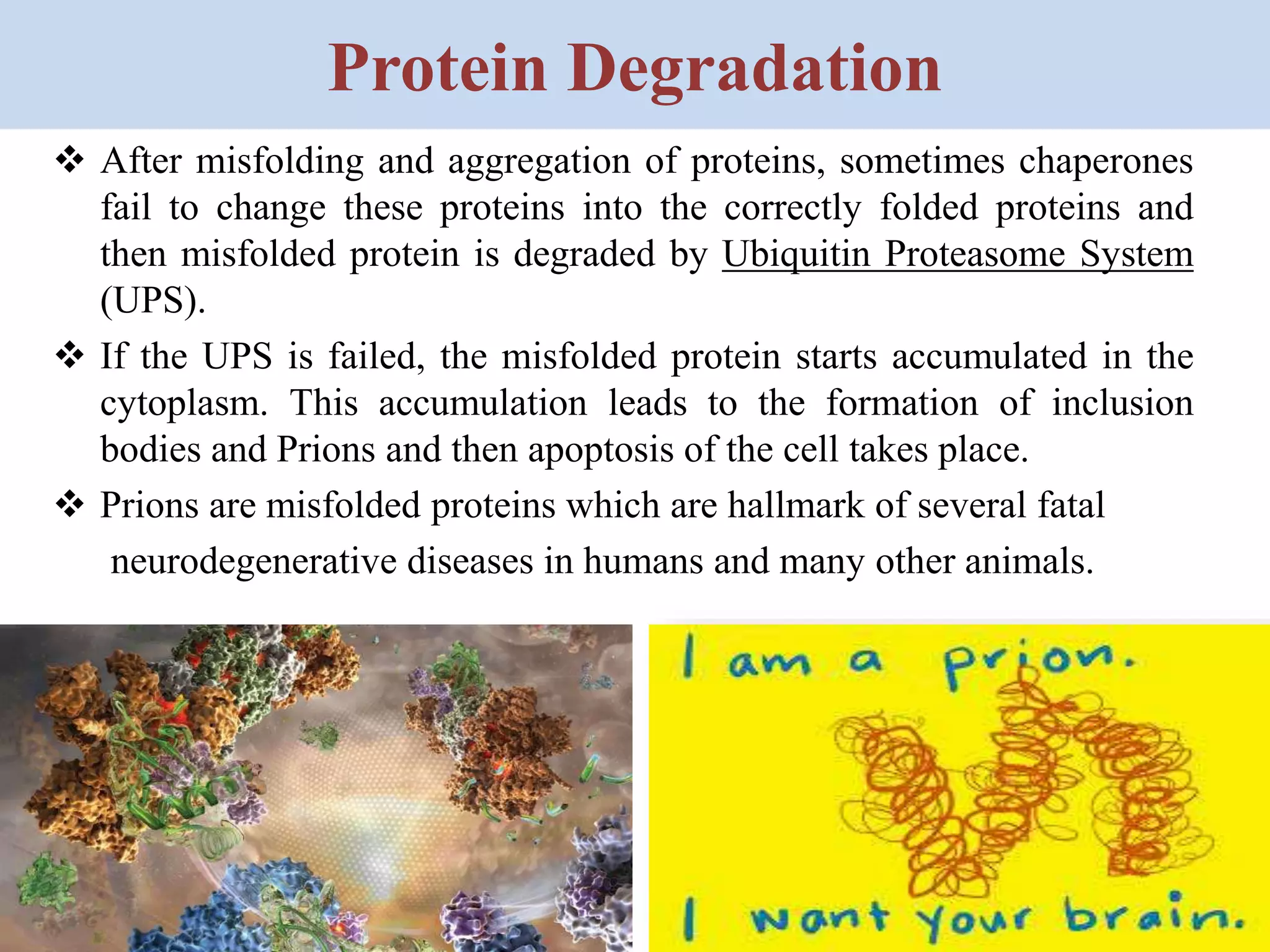
HEAT SHOCK PROTEINS
(HSPs)
POOJA CHAUDHARY
Self study: ZOM -953
Enrolment No. 184243
Department of Zoology
Faculty of Science
Dayalbagh Educational Institute
HEAT SHOCK PROTEINS
 There are several factors responsible for the production of heat shock
proteins, out of these, stress can be a major one. It may be in the form
of heat, cold, UV radiation & chemical toxicity. Due to these factors,
either protein synthesis may be altered or protein may be misfolded.
 HSPs had been first discovered by F. Ritossa (1960), in the fruit fly
(Drosophila melanogaster). When the isolated tissues or whole flies
were subjected to a heat shock, new proteins (HSPs) were detected,
however such proteins were not detectable in unshocked cells.
F. Ritossa
 The self-explanatory name as HSP was
initially believed due to exposure of
elevated temperature, but now its
proven fact that not only temperature
but any other kind of stress is
responsible for the production for HSPs.
 HSPs are a group of highly conserved proteins found from bacteria
to humans. They are involved in a wide range of cellular processes
such as assisting in protein folding and degradation of misfolded
proteins.
 Different types of Heat Shock Proteins
-Heat Shock Proteins are classified on the basis of their molecular
weight.
-They are divided into several families, namely
1. HSP90
2. HSP70
3. HSP60
4. HSP40
5. Ubiquitin- ̴ 8.5 KDa smallest protein
Most widely
studied HSPs
CHAPERONES CLASSES
 Chaperones have mainly three classes:
1. HOLDASES- Prevent protein damage by providing favourable
conditions. e.g. HSP-40
2. FOLDASES- Misfolded protein back to native conformation.
- Shows ATP dependent activity. e.g. HSP-60
3. DISAGGREGASES - Prevent aggregation of proteins.
- Prevent inclusion body formation and PRION formation.
- Rescues proteins from aggregated states.
e.g HSP-104
MOLECULAR CHAPERONES
↓
Quality control system
“ protein control system’’
↓
Resume Protein from
Misfolding
Prevent
Degradation
Protein Folding
Assistance
Prevent
Aggregations
Prevent Prions
Formation
Transcription and Modulation of HSP
 HSP expression is increased when cells are exposed to stresses like
elevated temperature. This increase in expression is transcriptionally
regulated. The dramatic up-regulation of the HSP is a key part of the
heat shock response and is induced primarily by heat shock factors
(HSFs).
 The mechanism by which stresses like heat-shock (or other
environmental stressors) activates the HSF, has been determined in the
bacteria. During heat stress, outer membrane proteins (OMPs) do not
fold and can not insert correctly into the outer membrane. These
misfolded OMPs accumulate in the periplasmic space and can be
detected by DegS, an inner membrane protease that passes the signal to
the sigma E transcription factor. Then, the HSE (factor of transcription)
and HSF use to transcribe the more HSP.
Transcription and Modulation of HSP
How do HSPs function?
 One major function of chaperones is to prevent both newly
synthesized polypeptide chains and assembled subunits from
aggregating into non functional structures.
1. HSPs helps in protein folding (Chaperonins)
2.If protein is not folded properly by chaperones
then Ubiqitination takes place by UPS
cell produce more HSPs
stress
↓
Cell
 High temperatures and other types of stresses, such as altered pH
and oxygen deprivation, make it more difficult for proteins to form
their proper structures and cause some already structured proteins to
unfold.
 Heat Shock Proteins are induced rapidly at high levels to deal with
these problems.
Stability of Proteins
Forces involved in Protein stabilization-
• Hydrogen bonding
• Vander Waals interaction
• Disulfide bonds
• Hydrophobicity: the dominant force in protein folding .
A stably folded proteins has…..
• Hydrophobic side chains buried inside
• Charged side chains on the surface
• Pack as close together as possible
• Minimize contacts between hydrophobic groups and water.
Chaperones Helps in Protein Folding
 HSPs function in the stabilization of proteins and assist protein refolding
under stress conditions.
 When a protein is not folded properly,
their hydrophobic groups are present on
the surface, then HSP-70 and HSP-40 bind
to the surface of substrate, then this
substrate must be transferred to a
chaperonin complex for productive
folding.
 In the cell, protein folding process is aided
by molecular chaperones, which act in
preventing protein misfolding and
aggregation.
Protein Folding is Assisted by
Chaperones & Chaperonins
HSP-70,40
Protein Degradation
 After misfolding and aggregation of proteins, sometimes chaperones
fail to change these proteins into the correctly folded proteins and
then misfolded protein is degraded by Ubiquitin Proteasome System
(UPS).
 If the UPS is failed, the misfolded protein starts accumulated in the
cytoplasm. This accumulation leads to the formation of inclusion
bodies and Prions and then apoptosis of the cell takes place.
 Prions are misfolded proteins which are hallmark of several fatal
neurodegenerative diseases in humans and many other animals.
Polyubiquitination
 Many Ubiquitin molecules bind to the same target protein. Once a
protein is tagged with a single Ub molecule, this is a signal to other
ligases to attach additional Ub molecules.
 Polyubiquitination of proteins is the
triggering signal that leads to
degradation of the misfolded
protein in the proteasome.
 Ubiquitin is a small regulatory
protein with 76 aa and its
uniqueness lies as glycine (G) at
76th position.
 Molecular weight of Ubiquitin is
~8.5 KDa.
 Misfolded protein contains different types of amino acids in their
structure in which lysine (K) is specific because lysine and glycine (G)
make isopeptide bond between misfolded protein and ubiquitin
molecules with the help of isopeptide transferase enzyme (E3).
 An Isopeptide bond is formed between the carboxyl group (COO−) of the
glycine of ubiquitin and the amino group (NH+
3) of the lysine of
substrate.
 Ubiquitination process requires three types of enzymes:-
E1 = Ubiquitin-activating enzymes
E2 = Ubiquitin-conjugating enzymes
E3 = Ubiquitin ligating enzymes
1. Activation- Ub is activated by E1 because they are present in the inert
form, This is the ATP-dependent process. The E1 binds both ATP and Ub
molecule. After this E1 transfer AMP on Ub.
2. Conjugation- E2 Ubiquitin-conjugating enzymes catalyze the transfer of
Ub from E1 to the E2 In order to perform this reaction, the E2 binds to
activated Ub. There are ~35 different types of E2 enzymes in humans.
3. Ligation- E3 Ubiquitin ligases catalyses the final step of the Ubiquitination
cascade. They create an isopeptide bond between a lysine of the target
protein and the C-terminal glycine of Ub.
Ubiquitination process involves 3 steps
Ubiquitination- Role of Enzymes
1. Activation
2. Conjugation
3. Ligation
Proteosome - Degradation of Misfolded
Protein
Proteasome
Discharge of
Peptides
Lid 19S
Wall 20S
Why don‫׳‬t Heat Shock Proteins
Denature ?
 Better Hydrogen Bond
 Better Hydrophobic Internal Packing
 Stabilized Secondary Structure and tertiary structure
Conclusion
 HSPs plays very important role in protein folding and degradation of
misfolded protein, if HSPs and UPS both functions are fail then the
misfolded proteins starts accumulated in the cytoplasm and form inclusion
bodies , this leads to apoptosis(death) of cells.
 Major applications of HSPs are–
1. Cancer vaccines
2. Agriculture
3. Autoimmunity treatment
4. Function in antigen presentation by MHC-I and MHC-II :
HSPs play an important part in
transfer of unfolded proteins to
proteasome and generated peptides
to MHC.
Heat shock proteins presentation

Heat shock proteins presentation

  • 1.
    HEAT SHOCK PROTEINS (HSPs) POOJACHAUDHARY Self study: ZOM -953 Enrolment No. 184243 Department of Zoology Faculty of Science Dayalbagh Educational Institute
  • 2.
    HEAT SHOCK PROTEINS There are several factors responsible for the production of heat shock proteins, out of these, stress can be a major one. It may be in the form of heat, cold, UV radiation & chemical toxicity. Due to these factors, either protein synthesis may be altered or protein may be misfolded.  HSPs had been first discovered by F. Ritossa (1960), in the fruit fly (Drosophila melanogaster). When the isolated tissues or whole flies were subjected to a heat shock, new proteins (HSPs) were detected, however such proteins were not detectable in unshocked cells. F. Ritossa  The self-explanatory name as HSP was initially believed due to exposure of elevated temperature, but now its proven fact that not only temperature but any other kind of stress is responsible for the production for HSPs.
  • 3.
     HSPs area group of highly conserved proteins found from bacteria to humans. They are involved in a wide range of cellular processes such as assisting in protein folding and degradation of misfolded proteins.  Different types of Heat Shock Proteins -Heat Shock Proteins are classified on the basis of their molecular weight. -They are divided into several families, namely 1. HSP90 2. HSP70 3. HSP60 4. HSP40 5. Ubiquitin- ̴ 8.5 KDa smallest protein Most widely studied HSPs
  • 4.
    CHAPERONES CLASSES  Chaperoneshave mainly three classes: 1. HOLDASES- Prevent protein damage by providing favourable conditions. e.g. HSP-40 2. FOLDASES- Misfolded protein back to native conformation. - Shows ATP dependent activity. e.g. HSP-60 3. DISAGGREGASES - Prevent aggregation of proteins. - Prevent inclusion body formation and PRION formation. - Rescues proteins from aggregated states. e.g HSP-104
  • 5.
    MOLECULAR CHAPERONES ↓ Quality controlsystem “ protein control system’’ ↓ Resume Protein from Misfolding Prevent Degradation Protein Folding Assistance Prevent Aggregations Prevent Prions Formation
  • 6.
    Transcription and Modulationof HSP  HSP expression is increased when cells are exposed to stresses like elevated temperature. This increase in expression is transcriptionally regulated. The dramatic up-regulation of the HSP is a key part of the heat shock response and is induced primarily by heat shock factors (HSFs).  The mechanism by which stresses like heat-shock (or other environmental stressors) activates the HSF, has been determined in the bacteria. During heat stress, outer membrane proteins (OMPs) do not fold and can not insert correctly into the outer membrane. These misfolded OMPs accumulate in the periplasmic space and can be detected by DegS, an inner membrane protease that passes the signal to the sigma E transcription factor. Then, the HSE (factor of transcription) and HSF use to transcribe the more HSP.
  • 7.
  • 8.
    How do HSPsfunction?  One major function of chaperones is to prevent both newly synthesized polypeptide chains and assembled subunits from aggregating into non functional structures. 1. HSPs helps in protein folding (Chaperonins) 2.If protein is not folded properly by chaperones then Ubiqitination takes place by UPS cell produce more HSPs stress ↓ Cell
  • 9.
     High temperaturesand other types of stresses, such as altered pH and oxygen deprivation, make it more difficult for proteins to form their proper structures and cause some already structured proteins to unfold.  Heat Shock Proteins are induced rapidly at high levels to deal with these problems.
  • 10.
    Stability of Proteins Forcesinvolved in Protein stabilization- • Hydrogen bonding • Vander Waals interaction • Disulfide bonds • Hydrophobicity: the dominant force in protein folding . A stably folded proteins has….. • Hydrophobic side chains buried inside • Charged side chains on the surface • Pack as close together as possible • Minimize contacts between hydrophobic groups and water.
  • 11.
    Chaperones Helps inProtein Folding  HSPs function in the stabilization of proteins and assist protein refolding under stress conditions.  When a protein is not folded properly, their hydrophobic groups are present on the surface, then HSP-70 and HSP-40 bind to the surface of substrate, then this substrate must be transferred to a chaperonin complex for productive folding.  In the cell, protein folding process is aided by molecular chaperones, which act in preventing protein misfolding and aggregation.
  • 12.
    Protein Folding isAssisted by Chaperones & Chaperonins HSP-70,40
  • 13.
    Protein Degradation  Aftermisfolding and aggregation of proteins, sometimes chaperones fail to change these proteins into the correctly folded proteins and then misfolded protein is degraded by Ubiquitin Proteasome System (UPS).  If the UPS is failed, the misfolded protein starts accumulated in the cytoplasm. This accumulation leads to the formation of inclusion bodies and Prions and then apoptosis of the cell takes place.  Prions are misfolded proteins which are hallmark of several fatal neurodegenerative diseases in humans and many other animals.
  • 14.
    Polyubiquitination  Many Ubiquitinmolecules bind to the same target protein. Once a protein is tagged with a single Ub molecule, this is a signal to other ligases to attach additional Ub molecules.  Polyubiquitination of proteins is the triggering signal that leads to degradation of the misfolded protein in the proteasome.  Ubiquitin is a small regulatory protein with 76 aa and its uniqueness lies as glycine (G) at 76th position.  Molecular weight of Ubiquitin is ~8.5 KDa.
  • 15.
     Misfolded proteincontains different types of amino acids in their structure in which lysine (K) is specific because lysine and glycine (G) make isopeptide bond between misfolded protein and ubiquitin molecules with the help of isopeptide transferase enzyme (E3).  An Isopeptide bond is formed between the carboxyl group (COO−) of the glycine of ubiquitin and the amino group (NH+ 3) of the lysine of substrate.  Ubiquitination process requires three types of enzymes:- E1 = Ubiquitin-activating enzymes E2 = Ubiquitin-conjugating enzymes E3 = Ubiquitin ligating enzymes
  • 16.
    1. Activation- Ubis activated by E1 because they are present in the inert form, This is the ATP-dependent process. The E1 binds both ATP and Ub molecule. After this E1 transfer AMP on Ub. 2. Conjugation- E2 Ubiquitin-conjugating enzymes catalyze the transfer of Ub from E1 to the E2 In order to perform this reaction, the E2 binds to activated Ub. There are ~35 different types of E2 enzymes in humans. 3. Ligation- E3 Ubiquitin ligases catalyses the final step of the Ubiquitination cascade. They create an isopeptide bond between a lysine of the target protein and the C-terminal glycine of Ub. Ubiquitination process involves 3 steps
  • 17.
    Ubiquitination- Role ofEnzymes 1. Activation 2. Conjugation 3. Ligation
  • 18.
    Proteosome - Degradationof Misfolded Protein Proteasome Discharge of Peptides Lid 19S Wall 20S
  • 19.
    Why don‫׳‬t HeatShock Proteins Denature ?  Better Hydrogen Bond  Better Hydrophobic Internal Packing  Stabilized Secondary Structure and tertiary structure
  • 20.
    Conclusion  HSPs playsvery important role in protein folding and degradation of misfolded protein, if HSPs and UPS both functions are fail then the misfolded proteins starts accumulated in the cytoplasm and form inclusion bodies , this leads to apoptosis(death) of cells.  Major applications of HSPs are– 1. Cancer vaccines 2. Agriculture 3. Autoimmunity treatment 4. Function in antigen presentation by MHC-I and MHC-II : HSPs play an important part in transfer of unfolded proteins to proteasome and generated peptides to MHC.