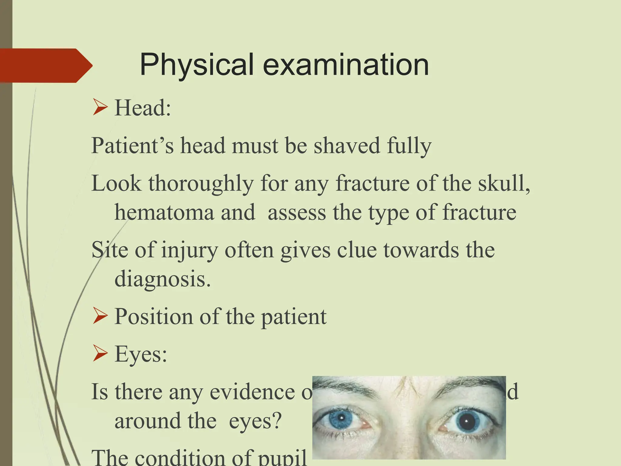
Head Injuries
Definition - Traumatic brain injury (TBI)
 Trauma to the scalp, skull or brain.
 encompasses a broad range of pathologic
injuries to the brain
 Head injury = traumatic brain injury
Etiology
 MotorV
ehicleaccident-44%
 Falls-26%
 Other/Unknown-13%
 Non-FirearmAssaults-9%
 Firearms-8%
Pathophysiology/ classification of TBI
Impact injuries: It results from an object
striking the head or the head striking an
object.
It includes:
Scalp injuries - scalp contusion, abrasion and/or
lacerations, excessive bleeding,
osteomyletis.
Skull fracture – linear.
- depressed blunt trauma,
hemorrhage , ICP, compressed delicate tissues,
contamination and infection, cortical damage and
epilepsy.
Base of skull fracture
Anterior cranial fossa fracture:
subconjunctival haematoma, epistaxis, CSF rhinorrhea
Middle cranial fossa fracture:
CSF otorrhoea, haemotympanium, ossicular disruption,
Battle sign, 7th and 8th cranial nerve palsy.
Cerebral contusion and laceration
Epidural hematoma
Acceleration and deceleration injuries:
 result of differential movement between skull and
cranial content.
 It includes:
Diffuse axonal injury
Sub-dural hematoma
Coupinjury
Contracoupinjury
Coupinjury:
 It occurs at the site of the impact to the head .
 compression of brain due to inward movement of the
bone
Contre-coup:
Injury occurs directly opposite to
the point of impact
common in frontal and temporal
6
lobe.
Produced by the head in motion
impacting on a stationary object
Brain injury
Primary brain injury:
Injury caused at the time of impact
Irreversible Secondary brain injury
progressive brain damage
Primarybraininjury Secondarybraininjury
Concussion Intracranialhaematoma
Corticallaceration/contusion Cerebraloedema
Diffuseaxonalinjury Ischaemia
Bonefragmentation Infection
Metabolic or endocrine
disturbances
Diffuse Axonal Injury
 Results from mechanical shearing at grey- white
interface due to severe acceleration and
deceleration force
 No obvious structural damage
 Severity - mild damage with confusion to coma and
even death
 Major cause of unconsciousness and persistent
vegetative state after head trauma
Cerebral Concussion
 It is the condition of temporary dysfunction
of brain without any structural damage
following head injury
 It is manifested as:
Transient loss of consciousness
Transient loss of memory
Autonomic dysfunction like bradycardia,
hypotension and sweating
Cerebral Contusion
 It is more severe degree of brain injury
manifested by areas of hemorrhage in the
brain parenchyma but without surface
laceration
 Neurological deficit which persists more than
24 hour
 Associated cerebral edema and defects in the
blood brain barrier
Cerebral laceration
 Severe degree of brain injury associated with a
breach in the surface parenchyma
 Tearing of brain surface may be due to skull
fracture or due to shearing forces
 Focal neurological defecit may be present
Extradural haematoma
 Collection of blood between the cranial bones
and duramater
 It is associated with the fracture of temporo-
parietal region
 Commonest vessel: Middle meningeal artery
 Confusion, irritability, drowsiness, hemiparesis
to the same side of injury
 Features of raised ICP: hypertension,
bradycardia, vomiting
 CT scan: Biconvex
lesion
 It is the surgical
emergency
 Craniotomy and
evacuation of clot
is done.
Sub Dural Haematoma
 Collection of blood
between brain and
duramater
 Common intracranial
mass lesion resulting
from trauma
Acute: <3 days
Sub-acute: 4-21 days
Chronic: >21 days
Sub Dural Haemotama
 Loss of consciousness occurs immediately
after trauma and is progressive
 Features of raised ICP and focal neurological
defecits
 CT Scan: Concavo-convex lesion
 T/t: surgical decompression by craniotomy
 Antibiotics
Cerebral Herniation
 Increased ICP or presence of
intracranial mass may
predispose to cerebral
herniation.
 Herniation of contents of
supratentorial compartment
through the tentorial hiatus
 Herniation of the contents of
the infraintentorial
compartment through the
foramen magnum
Brain swelling
It follows significant
head injury
Occurs due to active
hyperaemia and
edema
Infection, Seizure, Hydrocephalus
Approach to Head Trauma
 Detailed history should be sought in all cases
of head trauma.
 If the patient is unconscious which is usually
the condition, history should be obtained
from the attendant.
 While one care provider is taking history,
resuscitation should be carried out
simultaneously by other care provider.
Ask about:
 Type of accident: ?RTA, ?fall from height
?acceleration/deceleration injury during driving
a motor car
 Level of consciousness: ?Unconscious,
?semiconscious
 If unconscious: duration of unconsciousness,
?immediately after trauma ?lucid interval
 Post traumatic amnesia
Ask about:
 Vomiting: ?blood in vomitus ?persistent
vomiting ?sign of recovery from cerebral
concussion
 Epileptic fits or seizure: Its nature may give
clue to localization of the site of trauma
 Swelling and pain in the head
 Other complaints: ?bleeding or watery
discharge from ear, nose and mouth
Ask about:
 Past history: ?fits or similar head injury in
the past
?Hypertension ?DM ?Renal diseases
 Personal history: ?unconsciousness due to
other cause (alcohol, opium poisoning,
diabetic coma)
 Family history: History of diabetes, HTN,
epilepsy in the family
Immediate management
 Initial assessment of head injuries must
follow advanced trauma and life support.
(ALTS)
 It includes:
Maintenance of airway along with cervical
spine control
Cervical spine immobilized in neutral
position using neck brace, sand bags,
forehead tape
Suction of airway to clear blood, vomitus
Chin lift, jaw thrust
 Maintenance of breathing
 Assessment of circulation and control of
haemorrhage
Establish iv access with two large bore
iv cannulas
IV infusion of NS (Avoid 5% Dextrose
as it may precipitate cerebral edema)
 Assessment of dysfunction of CNS
 Exposure in a controlled environment
Remove all clothes and look for any
Physical examination
 Pulse and blood pressure:
Pulse will be rapid, thready with low BP in
case of cerebral concussion
Pulse becomes slow and bounding with high
BP in case of cerebral irritation
Rapid pulse in deeply unconscious heralds
impeding death
 Temperature:
In cerebral concussion, contusion
temperature may remain subnormal
With appearance of cerebral compression,
temperature may rise upto 100˚F
Victor Horsley’s sign
Physical examination
 Head:
Patient’s head must be shaved fully
Look thoroughly for any fracture of the skull,
hematoma and assess the type of fracture
Site of injury often gives clue towards the
diagnosis.
 Position of the patient
 Eyes:
Is there any evidence of haemorrhage in and
around the eyes?
The condition of pupil
Neurological assessment
NeurologicalassessmentcanbedonebyusingGlasgowcomascale.
 Minor head injury: GCS 15 with no loss
of consciousness (LOC)
 Mild head injury: GCS 14 or 15 with
LOC
 Moderate head injury: GCS 9–13
 Severe head injury: GCS 3–8.
 Presence of neurological deficits:
Check for power, tone, superficial and deep
tendon reflexes
 Rigidity of the neck:
May be present in the case of subarachnoid
hemorrhage, fracture dislocation of cervical
spine
 Cranial nerve examination
Cranial nerve should be examined one after
another Of these most important is the third
General Examination
Examine
Chest for the fracture of the ribs, surgical
emphysema
Spine, pelvis and limbs for the presence of
fracture
Exclude abdomen for rupture of any hollow
viscus, internal haemorrhage form injury to
any solid viscus eg: liver, spleen
Management:
 Place a Nasogastric tube to decompress the
stomach and reduce the risk of vomiting as
aspiration
 Avoid NG tube for the patients with facial
injuries as the tube could enter the brain
through bony fracture
 Insert an dwelling urinary catheter for
hourly urine output monitoring
 Avoid insertion if urethral injury suspected
Treatment of raised ICP
 IV Mannitol
 IV furosemide
 Reverse Trendelenburg if no
counter indications like
hypovolaemia, spine injury
 If significant agitation and if
hypoxia, hypovolaemia or pain
is excluded as the cause of
agitation: give IV Midazolam
 Analgesics for the pain
management
Monitor
 Blood pressure
 Heart rate
 Respiratory rate
 Spo2
 ECG
 Blood samples for serum electrolyte
Arterial blood gas hyper/
hypoglycaemia
Special investigations!!!!
Definitive treatment!!!!
Complications
 Personality
Changes
 Hypopituitarism
e.g. DI
 Post-Traumatic
Seizures
 Infections e.g.
Meningitis
 Vasospasm,
Long-Termeffects
 Parkinson’s
 Alzheimer’sDementia
Rehabilitation
Physiotherapy
OccupationalTherapy
SpeechandLanguageTherapy
Psychologists/Psychiatrists
References
SHOR
TPRACTICEofS
U
R
G
E
R
Y
Bailey&
Love’
s25thEdition
S
R
BManualOfSurgery
Death&
DeductionForensicMedicine
A
ManualOnClinicalSurgery
headinjuries -  types, causes, management

headinjuries - types, causes, management

  • 1.
  • 2.
    Definition - Traumaticbrain injury (TBI)  Trauma to the scalp, skull or brain.  encompasses a broad range of pathologic injuries to the brain  Head injury = traumatic brain injury
  • 3.
    Etiology  MotorV ehicleaccident-44%  Falls-26% Other/Unknown-13%  Non-FirearmAssaults-9%  Firearms-8%
  • 4.
    Pathophysiology/ classification ofTBI Impact injuries: It results from an object striking the head or the head striking an object. It includes: Scalp injuries - scalp contusion, abrasion and/or lacerations, excessive bleeding, osteomyletis.
  • 5.
    Skull fracture –linear. - depressed blunt trauma, hemorrhage , ICP, compressed delicate tissues, contamination and infection, cortical damage and epilepsy.
  • 7.
    Base of skullfracture Anterior cranial fossa fracture: subconjunctival haematoma, epistaxis, CSF rhinorrhea Middle cranial fossa fracture: CSF otorrhoea, haemotympanium, ossicular disruption, Battle sign, 7th and 8th cranial nerve palsy.
  • 8.
    Cerebral contusion andlaceration Epidural hematoma
  • 9.
    Acceleration and decelerationinjuries:  result of differential movement between skull and cranial content.  It includes: Diffuse axonal injury Sub-dural hematoma
  • 10.
    Coupinjury Contracoupinjury Coupinjury:  It occursat the site of the impact to the head .  compression of brain due to inward movement of the bone Contre-coup: Injury occurs directly opposite to the point of impact common in frontal and temporal 6 lobe. Produced by the head in motion impacting on a stationary object
  • 11.
    Brain injury Primary braininjury: Injury caused at the time of impact Irreversible Secondary brain injury progressive brain damage
  • 12.
    Primarybraininjury Secondarybraininjury Concussion Intracranialhaematoma Corticallaceration/contusionCerebraloedema Diffuseaxonalinjury Ischaemia Bonefragmentation Infection Metabolic or endocrine disturbances
  • 13.
    Diffuse Axonal Injury Results from mechanical shearing at grey- white interface due to severe acceleration and deceleration force  No obvious structural damage  Severity - mild damage with confusion to coma and even death  Major cause of unconsciousness and persistent vegetative state after head trauma
  • 15.
    Cerebral Concussion  Itis the condition of temporary dysfunction of brain without any structural damage following head injury  It is manifested as: Transient loss of consciousness Transient loss of memory Autonomic dysfunction like bradycardia, hypotension and sweating
  • 16.
    Cerebral Contusion  Itis more severe degree of brain injury manifested by areas of hemorrhage in the brain parenchyma but without surface laceration  Neurological deficit which persists more than 24 hour  Associated cerebral edema and defects in the blood brain barrier
  • 17.
    Cerebral laceration  Severedegree of brain injury associated with a breach in the surface parenchyma  Tearing of brain surface may be due to skull fracture or due to shearing forces  Focal neurological defecit may be present
  • 18.
    Extradural haematoma  Collectionof blood between the cranial bones and duramater  It is associated with the fracture of temporo- parietal region  Commonest vessel: Middle meningeal artery  Confusion, irritability, drowsiness, hemiparesis to the same side of injury  Features of raised ICP: hypertension, bradycardia, vomiting
  • 19.
     CT scan:Biconvex lesion  It is the surgical emergency  Craniotomy and evacuation of clot is done.
  • 20.
    Sub Dural Haematoma Collection of blood between brain and duramater  Common intracranial mass lesion resulting from trauma Acute: <3 days Sub-acute: 4-21 days Chronic: >21 days
  • 21.
    Sub Dural Haemotama Loss of consciousness occurs immediately after trauma and is progressive  Features of raised ICP and focal neurological defecits  CT Scan: Concavo-convex lesion  T/t: surgical decompression by craniotomy  Antibiotics
  • 22.
    Cerebral Herniation  IncreasedICP or presence of intracranial mass may predispose to cerebral herniation.  Herniation of contents of supratentorial compartment through the tentorial hiatus  Herniation of the contents of the infraintentorial compartment through the foramen magnum
  • 23.
    Brain swelling It followssignificant head injury Occurs due to active hyperaemia and edema Infection, Seizure, Hydrocephalus
  • 24.
    Approach to HeadTrauma  Detailed history should be sought in all cases of head trauma.  If the patient is unconscious which is usually the condition, history should be obtained from the attendant.  While one care provider is taking history, resuscitation should be carried out simultaneously by other care provider.
  • 25.
    Ask about:  Typeof accident: ?RTA, ?fall from height ?acceleration/deceleration injury during driving a motor car  Level of consciousness: ?Unconscious, ?semiconscious  If unconscious: duration of unconsciousness, ?immediately after trauma ?lucid interval  Post traumatic amnesia
  • 26.
    Ask about:  Vomiting:?blood in vomitus ?persistent vomiting ?sign of recovery from cerebral concussion  Epileptic fits or seizure: Its nature may give clue to localization of the site of trauma  Swelling and pain in the head  Other complaints: ?bleeding or watery discharge from ear, nose and mouth
  • 27.
    Ask about:  Pasthistory: ?fits or similar head injury in the past ?Hypertension ?DM ?Renal diseases  Personal history: ?unconsciousness due to other cause (alcohol, opium poisoning, diabetic coma)  Family history: History of diabetes, HTN, epilepsy in the family
  • 28.
    Immediate management  Initialassessment of head injuries must follow advanced trauma and life support. (ALTS)  It includes: Maintenance of airway along with cervical spine control Cervical spine immobilized in neutral position using neck brace, sand bags, forehead tape Suction of airway to clear blood, vomitus Chin lift, jaw thrust
  • 29.
     Maintenance ofbreathing  Assessment of circulation and control of haemorrhage Establish iv access with two large bore iv cannulas IV infusion of NS (Avoid 5% Dextrose as it may precipitate cerebral edema)  Assessment of dysfunction of CNS  Exposure in a controlled environment Remove all clothes and look for any
  • 30.
    Physical examination  Pulseand blood pressure: Pulse will be rapid, thready with low BP in case of cerebral concussion Pulse becomes slow and bounding with high BP in case of cerebral irritation Rapid pulse in deeply unconscious heralds impeding death
  • 31.
     Temperature: In cerebralconcussion, contusion temperature may remain subnormal With appearance of cerebral compression, temperature may rise upto 100˚F Victor Horsley’s sign
  • 32.
    Physical examination  Head: Patient’shead must be shaved fully Look thoroughly for any fracture of the skull, hematoma and assess the type of fracture Site of injury often gives clue towards the diagnosis.  Position of the patient  Eyes: Is there any evidence of haemorrhage in and around the eyes? The condition of pupil
  • 33.
  • 34.
     Minor headinjury: GCS 15 with no loss of consciousness (LOC)  Mild head injury: GCS 14 or 15 with LOC  Moderate head injury: GCS 9–13  Severe head injury: GCS 3–8.
  • 35.
     Presence ofneurological deficits: Check for power, tone, superficial and deep tendon reflexes  Rigidity of the neck: May be present in the case of subarachnoid hemorrhage, fracture dislocation of cervical spine  Cranial nerve examination Cranial nerve should be examined one after another Of these most important is the third
  • 36.
    General Examination Examine Chest forthe fracture of the ribs, surgical emphysema Spine, pelvis and limbs for the presence of fracture Exclude abdomen for rupture of any hollow viscus, internal haemorrhage form injury to any solid viscus eg: liver, spleen
  • 37.
    Management:  Place aNasogastric tube to decompress the stomach and reduce the risk of vomiting as aspiration  Avoid NG tube for the patients with facial injuries as the tube could enter the brain through bony fracture  Insert an dwelling urinary catheter for hourly urine output monitoring  Avoid insertion if urethral injury suspected
  • 38.
    Treatment of raisedICP  IV Mannitol  IV furosemide  Reverse Trendelenburg if no counter indications like hypovolaemia, spine injury  If significant agitation and if hypoxia, hypovolaemia or pain is excluded as the cause of agitation: give IV Midazolam  Analgesics for the pain management
  • 39.
    Monitor  Blood pressure Heart rate  Respiratory rate  Spo2  ECG  Blood samples for serum electrolyte Arterial blood gas hyper/ hypoglycaemia
  • 40.
  • 41.
    Complications  Personality Changes  Hypopituitarism e.g.DI  Post-Traumatic Seizures  Infections e.g. Meningitis  Vasospasm, Long-Termeffects  Parkinson’s  Alzheimer’sDementia
  • 42.
  • 43.