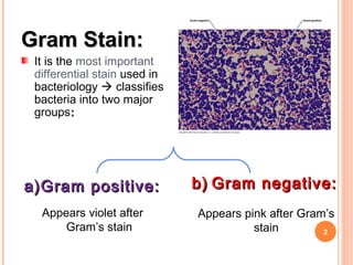
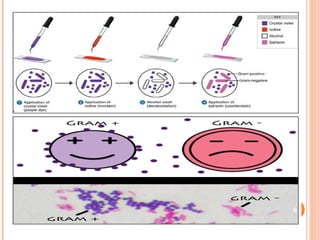
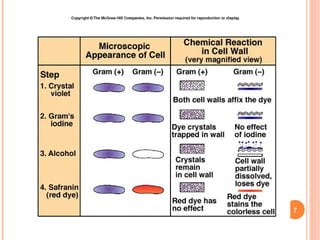
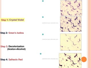
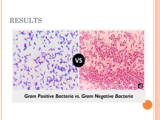
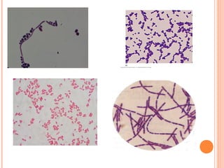
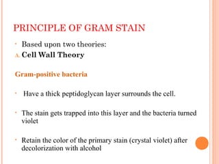
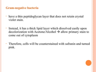
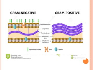
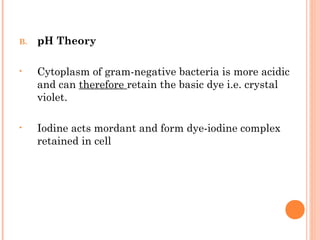
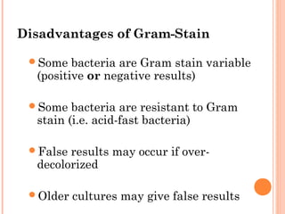
GRAM-STAIN PRACTICAL BRIEFING
1
Ms. Clemencia Tjazuko
MSc Medical Microbiology
2
Gram Stain:Gram Stain:
It is the most important
differential stain used in
bacteriology  classifies
bacteria into two major
groups::
a)a)Gram positive:Gram positive:
Appears violet after
Gram’s stain
b)b) Gram negative:Gram negative:
Appears pink after Gram’s
stain
 Developed by Hans Christian Gram (1884)
 Differential staining technique two stains
are used to impart different colours to different
bacteria
 Other differential stains:
 Acid-fast Stain
 Albert Stain
3
PROCEDURE OF GRAM STAIN
1. Fixation: smear made on slide from bacterial culture air
dried heat fixed
2. Primary stain: stained with crystal violet. Slide rinsed with
water. All bacteria stained violet
3. Mordant: gram’s iodine poured over slide and kept for 1
min. Slide rinsed with water.
4. Decolourization: few drops of decolourizer to smear i.e.
ethanol (20-30 sec). Slide rinsed with water.
4
• Removes the primary stain from gram-negative bacteria. Most
important step
5. Counter stain: secondary stains  safranin added for 30 sec.
Gives pink/red colour to gram-negative bacteria.
Slide rinsed with water, dried and examined under oil
immersion objective
5
6
7
8
Step 1:Step 1: Crystal Violet
Step 2:Step 2: Gram’s IodineGram’s Iodine
Step 3:Step 3: DecolorizationDecolorization
(Aceton-Alcohol)(Aceton-Alcohol)
Step 4:Step 4: Safranin RedSafranin Red
RESULTS
9
10
PRINCIPLE OF GRAM STAIN
• Based upon two theories:
A. Cell Wall Theory
Gram-positive bacteria
• Have a thick peptidoglycan layer surrounds the cell.
• The stain gets trapped into this layer and the bacteria turned
violet
• Retain the color of the primary stain (crystal violet) after
decolorization with alcohol
11
Gram-negative bacteria
• have a thin peptidoglycan layer that does not retain crystal
violet stain.
• Instead, it has a thick lipid layer which dissolved easily upon
decolorization with Acetone/Alcohol  allow primary stain to
come out of cytoplasm
• Therefore, cells will be counterstained with safranin and turned
pink.
12
13
B. pH Theory
• Cytoplasm of gram-negative bacteria is more acidic
and can therefore retain the basic dye i.e. crystal
violet.
• Iodine acts mordant and form dye-iodine complex
retained in cell
14
Disadvantages of Gram-Stain
Some bacteria are Gram stain variable
(positive or negative results)
Some bacteria are resistant to Gram
stain (i.e. acid-fast bacteria)
False results may occur if over-
decolorized
Older cultures may give false results15
MODIFICATIONS OF GRAM-STAIN
 Kopeloff and Beerman’s modification
 Primary stain  methyl red and counterstain  basic
fuschin
 Jensen’s Modification
 Absolute alcohol used as declourizer and neutral red as
counterstain
 Used for meningococci and gonnococci
 Weigert’s Modification
 Aniline-xylol used as decolourizer. Used for staining
tissue sections
 Preston and Morrell’s Modification
 Iodine-acetone used as decolourizer 16
APPLICATION/ USES OF GRAM-STAIN
1. Differentiation of bacteria into gram-positive and
gram-negative first step of identification
2. Helps with rapid diagnosis in critical conditions such
as meningitis
3. Choice of medicine gram-stain gives possible clue
of what the organism might be (gram-negative/gram-
positive). Doctor can start treament with broad
spectrum antiobiotic
4. Fastidious take time to grow organisms but gram-
stain help in presumptive identification
17
5. Anaerobic organisms do not grow in routine culture, gives
clue to put for anerobic culture e.g. Clostriduim
6. Yeasts  gram-stain useful for staining certain fungi e.g.
Candida and Cryptococcus
18

Gram Stain Practical Briefing

  • 1.
    GRAM-STAIN PRACTICAL BRIEFING 1 Ms.Clemencia Tjazuko MSc Medical Microbiology
  • 2.
    2 Gram Stain:Gram Stain: Itis the most important differential stain used in bacteriology  classifies bacteria into two major groups:: a)a)Gram positive:Gram positive: Appears violet after Gram’s stain b)b) Gram negative:Gram negative: Appears pink after Gram’s stain
  • 3.
     Developed byHans Christian Gram (1884)  Differential staining technique two stains are used to impart different colours to different bacteria  Other differential stains:  Acid-fast Stain  Albert Stain 3
  • 4.
    PROCEDURE OF GRAMSTAIN 1. Fixation: smear made on slide from bacterial culture air dried heat fixed 2. Primary stain: stained with crystal violet. Slide rinsed with water. All bacteria stained violet 3. Mordant: gram’s iodine poured over slide and kept for 1 min. Slide rinsed with water. 4. Decolourization: few drops of decolourizer to smear i.e. ethanol (20-30 sec). Slide rinsed with water. 4
  • 5.
    • Removes theprimary stain from gram-negative bacteria. Most important step 5. Counter stain: secondary stains  safranin added for 30 sec. Gives pink/red colour to gram-negative bacteria. Slide rinsed with water, dried and examined under oil immersion objective 5
  • 6.
  • 7.
  • 8.
    8 Step 1:Step 1:Crystal Violet Step 2:Step 2: Gram’s IodineGram’s Iodine Step 3:Step 3: DecolorizationDecolorization (Aceton-Alcohol)(Aceton-Alcohol) Step 4:Step 4: Safranin RedSafranin Red
  • 9.
  • 10.
  • 11.
    PRINCIPLE OF GRAMSTAIN • Based upon two theories: A. Cell Wall Theory Gram-positive bacteria • Have a thick peptidoglycan layer surrounds the cell. • The stain gets trapped into this layer and the bacteria turned violet • Retain the color of the primary stain (crystal violet) after decolorization with alcohol 11
  • 12.
    Gram-negative bacteria • havea thin peptidoglycan layer that does not retain crystal violet stain. • Instead, it has a thick lipid layer which dissolved easily upon decolorization with Acetone/Alcohol  allow primary stain to come out of cytoplasm • Therefore, cells will be counterstained with safranin and turned pink. 12
  • 13.
  • 14.
    B. pH Theory •Cytoplasm of gram-negative bacteria is more acidic and can therefore retain the basic dye i.e. crystal violet. • Iodine acts mordant and form dye-iodine complex retained in cell 14
  • 15.
    Disadvantages of Gram-Stain Somebacteria are Gram stain variable (positive or negative results) Some bacteria are resistant to Gram stain (i.e. acid-fast bacteria) False results may occur if over- decolorized Older cultures may give false results15
  • 16.
    MODIFICATIONS OF GRAM-STAIN Kopeloff and Beerman’s modification  Primary stain  methyl red and counterstain  basic fuschin  Jensen’s Modification  Absolute alcohol used as declourizer and neutral red as counterstain  Used for meningococci and gonnococci  Weigert’s Modification  Aniline-xylol used as decolourizer. Used for staining tissue sections  Preston and Morrell’s Modification  Iodine-acetone used as decolourizer 16
  • 17.
    APPLICATION/ USES OFGRAM-STAIN 1. Differentiation of bacteria into gram-positive and gram-negative first step of identification 2. Helps with rapid diagnosis in critical conditions such as meningitis 3. Choice of medicine gram-stain gives possible clue of what the organism might be (gram-negative/gram- positive). Doctor can start treament with broad spectrum antiobiotic 4. Fastidious take time to grow organisms but gram- stain help in presumptive identification 17
  • 18.
    5. Anaerobic organismsdo not grow in routine culture, gives clue to put for anerobic culture e.g. Clostriduim 6. Yeasts  gram-stain useful for staining certain fungi e.g. Candida and Cryptococcus 18