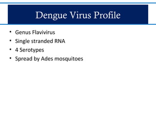
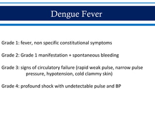
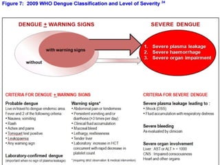
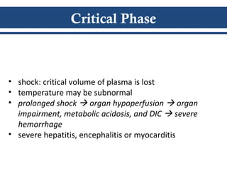
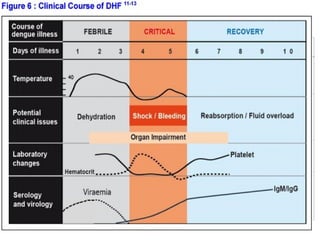
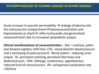
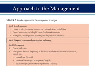
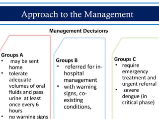
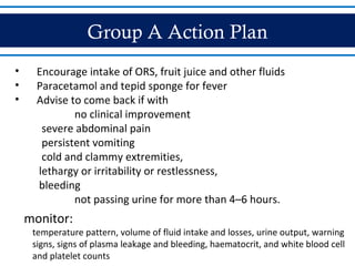
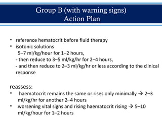
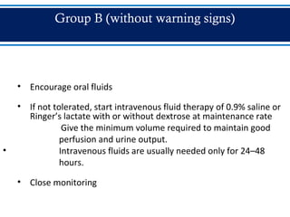
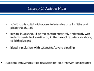
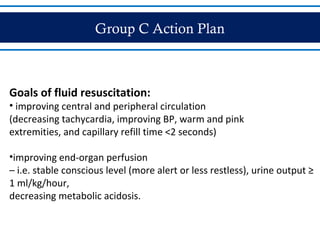
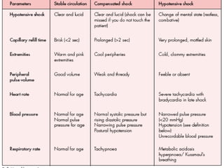
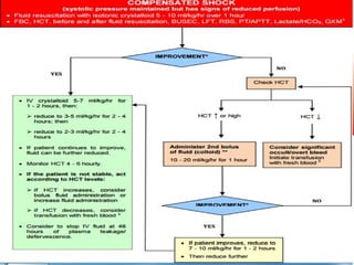
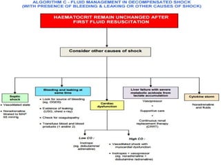
DENGUE
Dr Abhijeet Deshmukh
EPIDEMIOLOGY
• most rapidly spreading mosquito-borne viral disease in the world
• 50 million dengue infections annually
EPIDEMIOLOGY
Dengue Virus Profile
• Genus Flavivirus
• Single stranded RNA
• 4 Serotypes
• Spread by Ades mosquitoes
Host Factors
• Incubation period: 4-10 days
• Primary infection induce lifelong immunity to the
infecting serotype
• Protection from different serotype within 2-3 months
of primary infection
• No long-term cross-protective immunity
• Individual risk factors:
- Age, ethnicity, chronic diseases
Dengue Fever
• Three phases
• Febrile phase
• Critical phase
• Recovery phase
• Previously classified into
• undifferentiated fever, dengue fever and DHF
• Grade 1-IV
Dengue Fever
Grade 1: fever, non specific constitutional symptoms
Grade 2: Grade 1 manifestation + spontaneous bleeding
Grade 3: signs of circulatory failure (rapid weak pulse, narrow pulse
pressure, hypotension, cold clammy skin)
Grade 4: profound shock with undetectable pulse and BP
3 Phases
• Febrile Phase
• Critical Phase
• Recovery Phase
Febrile Phase
 facial flushing
 skin erythema
 generalized body ache
 myalgia and arthralgia
 headache
 sorethroat, injected pharynx,
and conjunctival injection
 anorexia, nausea and vomiting
 earliest abnormality -
progressive decrease in total wbc
• Sudden onset of
high-grade fever
• Lasts for 2-7 days
Critical Phase
• temperature drops to 37.5-38 (days 3-7)
• increase in capillary permeability with increasing
hematocrit levels
• significant plasma leakage lasts for 24-48 hours
• progressive leukopenia followed by rapid decrease in
platelet precedes plasma leakage
Critical Phase
• if (-) increase in capillary permeability  improve
• if (+) increase in capillary permeability  pleural
effusion and ascites
• degree of increase above the baseline hematocrit
reflects the severity of plasma leakage
Critical Phase
• shock: critical volume of plasma is lost
• temperature may be subnormal
• prolonged shock  organ hypoperfusion  organ
impairment, metabolic acidosis, and DIC  severe
hemorrhage
• severe hepatitis, encephalitis or myocarditis
Recovery Phase
• gradual reabsorption of extravascular compartment
fluid (48-72 hours)
• general well-being improves, appetite returns, GI
symptoms abate, hemodynamic status stabilizes and
diuresis ensues
Recovery Phase
• hematocrit stabilizes or may be lower due to dilutional
effect of reabsorbed fluid
• wbc starts to rise
• recovery of platelet count occurs later
PATHOPHYSIOLOGY OF PLASMA LEAKAGE IN SEVERE DENGUE :
Acute increase in vascular permeability  leakage of plasma into
the extravascular compartmenthaemoconcentration and
hypovolaemia or shock reflex tachycardia and generalised
vasoconstriction due to increased sympathetic output.
Clinical manifestations of vasoconstriction : Skin - coolness, pallor
and delayed capillary refill time. CVS- raised diastolic blood pressure
and a narrowing of pulse pressure . Renal system - reducing urine
output. GI- persistent vomiting, persistent diarrhoea and
abdominal pain. CNS– lethargy, restlessness, apprehension,
reduced level of consciousness. RS– tachypnoea (respiratory rate
>20/min)
Approach to the Management
•Triage
- Severe dengue
- With warning signs
-Non-urgent cases
Approach to the Management
Disease notification
• In dengue-endemic countries, cases of suspected, probable and
confirmed dengue should be notified
• suspected cases
• lives in or has travelled to a dengue-endemic area
• fever for three days or more
• low or decreasing white cell counts
• thrombocytopaenia ± positive tourniquet test.
Approach to the Management
Groups A
• may be sent
home
• tolerate
adequate
volumes of oral
fluids and pass
urine at least
once every 6
hours
• no warning signs
Groups B
• referred for in-
hospital
management
• with warning
signs, co-
existing
conditions,
Groups C
• require
emergency
treatment and
urgent referral
• severe
dengue (in
critical phase)
Management Decisions
Group A Action Plan
• Encourage intake of ORS, fruit juice and other fluids
• Paracetamol and tepid sponge for fever
• Advise to come back if with
no clinical improvement
severe abdominal pain
persistent vomiting
cold and clammy extremities,
lethargy or irritability or restlessness,
bleeding
not passing urine for more than 4–6 hours.
monitor:
temperature pattern, volume of fluid intake and losses, urine output, warning
signs, signs of plasma leakage and bleeding, haematocrit, and white blood cell
and platelet counts
Group B (with warning signs)
Action Plan
• reference hematocrit before fluid therapy
• isotonic solutions
5–7 ml/kg/hour for 1–2 hours,
- then reduce to 3–5 ml/kg/hr for 2–4 hours,
- and then reduce to 2–3 ml/kg/hr or less according to the clinical
response
reassess:
• haematocrit remains the same or rises only minimally  2–3
ml/kg/hr for another 2–4 hours
• worsening vital signs and rising haematocrit rising  5–10
ml/kg/hour for 1–2 hours
Group B (with warning signs)
Action Plan
Give minimum intravenous fluid volume: maintain good perfusion
and urine output of about 1 ml/kg/hr
• Intravenous fluids are usually needed for only 24–48 hours.
• Reduce intravenous fluids gradually when the rate of plasma
leakage decreases towards the end of the critical phase.
monitor:
• vital signs and peripheral perfusion (1–4 hourly until the
patient is out of the critical phase)
• urine output (4–6 hourly)
• hematocrit (before and after fluid replacement, then 6–12
hourly)
• blood glucose
• organ functions (renal profile, liver profile, coagulation profile)
Group B (without warning signs)
• Encourage oral fluids
• If not tolerated, start intravenous fluid therapy of 0.9% saline or
Ringer’s lactate with or without dextrose at maintenance rate
Give the minimum volume required to maintain good
perfusion and urine output.
• Intravenous fluids are usually needed only for 24–48
hours.
• Close monitoring
Group C Action Plan
• admit to a hospital with access to intensive care facilities and
blood transfusion
• plasma losses should be replaced immediately and rapidly with
isotonic crystalloid solution or, in the case of hypotensive shock,
colloid solutions
• blood transfusion: with suspected/severe bleeding
• judicious intravenous fluid resuscitation: sole intervention required
Group C Action Plan
Goals of fluid resuscitation:
• improving central and peripheral circulation
(decreasing tachycardia, improving BP, warm and pink
extremities, and capillary refill time <2 seconds)
•improving end-organ perfusion
– i.e. stable conscious level (more alert or less restless), urine output ≥
1 ml/kg/hour,
decreasing metabolic acidosis.
Common pitfalls in fluid therapy
• Treating patient with unnecessary fluid bolus based on raised
HCT as the sole parameter without considering other clinical
parameters.
• Excessive and prolonged fixed fluid regime in stable patients.
• Infrequent monitoring and adjustment of infusion rate.
• Continuation of intravenous fluid during the recovery phase.
• Inappropriate fluid therapy in patients with co-morbidities
(such as heart disease and renal disease).
Fluid Management
Treatment of Hemorrhagic Complications
Patients at risk of major bleeding are those who:
• prolonged/refractory shock;
• hypotensive shock and renal or liver failure and/or severe and
persistent metabolic acidosis
•given non-steroidal anti-inflammatory agents
• pre-existing peptic ulcer disease
• anticoagulant therapy
• any form of trauma
Treatment of Hemorrhagic Complications
• Blood transfusion is life-saving and should be given as soon as
severe bleeding is suspected or recognized
• Do not wait for the haematocrit to drop too low before deciding on
blood transfusion
• Risk of fluid overload.
Treatment of Hemorrhagic Complications
• Blood transfusion :
• 10-15 ml/kg of PRBC or 10-20 ml/kg FWB
• repeat if with further blood loss or no rise in hematocrit after
transfusion
• little evidence to support transfusion of platelet concentrate and
FFP
• massive bleeding not managed by FWB/PRBC
• may exacerbate fluid overload
Complications
•Fluid Overload
Causes:
– excessive and/or too rapid intravenous fluids;
– incorrect use of hypotonic rather than isotonic crystalloid
solutions;
– inappropriate use of large volumes of intravenous fluids in
patients with unrecognized severe bleeding;
– inappropriate transfusion of FFP, platelet concentrates and
cryoprecipitates;
– continuation of IVF after plasma leakage has resolved
– co-morbid conditions such as congenital heart disease, chronic
lung and renal diseases
Complications
Clinical Features:
– respiratory distress, difficulty
in breathing;
– Tachypnea
– chest wall in-drawing;
– wheezing
– large pleural effusions;
– tense ascites;
Other investigations:
• CXR
•ECG/Echo
•ABG
•When to discontinue IVF:
– stable blood pressure, pulse and peripheral perfusion;
– haematocrit decreases in the presence of a good pulse
volume;
– afebrile for more than 24–48 days (without the use of
antipyretics);
– resolving bowel/abdominal symptoms;
– improving urine output
•If necessary, give oral or intravenous furosemide 0.1–0.5
mg/kg/dose once or twice daily, or continuous infusion of
furosemide 0.1 mg/kg/hour.
• If the patient has stable haemodynamic status but is still within the
critical phase, reduce the intravenous fluid accordingly. Avoid
diuretics during the plasma leakage phase
• Patients who remain in shock with low or normal haematocrit
levels but show signs of fluid overload may have occult
haemorrhage.
• Careful fresh whole blood transfusion
• repeated small boluses
Criteria for Discharge
• Improved general well-being
• Afebrile for 24-48 hours
• Rising white cell count followed by platelet count
• Stable haematocrit
• Resolution/recovery of organ dysfunction
Thank You !!!
Dengue

Dengue

  • 1.
  • 2.
    EPIDEMIOLOGY • most rapidlyspreading mosquito-borne viral disease in the world • 50 million dengue infections annually
  • 3.
  • 4.
    Dengue Virus Profile •Genus Flavivirus • Single stranded RNA • 4 Serotypes • Spread by Ades mosquitoes
  • 5.
    Host Factors • Incubationperiod: 4-10 days • Primary infection induce lifelong immunity to the infecting serotype • Protection from different serotype within 2-3 months of primary infection • No long-term cross-protective immunity • Individual risk factors: - Age, ethnicity, chronic diseases
  • 6.
    Dengue Fever • Threephases • Febrile phase • Critical phase • Recovery phase • Previously classified into • undifferentiated fever, dengue fever and DHF • Grade 1-IV
  • 7.
    Dengue Fever Grade 1:fever, non specific constitutional symptoms Grade 2: Grade 1 manifestation + spontaneous bleeding Grade 3: signs of circulatory failure (rapid weak pulse, narrow pulse pressure, hypotension, cold clammy skin) Grade 4: profound shock with undetectable pulse and BP
  • 9.
    3 Phases • FebrilePhase • Critical Phase • Recovery Phase
  • 10.
    Febrile Phase  facialflushing  skin erythema  generalized body ache  myalgia and arthralgia  headache  sorethroat, injected pharynx, and conjunctival injection  anorexia, nausea and vomiting  earliest abnormality - progressive decrease in total wbc • Sudden onset of high-grade fever • Lasts for 2-7 days
  • 11.
    Critical Phase • temperaturedrops to 37.5-38 (days 3-7) • increase in capillary permeability with increasing hematocrit levels • significant plasma leakage lasts for 24-48 hours • progressive leukopenia followed by rapid decrease in platelet precedes plasma leakage
  • 12.
    Critical Phase • if(-) increase in capillary permeability  improve • if (+) increase in capillary permeability  pleural effusion and ascites • degree of increase above the baseline hematocrit reflects the severity of plasma leakage
  • 13.
    Critical Phase • shock:critical volume of plasma is lost • temperature may be subnormal • prolonged shock  organ hypoperfusion  organ impairment, metabolic acidosis, and DIC  severe hemorrhage • severe hepatitis, encephalitis or myocarditis
  • 14.
    Recovery Phase • gradualreabsorption of extravascular compartment fluid (48-72 hours) • general well-being improves, appetite returns, GI symptoms abate, hemodynamic status stabilizes and diuresis ensues
  • 15.
    Recovery Phase • hematocritstabilizes or may be lower due to dilutional effect of reabsorbed fluid • wbc starts to rise • recovery of platelet count occurs later
  • 17.
    PATHOPHYSIOLOGY OF PLASMALEAKAGE IN SEVERE DENGUE : Acute increase in vascular permeability  leakage of plasma into the extravascular compartmenthaemoconcentration and hypovolaemia or shock reflex tachycardia and generalised vasoconstriction due to increased sympathetic output. Clinical manifestations of vasoconstriction : Skin - coolness, pallor and delayed capillary refill time. CVS- raised diastolic blood pressure and a narrowing of pulse pressure . Renal system - reducing urine output. GI- persistent vomiting, persistent diarrhoea and abdominal pain. CNS– lethargy, restlessness, apprehension, reduced level of consciousness. RS– tachypnoea (respiratory rate >20/min)
  • 18.
    Approach to theManagement •Triage - Severe dengue - With warning signs -Non-urgent cases
  • 19.
    Approach to theManagement
  • 20.
    Approach to theManagement Disease notification • In dengue-endemic countries, cases of suspected, probable and confirmed dengue should be notified • suspected cases • lives in or has travelled to a dengue-endemic area • fever for three days or more • low or decreasing white cell counts • thrombocytopaenia ± positive tourniquet test.
  • 21.
    Approach to theManagement Groups A • may be sent home • tolerate adequate volumes of oral fluids and pass urine at least once every 6 hours • no warning signs Groups B • referred for in- hospital management • with warning signs, co- existing conditions, Groups C • require emergency treatment and urgent referral • severe dengue (in critical phase) Management Decisions
  • 22.
    Group A ActionPlan • Encourage intake of ORS, fruit juice and other fluids • Paracetamol and tepid sponge for fever • Advise to come back if with no clinical improvement severe abdominal pain persistent vomiting cold and clammy extremities, lethargy or irritability or restlessness, bleeding not passing urine for more than 4–6 hours. monitor: temperature pattern, volume of fluid intake and losses, urine output, warning signs, signs of plasma leakage and bleeding, haematocrit, and white blood cell and platelet counts
  • 23.
    Group B (withwarning signs) Action Plan • reference hematocrit before fluid therapy • isotonic solutions 5–7 ml/kg/hour for 1–2 hours, - then reduce to 3–5 ml/kg/hr for 2–4 hours, - and then reduce to 2–3 ml/kg/hr or less according to the clinical response reassess: • haematocrit remains the same or rises only minimally  2–3 ml/kg/hr for another 2–4 hours • worsening vital signs and rising haematocrit rising  5–10 ml/kg/hour for 1–2 hours
  • 24.
    Group B (withwarning signs) Action Plan Give minimum intravenous fluid volume: maintain good perfusion and urine output of about 1 ml/kg/hr • Intravenous fluids are usually needed for only 24–48 hours. • Reduce intravenous fluids gradually when the rate of plasma leakage decreases towards the end of the critical phase. monitor: • vital signs and peripheral perfusion (1–4 hourly until the patient is out of the critical phase) • urine output (4–6 hourly) • hematocrit (before and after fluid replacement, then 6–12 hourly) • blood glucose • organ functions (renal profile, liver profile, coagulation profile)
  • 25.
    Group B (withoutwarning signs) • Encourage oral fluids • If not tolerated, start intravenous fluid therapy of 0.9% saline or Ringer’s lactate with or without dextrose at maintenance rate Give the minimum volume required to maintain good perfusion and urine output. • Intravenous fluids are usually needed only for 24–48 hours. • Close monitoring
  • 26.
    Group C ActionPlan • admit to a hospital with access to intensive care facilities and blood transfusion • plasma losses should be replaced immediately and rapidly with isotonic crystalloid solution or, in the case of hypotensive shock, colloid solutions • blood transfusion: with suspected/severe bleeding • judicious intravenous fluid resuscitation: sole intervention required
  • 27.
    Group C ActionPlan Goals of fluid resuscitation: • improving central and peripheral circulation (decreasing tachycardia, improving BP, warm and pink extremities, and capillary refill time <2 seconds) •improving end-organ perfusion – i.e. stable conscious level (more alert or less restless), urine output ≥ 1 ml/kg/hour, decreasing metabolic acidosis.
  • 28.
    Common pitfalls influid therapy • Treating patient with unnecessary fluid bolus based on raised HCT as the sole parameter without considering other clinical parameters. • Excessive and prolonged fixed fluid regime in stable patients. • Infrequent monitoring and adjustment of infusion rate. • Continuation of intravenous fluid during the recovery phase. • Inappropriate fluid therapy in patients with co-morbidities (such as heart disease and renal disease).
  • 30.
  • 34.
    Treatment of HemorrhagicComplications Patients at risk of major bleeding are those who: • prolonged/refractory shock; • hypotensive shock and renal or liver failure and/or severe and persistent metabolic acidosis •given non-steroidal anti-inflammatory agents • pre-existing peptic ulcer disease • anticoagulant therapy • any form of trauma
  • 35.
    Treatment of HemorrhagicComplications • Blood transfusion is life-saving and should be given as soon as severe bleeding is suspected or recognized • Do not wait for the haematocrit to drop too low before deciding on blood transfusion • Risk of fluid overload.
  • 36.
    Treatment of HemorrhagicComplications • Blood transfusion : • 10-15 ml/kg of PRBC or 10-20 ml/kg FWB • repeat if with further blood loss or no rise in hematocrit after transfusion • little evidence to support transfusion of platelet concentrate and FFP • massive bleeding not managed by FWB/PRBC • may exacerbate fluid overload
  • 37.
    Complications •Fluid Overload Causes: – excessiveand/or too rapid intravenous fluids; – incorrect use of hypotonic rather than isotonic crystalloid solutions; – inappropriate use of large volumes of intravenous fluids in patients with unrecognized severe bleeding; – inappropriate transfusion of FFP, platelet concentrates and cryoprecipitates; – continuation of IVF after plasma leakage has resolved – co-morbid conditions such as congenital heart disease, chronic lung and renal diseases
  • 38.
    Complications Clinical Features: – respiratorydistress, difficulty in breathing; – Tachypnea – chest wall in-drawing; – wheezing – large pleural effusions; – tense ascites; Other investigations: • CXR •ECG/Echo •ABG
  • 39.
    •When to discontinueIVF: – stable blood pressure, pulse and peripheral perfusion; – haematocrit decreases in the presence of a good pulse volume; – afebrile for more than 24–48 days (without the use of antipyretics); – resolving bowel/abdominal symptoms; – improving urine output •If necessary, give oral or intravenous furosemide 0.1–0.5 mg/kg/dose once or twice daily, or continuous infusion of furosemide 0.1 mg/kg/hour.
  • 40.
    • If thepatient has stable haemodynamic status but is still within the critical phase, reduce the intravenous fluid accordingly. Avoid diuretics during the plasma leakage phase • Patients who remain in shock with low or normal haematocrit levels but show signs of fluid overload may have occult haemorrhage. • Careful fresh whole blood transfusion • repeated small boluses
  • 41.
    Criteria for Discharge •Improved general well-being • Afebrile for 24-48 hours • Rising white cell count followed by platelet count • Stable haematocrit • Resolution/recovery of organ dysfunction
  • 42.

Editor's Notes

  • #5 The genome is cleaved by host and viral proteases in three structural proteins (capsid, C, prM, the precursor of membrane, M, protein and envelope, E) and seven nonstructural proteins (NS).
  • #6 The genome is cleaved by host and viral proteases in three structural proteins (capsid, C, prM, the precursor of membrane, M, protein and envelope, E) and seven nonstructural proteins (NS).
  • #9 the classifi cation into levels of severity has a high potential for being of practical use in the clinicians’ decision as to where and how intensively the patient should be observed and treated (i.e. triage, which is particularly useful in outbreaks), in more consistent reporting in the national and international surveillance system, and as an end-point measure in dengue vaccine and drug trials.
  • #10 Clinical features are indistinguishable bet wevere and non-severe dengue cases therefore monitoring for warning signs and other clinical parameters is crucial to recognizing progression to the critical phase
  • #11 Clinical features are indistinguishable bet wevere and non-severe dengue cases therefore monitoring for warning signs and other clinical parameters is crucial to recognizing progression to the critical phase
  • #32 Reassess the patient’s clinical condition, vital signs, pulse volume, capillary refill time, urine output and temperature of extremities. ** Colloid is preferable if the patient has already received previous bolus of crystalloid IV = intravenous ; HCT = haematocrit ↑= increased ; ↓ = decreased 1GXM: require 1st stage cross match or emergency O 2 fresh blood : less than 5 days, either packed cells or whole blood
  • #34 All the above types of shocks need to be supported by echocardiography and non-invasive cardiac output monitoring and treatments tailor to each patient. HCT = haematocrit ; MAP = mean arterial pressure ; CO = cardiac output; OGDS = oesophagogastroduodenoscopy USG = ultrasonography ↑= increased ; ↓ = decreased 1GXM: require 1st stage cross match or emergency O 2 fresh blood : less than 5 days