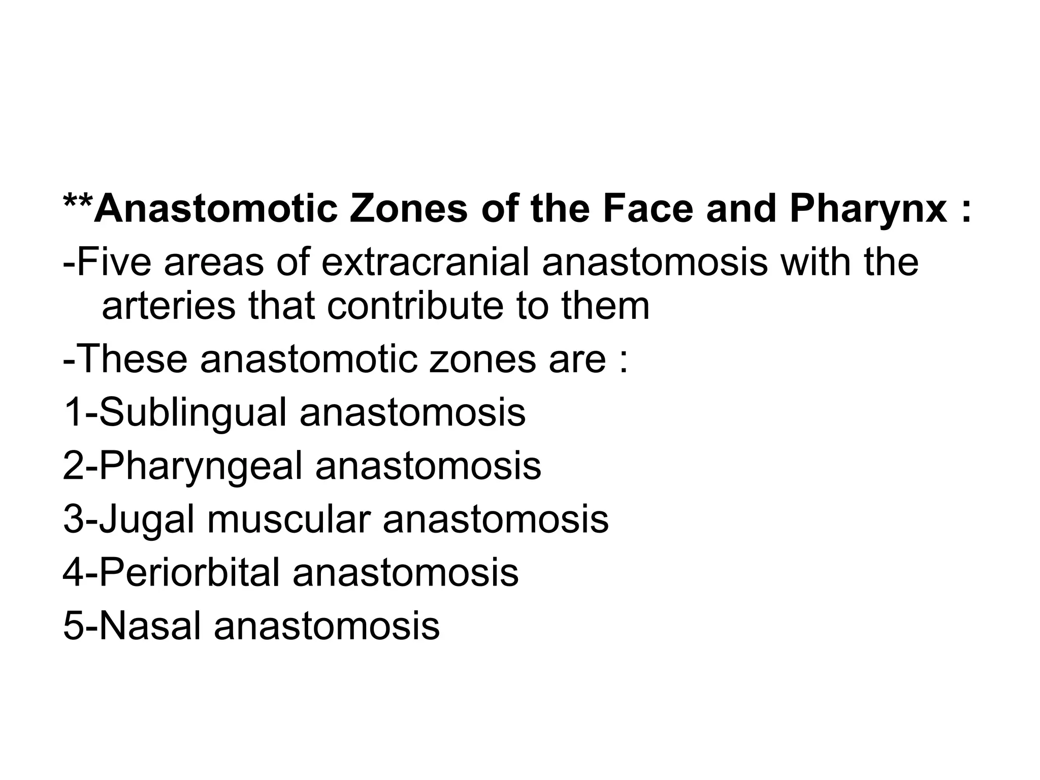
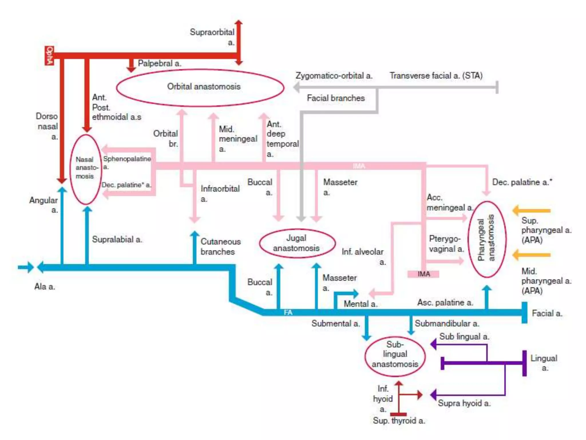
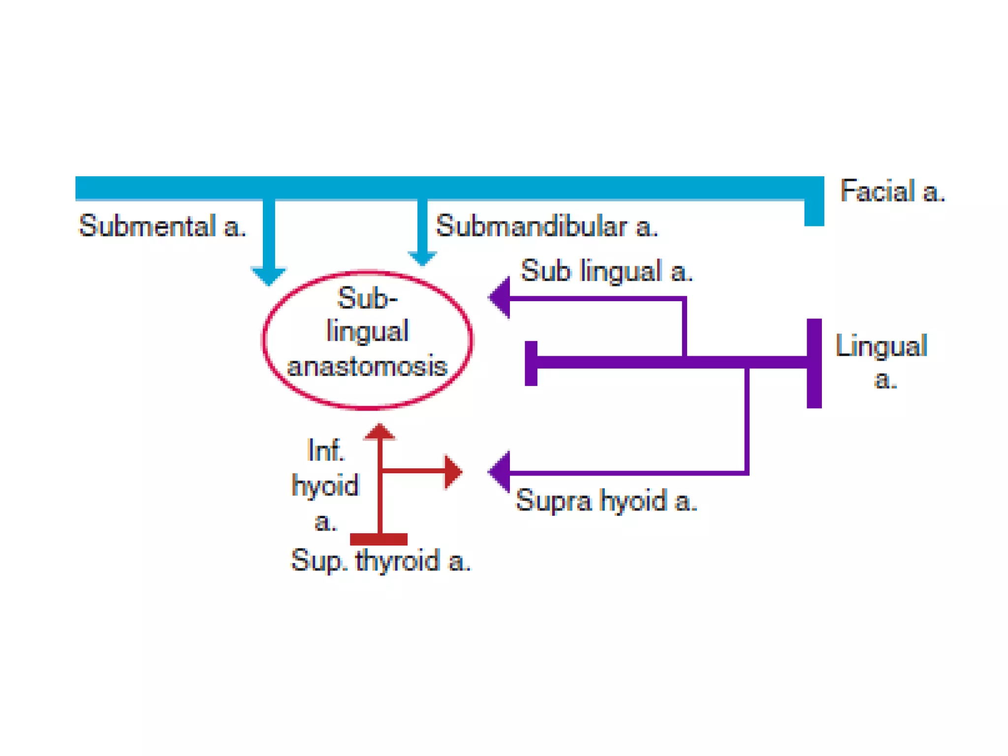
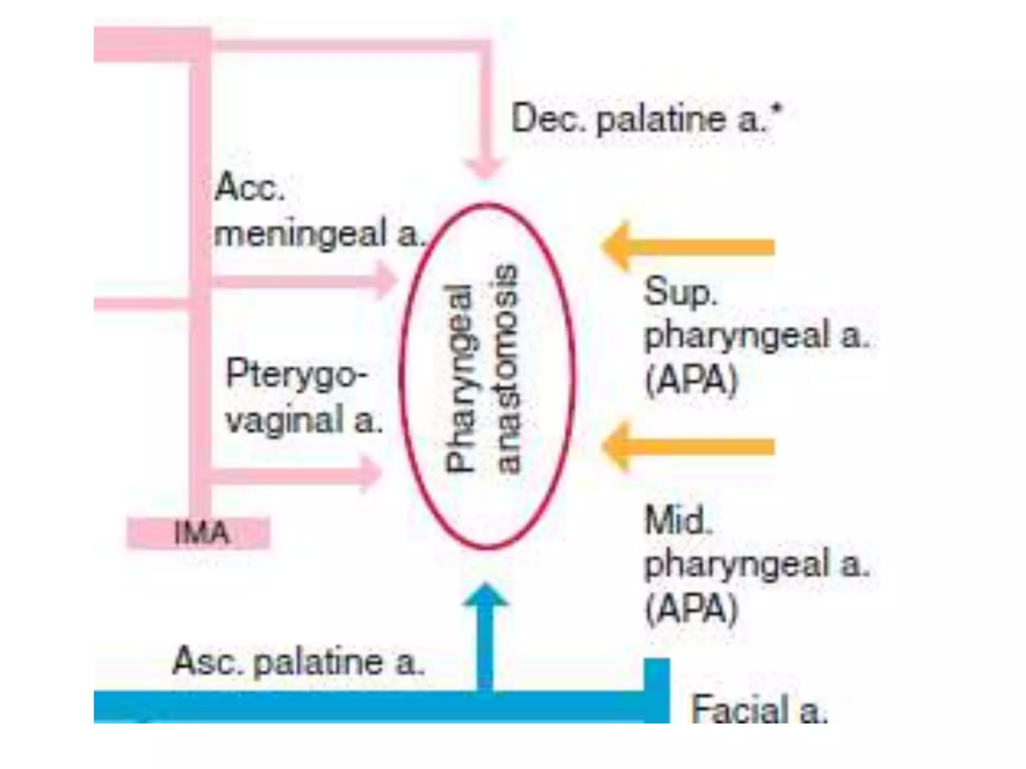
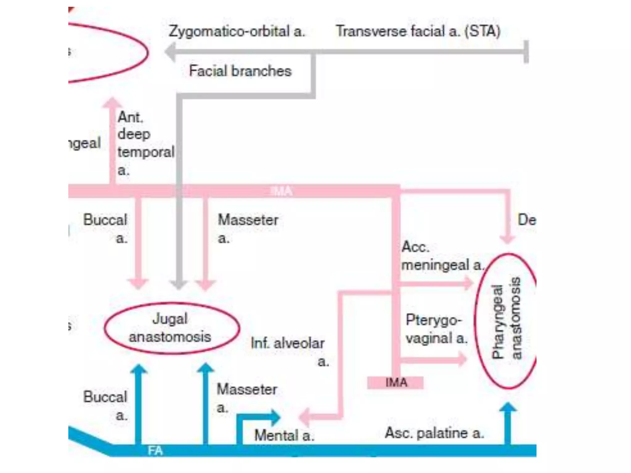
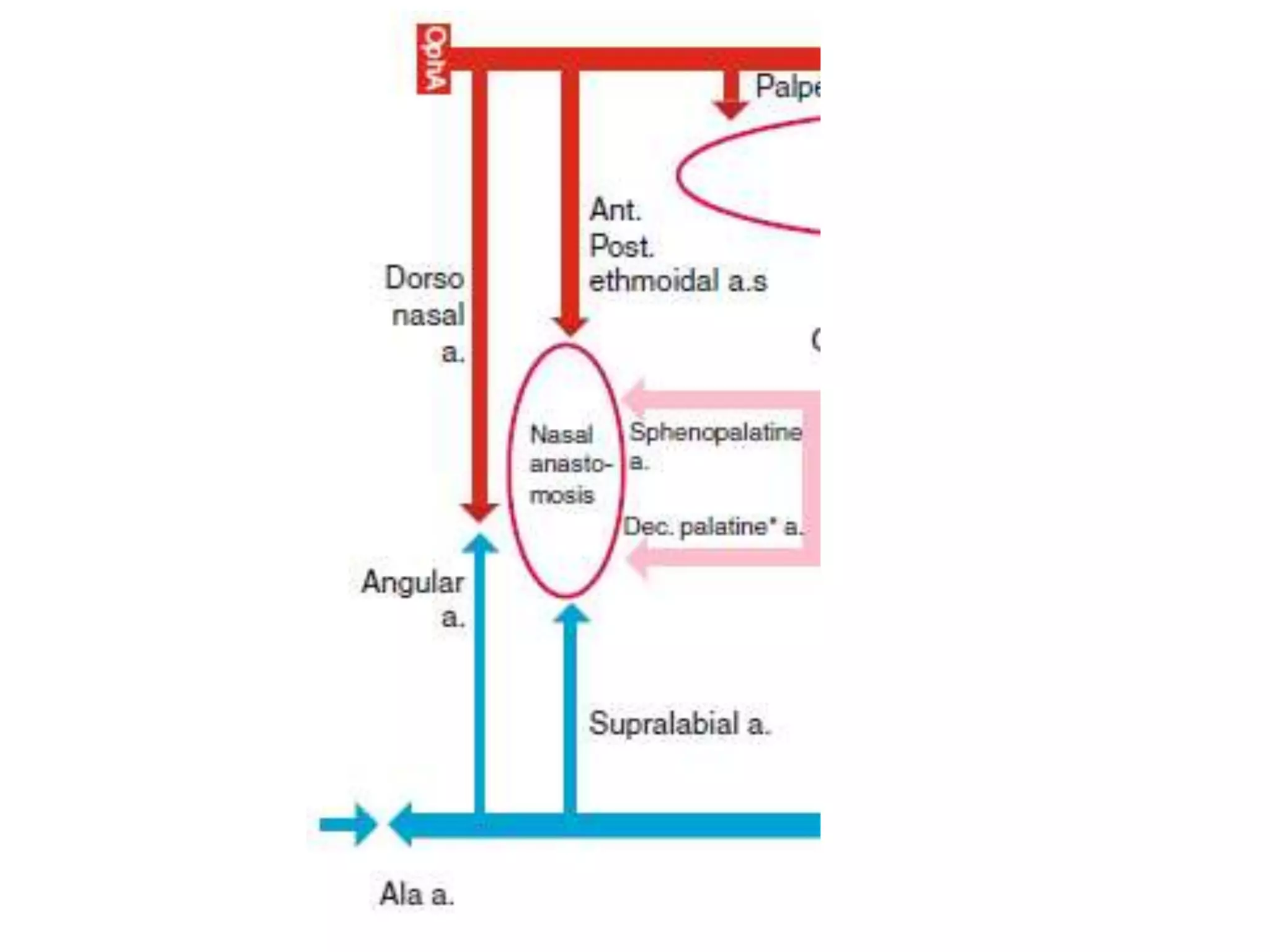
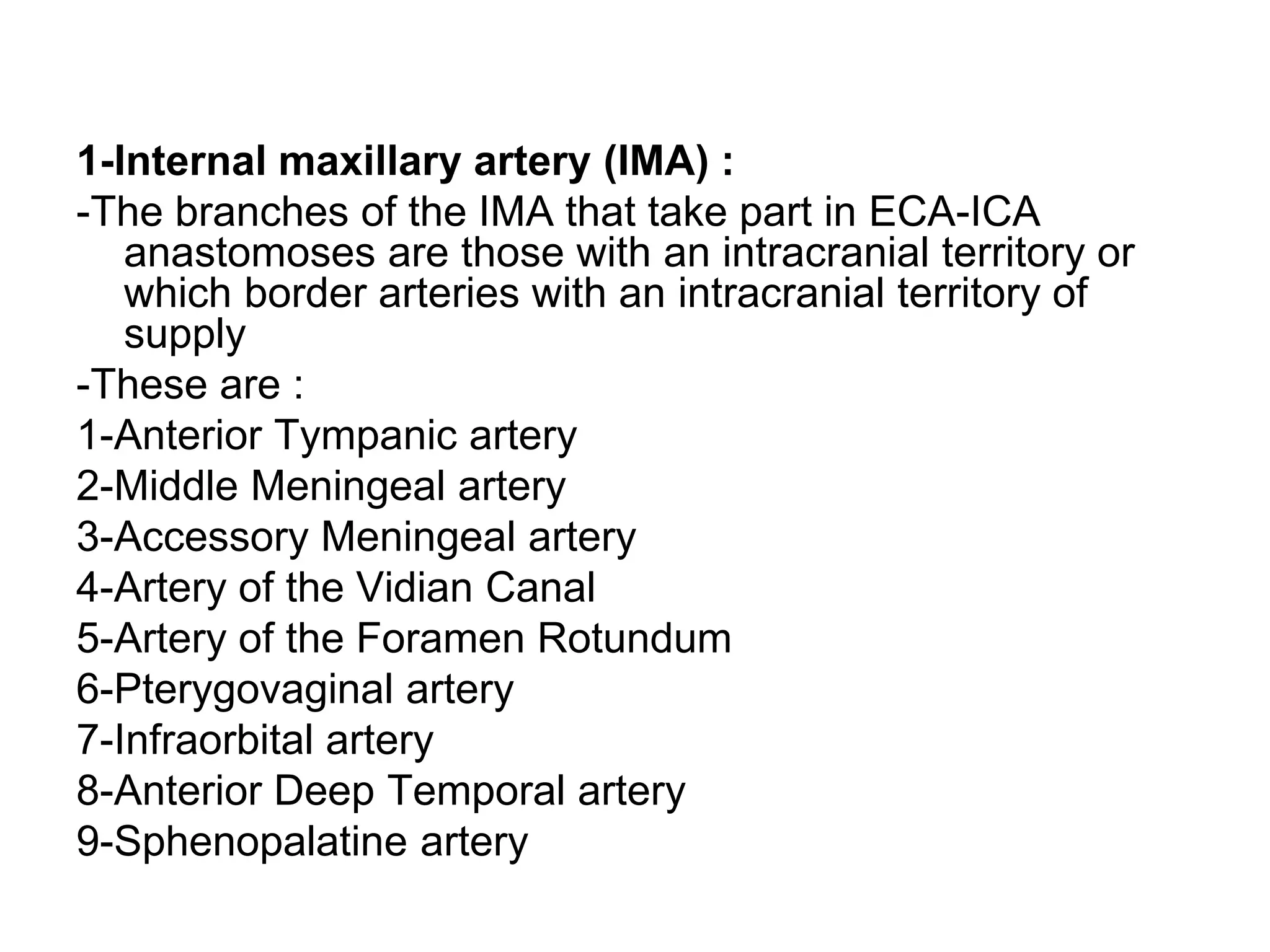
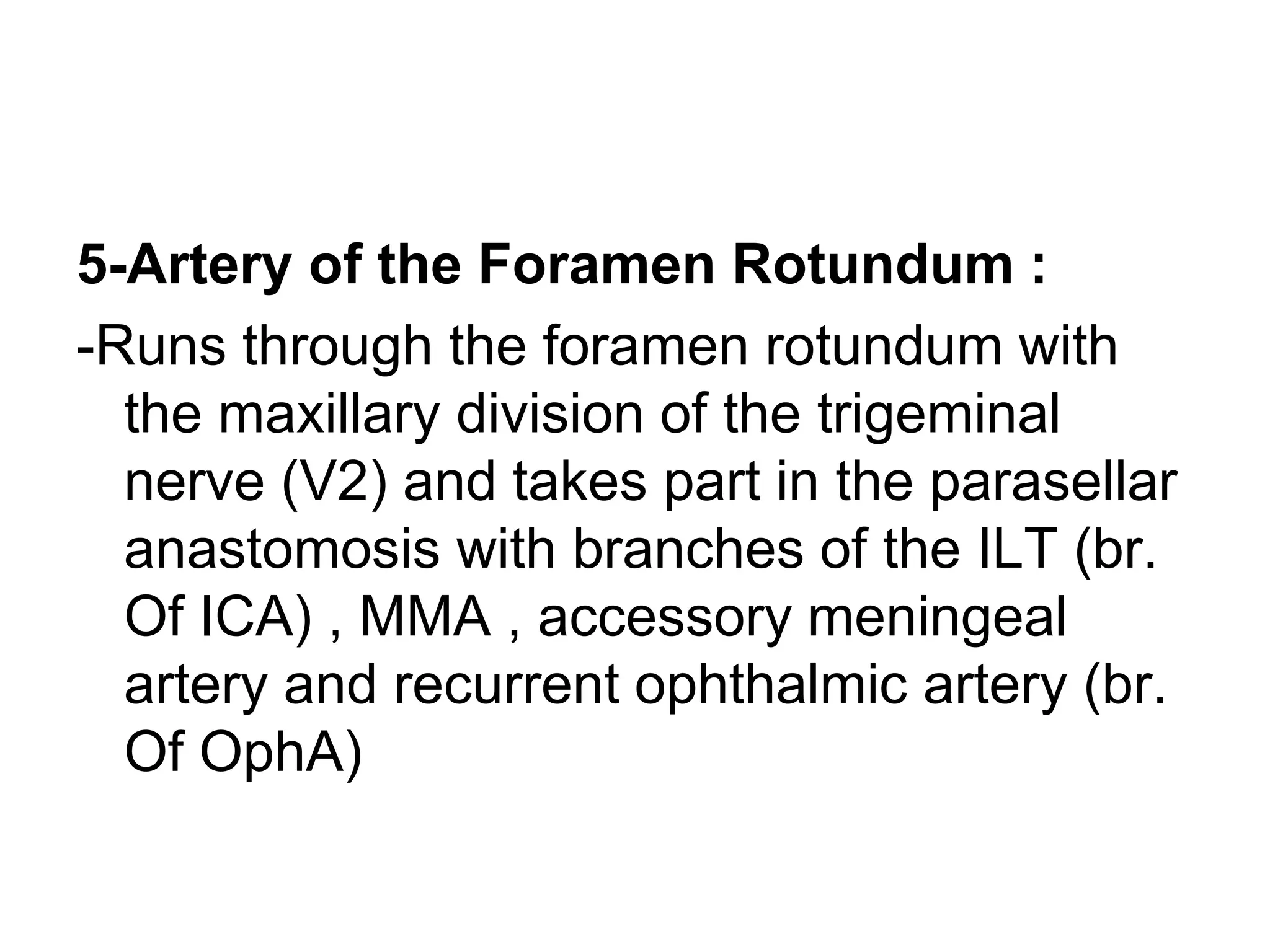
Interventional Neuroradiology
Cranial Anastomoses and
Dangerous Vascular Connections
Mohamed M.A. Zaitoun, MD
Interventional Radiology Consultant, Zagazig University Hospitals, Egypt
FINR-Switzerland
zaitoun82@gmail.com
Interventional Radiology Unit,
Zagazig University, Egypt
Knowing as much as possible
about your enemy precedes
successful battle and learning
about the disease process
precedes successful
management.
Cranial Anastomoses and Dangerous
Vascular Connections
a) Anastomoses Between Superficial Anterior
Branches of the ECA
b) Anastomoses Between Deep Anterior Branches
of the ECA
c) Anastomoses Between Posterior Branches of
the External Carotid Artery
d) Arterial Supply to the Cranial Nerves
a) Anastomoses Between Superficial
Anterior Branches of the ECA :
-The classic description of the ECA primary
branches in the order they arise (from
proximal to distal) is not helpful in
identifying their territorial borders because
branches directed posteriorly may arise
adjacent to an anterior directed branch
and that is the limit of their proximity
-Therefore , the ECA territories will be considered in
superficial and deep groups , separated into those
supplying anterior or posterior tissues
-To separate them into anterior or posterior groups , they
will be described as running either anterior or posterior to
an arbitrary line extending from the ECA origin to the
proximal portion of the superficial temporal artery (STA)
-This is a purely descriptive division which the paths of
some branches straddle (e.g. STA and APA) and others
cross (e.g. MMA)
-The superficial groups are less likely to involve dangerous
EC-IC anastomoses but the extensive anastomoses
between arteries supplying the face need to be
interpreted for treatment planning to avoid collateral
tissue damage
-Anastomoses between the anterior branches of
the ECA will be described based on the central
contribution of the facial artery (FA)
-The anterior branches involved are :
a) Superior thyroid artery
b) Lingual artery
c) Facial artery
d) Internal maxillary artery (IMA)
e) Ascending pharyngeal artery (APA)
a) Superior thyroid artery :
-This artery supplies the larynx and thyroid gland
-A branch (inferior hyoid artery) contributes to the
sublingual anastomosis and gives a branch to
anastomose with the suprahyoid artery (br. Of lingual
artery) around the hyoid
-It also anastomoses with the opposite side via another
branch (cricothyroid artery)
-Its terminal branches to the thyroid gland anastomose with
the inferior thyroid artery
-The infrahyoid artery is thus a potential bypass route
between FA and lingual artery
b) Lingual artery :
-The lingual artery territory lies between the
vascular territories of the FA and superior thyroid
arteries
-Its proximal branches , takes part in anastomoses
with these vessels in the sublingual and
suprahyoid regions
-It supplies the sublingual gland , floor of mouth
and the tongue
-It gives the suprahyoid artery which anastomoses
with the superior thyroid artery and the
sublingual artery which takes part in the
sublingual anastomosis around the sublingual
gland
c) Facial artery :
-The long course of the facial artery , from its origin
medial to the stylohyoid muscle and the
posterior belly of digastric muscle to its
termination medial to the orbit , borders several
territories of ECA and ophthalmic artery (OphA)
branches
-Its named branches that border and anastomose
with adjacent arterial territories are :
1-Ascending palatine artery to the
pharyngeal anastomosis
2-Submental and smaller submandibular
arteries to the sublingual anastomosis
3-Middle mental artery which anastomoses
with the mental artery over the chin , the
mental artery is a terminal branch of the
inferior alveolar artery (br. of IMA)
4-Branches to masseter and buccinator
muscles which anastomose with their
counterparts arising from the IMA over a
wide area of the cheek region in the jugal
muscular anastomosis
5-Cutaneous branches anastomose with
the inferior orbital artery (br. of IMA) and
branches of the transverse facial artery
(br. of STA) on the cheek
6-Cutaneous branches anastomose with inferior
orbital artery (br. of IMA) , transverse facial
artery (br. of STA) and the zygomatico-orbital
artery (br. of STA) in the periorbital anastomosis
7-Angular artery which anastomoses with the
dorsal nasal artery and supratrochlear artery
(extraorbital terminal branches of OphA) and the
inferior palpebral artery (br. of OphA) though the
latter usually anastomoses with the infraorbital
artery (br. of IMA)
8-Alar artery , which supplies the nostrils ,
anastomoses with the contralateral facial
artery and an extensive network of arteries
over the nose , it can take part in the nasal
(mucosal) anastomosis with terminal
branches of the sphenopalatine artery (br.
of IMA)
d) Internal maxillary artery (IMA) :
-The internal maxillary artery supplies the temporal
and pterygoid regions of the face , paranasal
sinuses , jaw and nose as well as giving
transosseous intracranial branches to supply
dura of the anterior cranium
-Its territory of supply can be divided into an
extracranial territory (which borders the
territories of the FA , APA , STA and OphA) and
an intracranial territory (which is the site of
potentially dangerous anastomoses with the
ICA)
-The branches of the IMA that take part in
anastomoses with adjacent anterior
arterial territories will be described
-These boundary connections are potential
routes for collateral support in the event of
a proximal arterial occlusion and pathways
for inadvertent spread of embolic agents
during endovascular treatments :
1-Inferior alveolar artery , via its mental
branch , anastomoses with the FA territory
over the chin
2-Masseter and buccal arteries to the
muscles and deep soft tissue of the cheek
take part in the jugal muscular
anastomosis with the FA branches and the
transverse facial artery (br. of STA)
3-Lesser descending palatine artery takes part
in the pharyngeal anastomosis , it supplies the
soft palate and oropharynx and anastomoses
principally with the middle pharyngeal artery (br.
of APA)
4-Infraorbital artery takes part in the cutaneous
periorbital anastomosis with cutaneous
branches of FA and transverse facial artery (br.
of STA) , it also participates in the orbital
anastomosis with the OphA
5-Sphenopalatine artery and greater
descending palatine artery are distal
branches of IMA that supply the nasal
mucosa and take part in the nasal
anastomosis with the alar and superior
labial arteries (br. Of FA)
e) Ascending pharyngeal artery (APA) :
-Anterior branches of the APA contribute to
the pharyngeal anastomosis but they are
deep branches which will be considered in
the next section
**Anastomotic Zones of the Face and Pharynx :
-Five areas of extracranial anastomosis with the
arteries that contribute to them
-These anastomotic zones are :
1-Sublingual anastomosis
2-Pharyngeal anastomosis
3-Jugal muscular anastomosis
4-Periorbital anastomosis
5-Nasal anastomosis
1-Sublingual anastomosis :
-An anastomosis around the sublingual gland and
base of the tongue which is usually dominated
by :
1-The submental artery and smaller
submandibular arteries (br. of FA) and links it
with
2-The infrahyoid artery (br. of superior thyroid
artery) and
3-Sublingual artery (br. of lingual artery)
2-Pharyngeal anastomosis :
-There is an extensive potential anastomotic network in the
mucosa of the pharynx which involves :
1-The ascending palatine artery (br. of FA)
2-The middle pharyngeal artery (br. of APA)
3-Lesser descending palatine artery (br. of IMA) in the
oropharynx
4-The superior pharyngeal artery (br. of APA)
5-Accessory meningeal artery (br. Of IMA)
6-The pterygovaginal artery (br. Of IMA) in the
nasopharynx
-It reflects the rich blood supply to the
mucosa , particularly of the soft palate and
uvula
-Thus is a potential site of EC-I
anastomoses since the accessory
meningeal artery has branches that
anastomose to the ICA
3-Jugal muscular anastomosis :
-The branches of the FA to masseter and buccinator
muscles reciprocate their territory of supply with the
branches of the IMA to the same muscles , thus
1-Masseter branches of the FA anastomose with masseter
branches of the IMA and the transverse facial artery (br.
of STA)
2-More superficial and anterior to these vessels on the
lateral angiogram , are anastomoses between the buccal
arteries of the IMA and muscular branches of the FA ,
this is a potential route for collaterals to develop in the
event of occlusion of IMA , FA and ECA
4-Periorbital anastomosis :
-The FA supplies the medial orbital margin , via the angular
artery which borders and anastomoses with the
territories of the supratrochlear artery and dorsal nasal
artery arising from OphA and supplying the
superomedial orbital margin
-The superior margin is supplied by the supraorbital artery
(br. of OphA) and borders the territory of the frontal
branch of the STA
-The inferior orbital margin is supplied by the inferior orbital
artery (br. of IMA) which border the cutaneous territory of
the FA on the cheek and the lateral margin by the
zygomatico-orbital artery (br. of STA)
-The inferior and superior palpebral arteries are branches
of the OphA and anastomose with the infraorbital artery
(br. of IMA) , frontal branch the STA and the angular
artery (br. of FA)
5-Nasal anastomosis :
-The principle source of blood supply to the nasal
mucosa is from the sphenopalatine artery (br. of
IMA) and descending palatine artery (br. of IMA)
-These arteries anastomose with nasal branches
of the alar artery and the superior labial artery
(brs. of FA) inferiorly and with the anterior and
posterior ethmoidal arteries (br. of OphA)
superiorly
-The anterior ethmoidal artery supplies the upper
nasal septum
b) Anastomoses Between Deep Anterior
Branches of the ECA :
-These involve the anterior deep branches
arising from the IMA and APA
-Since the majority of the APA branches are
directed posteriorly , the connections
described in this section principally involve
branches of IMA
-Arteries that supply dura , cranial nerves and the
organs of the special senses are sites of
anastomoses between the ECA and ICA , and
therefore particularly dangerous EC-IC
connections
-They generally enter the cranium through the
foramen in the skull base (though blood fl ow in
both directions occurs)
-The external carotid branches involved are :
1-Internal maxillary artery (IMA)
2-Ascending pharyngeal artery (APA)
1-Internal maxillary artery (IMA) :
-The branches of the IMA that take part in ECA-ICA
anastomoses are those with an intracranial territory or
which border arteries with an intracranial territory of
supply
-These are :
1-Anterior Tympanic artery
2-Middle Meningeal artery
3-Accessory Meningeal artery
4-Artery of the Vidian Canal
5-Artery of the Foramen Rotundum
6-Pterygovaginal artery
7-Infraorbital artery
8-Anterior Deep Temporal artery
9-Sphenopalatine artery
1-Anterior Tympanic artery :
-Supplies the middle ear which it reaches
through the petrotympanic fissure
accompanied by the chorda tympani
-It takes part in the anastomosis of the
middle ear which connects it with the
caroticotympanic trunk (CCT) of the ICA
2-Middle Meningeal artery :
-Is a route for several real and potential anastomoses with
the ICA and OphA
**ICA :
a) Petrous branch :
-Which arises soon after the artery passes through the
foramen spinosum) gives the superior tympanic artery to
supply the middle ear and participates in the middle ear
anastomosis with branches of CCT (br. Of ICA)
b) Cavernous branch :
-To supply dura in the parasellar and petrosal regions (ILT–
ICA) and lateral clival artery (br. Of meningohypophyseal
trunk)
**OphA :
c) Sphenoid branches :
-Run along the greater wing of the sphenoid to supply dura
and may enter the orbit via the superior orbital fissure to
anastomose with a recurrent branch of the OphA
d) The terminal territory of the frontal branch :
-Supplies the anterior falx and may anastomose with the
anterior ethmoidal artery (br. of OphA)
e) Branch to the lacrimal gland :
-Not infrequent variant (that enters the lateral orbit through
Hyrtl’s canal) termed the meningo-orbital or
meningolacrimal artery (depending on the extent of its
territory) and creates a potential collateral route to the
OphA
3-Accessory Meningeal artery :
-This small artery gives dural branches in the
middle cranial fossa which contribute to the :
a) Parasellar dural anastomosis with branches of
the ILT (br. of ICA)
b) To the anastomosis around the Eustachian tube
with the mandibular artery (br. of mandibulo-
Vidian trunk (MVT)) , the pterygovaginal artery
(br. of IMA) and the Eustachian branch of the
APA
4-Artery of the Vidian Canal :
-Anastomoses with the mandibular artery
and takes part in the Eustachian
anastomosis (MVT-ICA)
-Its origin is variable either from the
mandibular artery and running IC to EC ,
or its origin is from the IMA and it runs EC
to IC
5-Artery of the Foramen Rotundum :
-Runs through the foramen rotundum with
the maxillary division of the trigeminal
nerve (V2) and takes part in the parasellar
anastomosis with branches of the ILT (br.
Of ICA) , MMA , accessory meningeal
artery and recurrent ophthalmic artery (br.
Of OphA)
6-Pterygovaginal artery :
-Supplies the Eustachian tube and takes
part in the Eustachian anastomosis
7-Infraorbital artery :
-Whilst in the inferior orbital fissure , this artery
gives the inferomedial muscular artery or a
series of branches which supply the lacrimal sac
, nasolacrimal duct and extraocular muscles
through which it anastomoses with branches of
the OphA
-It also anastomoses with the recurrent ophthalmic
artery (OphA) which returns through the inferior
orbital fissure and takes part in the parasellar
anastomosis with MMA , accessory meningeal
artery and artery of the foramen rotundum (brs.
of IMA) and the ILT (br. of ICA)
-After emerging from the infraorbital foramen
, it anastomoses with the inferior palpebral
artery and dorsonasal artery which are
respectively terminal branches of the
OphA in the anterior orbit and cutaneous
branches of FA (see periorbital
anastomosis)
8-Anterior Deep Temporal artery :
-Runs on the surface of the skull , under
temporalis which , along with the middle deep
temporal artery (br. of IMA) and posterior deep
temporal artery (br. of STA) , it supplies
-Unlike them , it gives a branch (the lateral
muscular artery) that enters the lateral orbit and
anastomoses with the lacrimal artery (OphA)
-This branch either enters the orbit via the inferior
orbital fissure or directly through the lateral
orbital wall , in the latter case , it is then called
the transmalar branch
9-Sphenopalatine artery :
-Branches of this artery supply most of the
nasal mucosa and anastomose with
branches of the anterior ethmoidal artery
(br. of OphA) in the roof of the nose , the
septal branch of the descending palatine
(br. of IMA) and branches of FA at the
nostrils
2-Ascending Pharyngeal artery (APA) :
-The APA is the smallest and usually first branch
of ECA
-It divides early in its course into anterior and
posterior divisions
-The latter , with the inferior tympanic artery will be
considered in the next section
-The anterior division gives the inferior (a small
and usually unrecognized artery) , middle and
superior pharyngeal arteries
-These supply the mucosa of the pharynx and soft
palate , the middle and superior constrictor
muscles
-Both middle pharyngeal artery and superior
pharyngeal artery take part in the pharyngeal
anastomoses with branches of the ascending
palatine artery (br. of FA) and the superior
pharyngeal artery anastomoses with the
accessory meningeal artery (br. of IMA)
-It takes part in the Eustachian anastomosis to
which it gives a Eustachian branch or branches
and thus may connect to the ICA via branches of
the MVT or the ITL
**Anastomotic Zones of the Skull Base :
(i) Anterior Group
-There are three main anterior zones of
extracranial anastomosis between the deep
branches of the IMA and ICA or OphA
-These comprise :
1-Orbital Zone
2-Parasellar Zone
3-Eustachian Zone
1-Orbital Zone :
-The principle artery supplying structures found within the
orbit is the OphA
-Additional marginal supply arteries or anatomical variants
(as described before) are potential routes for the
inadvertent spread of embolic agents to the retina
-These involve :
1-The inferomedial muscular artery (br. Of infraorbital
artery) and the lateral muscular artery (br. of anterior
deep temporal artery) supply the medial and lateral orbit
respectively and anastomose with the OphA territory
-In the event of ICA or OphA narrowing or occlusion , the
anterior deep temporal artery route is more frequently
recruited than the former
2-The anterior branch of the ILT passes through the
superior orbital fissure to anastomose with the recurrent
ophthalmic artery (br. of OphA)
3-The MMA may contribution to the orbital
anastomosis via the meningoorbital artery (or
meningolacrimal artery)
-This artery , if present , passes through Hyrtl’s
canal and subsumes the territory of the lacrimal
branch of OphA
-It is the most common of the possible variants that
reflect the embryonic origin of the middle
meningeal territory from the hyoid / stapedial
system and a dangerous connection between
the OphA and the IMA
-The existence of a possible intraorbital supply
when embolising in the MMA should always be
anticipated
4-The anterior and posterior ethmoidal arteries
(brs. of OphA) supply the ethmoid sinuses
-The anterior ethmoidal artery gives a branch to
the falx cerebri through the foramen caecum and
both supply the roof of the nasal cavity and may
give branches through the cribriform plate to the
anterior fossa dura
-Their territories thus border the sphenopalatine
artery (br. of IMA) territory and the anterior
territory of MMA
-These anastomoses via the ethmoidal arteries
thus constitute potential routes for ECA-OphA-
ICA connections
-Rarely , the frontopolar artery (br. of ACA)
supplies the anterior falx via a transpial branch
and thus creates an additional ECA-ICA
connection
2-Parasellar Zone :
-In the parasellar region , the ILT and
meningohypophyseal trunk (MHT) arise
from the ICA
-They give branches which anastomose with
branches of IMA and APA
-The posterior branches of the ILT are
closely related to the dural branches of the
MHT
-The ILT is the key to understanding the real
and potential anastomoses that exist in the
parasellar region :
1-The Anterior Branch of the ILT :
-Anastomoses with the recurrent ophthalmic artery
(br. Of OphA) , the artery of the foramen
rotundum (IMA) and the accessory meningeal
artery (br. of IMA)
-The accessory meningeal artery may be directed
to the posterior branch of the ILT
-The anastomosis with the artery of the foramen
rotundum is a common collateral route when the
ICA is occluded proximal to the ILT
2-The Posterior Branch of the ILT :
-Anastomoses with the cavernous branch of the
MMA and gives the recurrent artery of the
foramen lacerum
-The recurrent artery of the foramen lacerum
usually arises from ILT but may arise from the
MHT
-It anastomoses with the superior pharyngeal
artery (br. of APA) around the foramen lacerum
3-Eustachian Zone :
-The Eustachian or pharyngotympanic tube lies in
the angle between the petrous and tympanic
parts of the temporal bone
-Its anterior cartilaginous portion pieces the
pharyngobasilar fascia above the superior
constrictor to open into the pharynx
-It is the focus for an anastomotic network between
IMA , APA branches and the mandibular artery
-The mandibular artery arises from the mandibulo-
Vidian trunk of the ICA but does not always
persist after birth
-If present , it is a potential EC-IC anastomotic
route :
1-The accessory meningeal artery (br. Of IMA) and
the carotid branch of the superior pharyngeal
artery (br. of APA) supply the Eustachian tube
and the mucosa at its pharyngeal opening
2-The pterygovaginal artery arises from the distal
IMA and anastomoses with the inferomedial
branch of the accessory meningeal artery at the
meatus of the Eustachian tube
3-The inconstant mandibular artery also takes part
in this anastomosis
-This artery arises from the petrous portion of the
ICA , divides in the foramen lacerum into a
branch which enters the pterygoid canal and
anastomoses with the artery of the pterygoid
canal (Vidian) and an inferior branch (the
mandibular artery) which takes part in the
anastomoses around the Eustachian tube
(ii) Posterior Group :
-There are two main posterior zones of
extracranial anastomosis between the
deep branches of the IMA and ICA
-These comprise the :
1-Clival Zone
2-Middle Ear Zone
1-Clival Zone :
-A series of dural arteries anastomose in the
region of the clivus
-They connect branches of the parasellar region
with those entering the cranium through the
foramen magnum and the hypoglossal and
jugular foramen
-The MHT arises from the proximal section of the
cavernous portion of the ICA
-It supplies the pituitary via the posterior
hypophyseal artery and gives branches to the
tentorium and dura over the clivus and petrous
margin of the posterior fossa
-The clivus and adjacent dura is supplied by IMA
(and vertebral artery) branches :
1-Basal Tentorial artery :
-Arising from the MHT , the basal tentorial artery
supplies the tentorium over its anteromedial
portion and borders the territory of the
petrosquamosal artery (br. of MMA)
2-Lateral Clival artery :
-The MHT also gives the lateral clival artery whose
inferomedial branch anastomoses with the
jugular artery (br. of APA) in the jugular fossa
3-The Medial Clival artery :
-The Posterior Inferior Hypophyseal artery (PIHA) arises
from the MHT and from it the medial clival artery usually
arises
-This is a dural artery with a territory medial to the lateral
clival artery
-It descends along the clivus and anastomoses with the
clival branches of the hypoglossal artery (br. Of APA)
and with branches of the small anterior meningeal artery
(br. of VA)
-This is a particularly dangerous ECA-ICA route that should
be considered when performing embolisation in the APA
-The anterior meningeal artery supplies the inferior clivus
and also takes part in the anastomoses forming the
odontoid arterial arcade
2-Middle Ear Zone :
-The inner ear is supplied by intracranial branches
of ICA , IMA , APA and the occipital artery (OA)
-Anastomoses , within the middle ear cavity , are
potential collateral routes that link branches of
the IMA , ICA and distal ECA
-The arterial arcade that follows the facial nerve
(VIIth) cranial nerve , links the stylomastoid
artery (br. of OA or posterior auricular artery
(PA)) with the anterior tympanic artery (br. Of
IMA) and the superior tympanic artery (br. Of
MMA)
-The superior tympanic artery and the inferior
tympanic artery (br. of APA) anastomose and
the inferior tympanic artery also anastomoses
with branches of the caroticotympanic trunk (br.
of ICA) :
1-Stylomastoid artery
2-Superior Tympanic artery
3-Anterior Tympanic artery
4-Inferior Tympanic artery
5-Labyrinthine artery
1-Stylomastoid artery :
-Which may arise from either OA or PA enters the middle
ear via the stylomastoid foramen and runs with the
postganglionic VIIth cranial nerve
2-The Superior Tympanic artery :
-Arises from the petrous branch of the MMA in the middle
fossa
-It enters the tympanic cavity with the greater superficial
petrosal nerve , supplies the geniculate ganglion of the
VIIth nerve and anastomoses with the stylomastoid
artery
3-The Anterior Tympanic artery :
-(br. of IMA)
-Runs posteriorly from its origin and enters
the inner ear cavity with the chorda
tympani (parasympathetic to salivary
glands) through the petrotympanic fissure
-It anastomoses with the stylomastoid artery
around the VIIth nerve and with branches
of the caroticotympanic trunk
4-The Inferior Tympanic artery :
-(br. of APA)
-Enters the middle ear from below , via the inferior
tympanic canaliculus accompanied by
Jacobson’s nerve (tympanic branch of IXth
cranial nerve) , and it branches to anastomose
with branches of the superior tympanic artery
(MMA) and those arising from the
caroticotympanic trunk (ICA)
-The caroticotympanic trunk (CCT) lies at the
junction of the ascending and horizontal petrous
ICA , i.e. proximal to the foramen lacerum
-It gives small branches which are described as
running posteriorly into the middle ear , but
these are, invisible on angiography
5-The Labyrinthine artery :
-(br. of basilar artery or the anterior inferior
cerebellar artery)
-Supplies the internal acoustic canal and
contributes to the subarcuate dural anastomosis
-It does not normally contribute to the anastomosis
in the tympanic cavity but rarely collateral flow
between branches of the labyrinthine artery and
the vessels supplying the tympanic cavity has
been described
c) Anastomoses Between Posterior Branches of the
External Carotid Artery :
-Some anastomoses between the posterior ECA branches
and the vertebral artery (VA) and upper cervical spinal
arteries are to be expected because they involve ECA
branch arteries whose embryonic ancestry involved
spinal somites , i.e. OA and APA
-The dangerous anastomoses between ECA and spinal
and posterior cerebral arteries will be described with
these arterial systems as the central players but , for
completeness , less important posterior cranial
anastomoses that involve posterior ECA branch arteries
are included
-The territories of the OA and APA border
those of the cervical branches of the
subclavian and vertebral arteries in the
neck :
a) Ascending Pharyngeal artery (APA)
b) Occipital artery (OA)
c) Posterior Auricular artery (PA)
d) Superficial Temporal artery (STA)
a) Ascending Pharyngeal artery (APA) :
-The importance of understanding the anatomy and
relationships of APA branches has been emphasized
previously
-This is evident in their participation in so many
anastomotic zones of the skull base , e.g. the parasellar,
middle ear and clival anastomoses , as described before
-Its anastomotic branches can be considered in three
groups and the inferior tympanic artery :
1-Anterior Division Group
2-Muscular Group
3-Posterior Division Group
4-Inferior Tympanic artery
1-Anterior Division Group :
-The middle pharyngeal artery and superior
pharyngeal artery take part in the
pharyngeal anastomoses with the
ascending palatine artery (br. of FA) and
the accessory meningeal artery (br. of
IMA)
2-Muscular Group :
-APA branches anastomose with branches
of the VA directly or via branches of the
ascending cervical artery at the C2 and C3
levels
-Direct muscular branches of the ECA or
ICA (rarely) may take part in this
anastomosis
3-Posterior Division Group :
a) The Hypoglossal artery :
-Supplies the XIIth cranial nerve and gives dural
branches to the posterior fossa meninges and
the upper spine
-Dural branches may include the posterior
meningeal artery and the artery to the falx
cerebelli
-It anastomoses with medial clival artery (br. of
ICA) and takes part in the clival anastomosis
-The prevertebral artery (i.e. descending branch)
contributes to the odontoid arcade at the C1/C2
levels and anastomose with the C3 branch of the
vertebral artery
b) The Jugular artery :
-Supplies the dura and cranial nerves VIth ,
IXth , Xth and XIth
-It anastomoses with the lateral clival artery
(br. of ICA) and also takes part in the clival
anastomosis
-It reciprocates its meningeal territory with
the hypoglossal artery and branches of the
OA , PA and MMA
4-Inferior Tympanic artery :
-This branch of the APA trunk or its posterior
division takes part in the middle ear
anastomosis along with the stylomastoid
artery (br. of OA) , superior tympanic
artery (br. of MMA) , anterior tympanic
artery (br. of IMA) and caroticotympanic
branches (br. of ICA)
b) Occipital artery (OA) :
-The occipital artery is the artery of the posterior cranium
-It supplies a mix of intracranial , spinal and extracranial
territories
-Its branches supply the scalp , parts of the occipital and
temporal bones , intracranial dura , VIIth cranial nerve ,
upper cervical muscles and spinal nerves
-Its territory borders those of the APA , VA , MMA , STA ,
PA and the contralateral OA
-It anastomoses with branches of these arteries and with
the deep cervical artery and AICA (br. of VA)
-Its proximal portion , with a muscular arterial
network and transmastoid intracranial branches ,
should always be regarded as particularly
dangerous sites of EC-IC anastomoses which
connect the OA to the VA , spinal arteries and
ICA
-They are potential collateral routes in the event of
occlusion of the VA
-The potential for dangerous anastomoses in the
territory of the distal OA is less
-Its branches are :
1-The Stylomastoid artery
2-Muscular branches
3-The Transmastoid artery
4-The Terminal Cutaneous branches
1-The Stylomastoid artery :
-Which supplies the VIIth cranial nerve and takes
part in the middle ear anastomosis , arises from
the OA or PA
2-Muscular branches :
-Which take part in the upper cervical anastomosis
from C1 to C4 with the VA , musculospinal
branches of the APA and the deep cervical
artery
-They also participate in the odontoid arcade with
the hypoglossal artery (APA) and VA and give
spinal radicular branches at C1 and C2 which
anastomose with spinal branches of VA
3-The Transmastoid artery :
-Enters the cranium through a transmastoid
foramen and takes part in anastomoses with the
jugular artery (br. of APA) around the jugular
foramen and with AICA (br. Of VA) in the
subarcuate arcade at the internal acoustic
meatus
-It anastomoses with the adjacent dural arterial
territories of the posterior meningeal artery (br.
Of VA , OA , or hypoglossal artery (APA)) and
the artery of the falx cerebelli (br. of VA, OA, or
hypoglossal artery (APA)) or AICA
4-The Terminal Cutaneous branches :
-Anastomose with branches of the STA , PA
and contralateral OA
-The scalp territory borders are not rigid and
their extent is reciprocal with these
‘neighbours’
c) Posterior Auricular artery (PA) :
-This artery is primarily a scalp vessel whose
territory lies between those of the OA and STA
-It supplies muscular branches to
sternocleidomastoid muscle along with OA , but
these are not a recognized focus for a
connecting anastomosis
-The site where it does take part in anastomoses
with potential consequences during embolisation
procedures are :
1-Stylomastoid artery
2-Cutaneous anastomoses
1-Stylomastoid artery :
-Arises slightly more commonly from the PA
than OA
-It contributes to the middle ear anastomosis
with the petrous branch of the middle
meningeal artery , the inferior tympanic
artery (br. of APA) , the anterior tympanic
artery (br. of IMA) and branches of the
caroticotympanic trunk (br. of ICA)
2-Cutaneous anastomoses :
-With the OA and STA
-On the scalp , there are extensive anastomoses
between the lateral branch of the OA and distal
branches of the PA
-The territories of these vessels are related and in
balance with those of the anterior auricular
branch(es) of the STA
-The OA and PA are so intimately related that they
may arise from the external carotid artery as a
common trunk
d) Superficial Temporal artery (STA) :
-The smaller terminal branch of the ECA , it
supplies skin and superficial muscles and
anastomoses with the contralateral STA and
adjacent PA and OA
-It provides a route for collateral supply to the
distal IMA and FA if either of these vessels are
occluded
-Its anastomoses are :
1-Transverse artery of Face
2-Zygomatico-orbital artery
3-Posterior Deep Temporal artery
4-Anterior Auricular artery
5-Frontal Temporal & Parietal Temporal arteries
1-Transverse artery of Face :
-Supplies a large area of the upper face
-It anastomoses with terminal branches of the
infraorbital artery (br. of IMA) and with the
adjacent zygomatico-orbital artery , taking part in
the superficial periorbital anastomosis
-Inferiorly it anastomoses with branches of the FA ,
over the cheek and with the buccal and
masseter branches in the jugal muscular
anastomosis (brs. of FA and IMA)
2-Zygomatico-orbital artery :
-Supplies the lateral margin of the orbit and
contributes to the superficial periorbital
anastomosis
3-Posterior Deep Temporal artery :
-Anastomoses with the middle deep
temporal artery (br. of IMA)
4-Anterior Auricular artery :
-Supplies the superficial tissue of the anterior
pinna and anastomoses with the PA
5-Frontal Temporal & Parietal Temporal
arteries :
-Supply the anterior scalp and anastomose
anteriorly with the periorbital anastomosis (OphA
, FA , IMA) and posteriorly with OA and PA
-They also anastomose across the midline
**Anastomotic Zones of the Craniocervical
Junction and Posterior Cranium :
-There are three main posterior zones of
extracranial anastomosis that have not been
covered
-These occur in the upper cervical spine and over
the posterior cranium and involve the OA , APA
and cervical arteries :
1-Odontoid Arcade
2-C3 and C4 anastomoses
3-Posterior Cervical anastomosis
1-Odontoid Arcade :
-This is a dural anastomosis in the anterior upper
cervical canal and reflects the embryonic
ancestry of the APA as part of the C3 metamere
-A descending branch of the hypoglossal artery
(br. of APA) anastomoses with the C3 spinal
artery (br. of VA) at the odontoid level
-It gives a series of prevertebral branch arteries
that ramify around the dens forming an arcade
of vessels , before running inferiorly to C3
-The C1 and C2 spinal arteries (VA) and
muscular branches (OA) contribute to this
system and thus a connection is
established at C1 and C2 between the VA
, OA and APA
-The term odontoid arcade is loosely used
for these anastomoses
AP view of right Hypoglossal division microcatheter injection with exquisite
demonstration of the odontoid arcade , a descending branch of the RT
hypoglossal division opacifies the arcade with visualization of the
contralateral AP anastomosis and vertebral arteries , the arcade is seen to
advantage because the remainder of right hypoglossal division has been
embolized , thereby diverting flow into the arcade
Dominant RT VA , an injection into the RT VA refluxes the LT VA , to interpret this
Townes projection , recall that one is looking at the posterior fossa from above ,
therefore , the PCOM (long arrows) course across the SCA bilaterally and
opacify the MCA (arrowheads) bilaterally , the arcade of the odontoid process is
well seen in this image filling from the C3 branches of the vertebral arteries
bilaterally (curved arrows)
2-C3 and C4 anastomoses :
-At the C3 level , the descending branch of the
hypoglossal artery and musculospinal artery
branches of the APA join the VA spinal arteries
-This anastomosis includes radicular branches
from the deep cervical artery (br. of costocervical
trunk)
-The ascending cervical artery and a
musculospinal artery (br. of APA) supply the C4
space
-This represents a potential collateral route
anterior to the spine (pretransverse) or to the
ECA from the subclavian artery
3-Posterior Cervical anastomosis :
-Descending muscle branches of the OA
anastomose with the deep cervical artery
between C1 and C4
-This represents a potential posterior
(posttransverse) collateral route to the
ECA if the common carotid artery is
occluded
d) Arterial Supply to the Cranial Nerves :
-The cranial nerves are clearly included in
the definition of tissues ‘at risk’ during
embolisation in the branches of the ECA
-In practice , the cranial nerves most
vulnerable to damage during embolization
are the optic nerve , the oculomotor nerve
, the facial nerve and the bulbar nerves
(IXth , Xth & XIth)
-Thus embolisation in the MMA and APA are more likely to
lead to cranial nerve palsies due to inadvertent migration
of particles or liquid agents
-The use of n-butyl cyanoacrylate has long been regarded
as increasing the risk of cranial nerve damage since its
relatively low viscosity combined with injections under
pressure (i.e. wedged-catheter technique) makes spread
to the small arterioles of the vasa nervorum more likely ,
this may or may not be true of higher viscosity liquid
agents
-Similarly , very small particles are liable to penetrate to
smaller vessels than larger particles , i.e. >150 m m ,
and their use is not advised in arteries whose branches
are likely to supply cranial nerves
-Finally , it is worth emphasizing that
positioning catheters distal to potentially
dangerous branches such as the petrous
branch on MMA which arises within 2-5
mm of the MMA after it enters the cranium
are simple precautions
-A trained therapist with a knowledge of
standard anatomy is able to plan
embolizations accordingly
Cranial anastomoses and dangerous vascular connections

Cranial anastomoses and dangerous vascular connections

  • 1.
    Interventional Neuroradiology Cranial Anastomosesand Dangerous Vascular Connections Mohamed M.A. Zaitoun, MD Interventional Radiology Consultant, Zagazig University Hospitals, Egypt FINR-Switzerland zaitoun82@gmail.com Interventional Radiology Unit, Zagazig University, Egypt
  • 3.
    Knowing as muchas possible about your enemy precedes successful battle and learning about the disease process precedes successful management.
  • 4.
    Cranial Anastomoses andDangerous Vascular Connections a) Anastomoses Between Superficial Anterior Branches of the ECA b) Anastomoses Between Deep Anterior Branches of the ECA c) Anastomoses Between Posterior Branches of the External Carotid Artery d) Arterial Supply to the Cranial Nerves
  • 5.
    a) Anastomoses BetweenSuperficial Anterior Branches of the ECA : -The classic description of the ECA primary branches in the order they arise (from proximal to distal) is not helpful in identifying their territorial borders because branches directed posteriorly may arise adjacent to an anterior directed branch and that is the limit of their proximity
  • 6.
    -Therefore , theECA territories will be considered in superficial and deep groups , separated into those supplying anterior or posterior tissues -To separate them into anterior or posterior groups , they will be described as running either anterior or posterior to an arbitrary line extending from the ECA origin to the proximal portion of the superficial temporal artery (STA) -This is a purely descriptive division which the paths of some branches straddle (e.g. STA and APA) and others cross (e.g. MMA) -The superficial groups are less likely to involve dangerous EC-IC anastomoses but the extensive anastomoses between arteries supplying the face need to be interpreted for treatment planning to avoid collateral tissue damage
  • 7.
    -Anastomoses between theanterior branches of the ECA will be described based on the central contribution of the facial artery (FA) -The anterior branches involved are : a) Superior thyroid artery b) Lingual artery c) Facial artery d) Internal maxillary artery (IMA) e) Ascending pharyngeal artery (APA)
  • 8.
    a) Superior thyroidartery : -This artery supplies the larynx and thyroid gland -A branch (inferior hyoid artery) contributes to the sublingual anastomosis and gives a branch to anastomose with the suprahyoid artery (br. Of lingual artery) around the hyoid -It also anastomoses with the opposite side via another branch (cricothyroid artery) -Its terminal branches to the thyroid gland anastomose with the inferior thyroid artery -The infrahyoid artery is thus a potential bypass route between FA and lingual artery
  • 9.
    b) Lingual artery: -The lingual artery territory lies between the vascular territories of the FA and superior thyroid arteries -Its proximal branches , takes part in anastomoses with these vessels in the sublingual and suprahyoid regions -It supplies the sublingual gland , floor of mouth and the tongue -It gives the suprahyoid artery which anastomoses with the superior thyroid artery and the sublingual artery which takes part in the sublingual anastomosis around the sublingual gland
  • 10.
    c) Facial artery: -The long course of the facial artery , from its origin medial to the stylohyoid muscle and the posterior belly of digastric muscle to its termination medial to the orbit , borders several territories of ECA and ophthalmic artery (OphA) branches -Its named branches that border and anastomose with adjacent arterial territories are :
  • 11.
    1-Ascending palatine arteryto the pharyngeal anastomosis 2-Submental and smaller submandibular arteries to the sublingual anastomosis 3-Middle mental artery which anastomoses with the mental artery over the chin , the mental artery is a terminal branch of the inferior alveolar artery (br. of IMA)
  • 12.
    4-Branches to masseterand buccinator muscles which anastomose with their counterparts arising from the IMA over a wide area of the cheek region in the jugal muscular anastomosis 5-Cutaneous branches anastomose with the inferior orbital artery (br. of IMA) and branches of the transverse facial artery (br. of STA) on the cheek
  • 13.
    6-Cutaneous branches anastomosewith inferior orbital artery (br. of IMA) , transverse facial artery (br. of STA) and the zygomatico-orbital artery (br. of STA) in the periorbital anastomosis 7-Angular artery which anastomoses with the dorsal nasal artery and supratrochlear artery (extraorbital terminal branches of OphA) and the inferior palpebral artery (br. of OphA) though the latter usually anastomoses with the infraorbital artery (br. of IMA)
  • 14.
    8-Alar artery ,which supplies the nostrils , anastomoses with the contralateral facial artery and an extensive network of arteries over the nose , it can take part in the nasal (mucosal) anastomosis with terminal branches of the sphenopalatine artery (br. of IMA)
  • 15.
    d) Internal maxillaryartery (IMA) : -The internal maxillary artery supplies the temporal and pterygoid regions of the face , paranasal sinuses , jaw and nose as well as giving transosseous intracranial branches to supply dura of the anterior cranium -Its territory of supply can be divided into an extracranial territory (which borders the territories of the FA , APA , STA and OphA) and an intracranial territory (which is the site of potentially dangerous anastomoses with the ICA)
  • 16.
    -The branches ofthe IMA that take part in anastomoses with adjacent anterior arterial territories will be described -These boundary connections are potential routes for collateral support in the event of a proximal arterial occlusion and pathways for inadvertent spread of embolic agents during endovascular treatments :
  • 17.
    1-Inferior alveolar artery, via its mental branch , anastomoses with the FA territory over the chin 2-Masseter and buccal arteries to the muscles and deep soft tissue of the cheek take part in the jugal muscular anastomosis with the FA branches and the transverse facial artery (br. of STA)
  • 18.
    3-Lesser descending palatineartery takes part in the pharyngeal anastomosis , it supplies the soft palate and oropharynx and anastomoses principally with the middle pharyngeal artery (br. of APA) 4-Infraorbital artery takes part in the cutaneous periorbital anastomosis with cutaneous branches of FA and transverse facial artery (br. of STA) , it also participates in the orbital anastomosis with the OphA
  • 19.
    5-Sphenopalatine artery andgreater descending palatine artery are distal branches of IMA that supply the nasal mucosa and take part in the nasal anastomosis with the alar and superior labial arteries (br. Of FA)
  • 20.
    e) Ascending pharyngealartery (APA) : -Anterior branches of the APA contribute to the pharyngeal anastomosis but they are deep branches which will be considered in the next section
  • 21.
    **Anastomotic Zones ofthe Face and Pharynx : -Five areas of extracranial anastomosis with the arteries that contribute to them -These anastomotic zones are : 1-Sublingual anastomosis 2-Pharyngeal anastomosis 3-Jugal muscular anastomosis 4-Periorbital anastomosis 5-Nasal anastomosis
  • 23.
    1-Sublingual anastomosis : -Ananastomosis around the sublingual gland and base of the tongue which is usually dominated by : 1-The submental artery and smaller submandibular arteries (br. of FA) and links it with 2-The infrahyoid artery (br. of superior thyroid artery) and 3-Sublingual artery (br. of lingual artery)
  • 25.
    2-Pharyngeal anastomosis : -Thereis an extensive potential anastomotic network in the mucosa of the pharynx which involves : 1-The ascending palatine artery (br. of FA) 2-The middle pharyngeal artery (br. of APA) 3-Lesser descending palatine artery (br. of IMA) in the oropharynx 4-The superior pharyngeal artery (br. of APA) 5-Accessory meningeal artery (br. Of IMA) 6-The pterygovaginal artery (br. Of IMA) in the nasopharynx
  • 27.
    -It reflects therich blood supply to the mucosa , particularly of the soft palate and uvula -Thus is a potential site of EC-I anastomoses since the accessory meningeal artery has branches that anastomose to the ICA
  • 28.
    3-Jugal muscular anastomosis: -The branches of the FA to masseter and buccinator muscles reciprocate their territory of supply with the branches of the IMA to the same muscles , thus 1-Masseter branches of the FA anastomose with masseter branches of the IMA and the transverse facial artery (br. of STA) 2-More superficial and anterior to these vessels on the lateral angiogram , are anastomoses between the buccal arteries of the IMA and muscular branches of the FA , this is a potential route for collaterals to develop in the event of occlusion of IMA , FA and ECA
  • 30.
    4-Periorbital anastomosis : -TheFA supplies the medial orbital margin , via the angular artery which borders and anastomoses with the territories of the supratrochlear artery and dorsal nasal artery arising from OphA and supplying the superomedial orbital margin -The superior margin is supplied by the supraorbital artery (br. of OphA) and borders the territory of the frontal branch of the STA -The inferior orbital margin is supplied by the inferior orbital artery (br. of IMA) which border the cutaneous territory of the FA on the cheek and the lateral margin by the zygomatico-orbital artery (br. of STA) -The inferior and superior palpebral arteries are branches of the OphA and anastomose with the infraorbital artery (br. of IMA) , frontal branch the STA and the angular artery (br. of FA)
  • 32.
    5-Nasal anastomosis : -Theprinciple source of blood supply to the nasal mucosa is from the sphenopalatine artery (br. of IMA) and descending palatine artery (br. of IMA) -These arteries anastomose with nasal branches of the alar artery and the superior labial artery (brs. of FA) inferiorly and with the anterior and posterior ethmoidal arteries (br. of OphA) superiorly -The anterior ethmoidal artery supplies the upper nasal septum
  • 34.
    b) Anastomoses BetweenDeep Anterior Branches of the ECA : -These involve the anterior deep branches arising from the IMA and APA -Since the majority of the APA branches are directed posteriorly , the connections described in this section principally involve branches of IMA
  • 35.
    -Arteries that supplydura , cranial nerves and the organs of the special senses are sites of anastomoses between the ECA and ICA , and therefore particularly dangerous EC-IC connections -They generally enter the cranium through the foramen in the skull base (though blood fl ow in both directions occurs) -The external carotid branches involved are : 1-Internal maxillary artery (IMA) 2-Ascending pharyngeal artery (APA)
  • 36.
    1-Internal maxillary artery(IMA) : -The branches of the IMA that take part in ECA-ICA anastomoses are those with an intracranial territory or which border arteries with an intracranial territory of supply -These are : 1-Anterior Tympanic artery 2-Middle Meningeal artery 3-Accessory Meningeal artery 4-Artery of the Vidian Canal 5-Artery of the Foramen Rotundum 6-Pterygovaginal artery 7-Infraorbital artery 8-Anterior Deep Temporal artery 9-Sphenopalatine artery
  • 37.
    1-Anterior Tympanic artery: -Supplies the middle ear which it reaches through the petrotympanic fissure accompanied by the chorda tympani -It takes part in the anastomosis of the middle ear which connects it with the caroticotympanic trunk (CCT) of the ICA
  • 38.
    2-Middle Meningeal artery: -Is a route for several real and potential anastomoses with the ICA and OphA **ICA : a) Petrous branch : -Which arises soon after the artery passes through the foramen spinosum) gives the superior tympanic artery to supply the middle ear and participates in the middle ear anastomosis with branches of CCT (br. Of ICA) b) Cavernous branch : -To supply dura in the parasellar and petrosal regions (ILT– ICA) and lateral clival artery (br. Of meningohypophyseal trunk)
  • 39.
    **OphA : c) Sphenoidbranches : -Run along the greater wing of the sphenoid to supply dura and may enter the orbit via the superior orbital fissure to anastomose with a recurrent branch of the OphA d) The terminal territory of the frontal branch : -Supplies the anterior falx and may anastomose with the anterior ethmoidal artery (br. of OphA) e) Branch to the lacrimal gland : -Not infrequent variant (that enters the lateral orbit through Hyrtl’s canal) termed the meningo-orbital or meningolacrimal artery (depending on the extent of its territory) and creates a potential collateral route to the OphA
  • 40.
    3-Accessory Meningeal artery: -This small artery gives dural branches in the middle cranial fossa which contribute to the : a) Parasellar dural anastomosis with branches of the ILT (br. of ICA) b) To the anastomosis around the Eustachian tube with the mandibular artery (br. of mandibulo- Vidian trunk (MVT)) , the pterygovaginal artery (br. of IMA) and the Eustachian branch of the APA
  • 41.
    4-Artery of theVidian Canal : -Anastomoses with the mandibular artery and takes part in the Eustachian anastomosis (MVT-ICA) -Its origin is variable either from the mandibular artery and running IC to EC , or its origin is from the IMA and it runs EC to IC
  • 42.
    5-Artery of theForamen Rotundum : -Runs through the foramen rotundum with the maxillary division of the trigeminal nerve (V2) and takes part in the parasellar anastomosis with branches of the ILT (br. Of ICA) , MMA , accessory meningeal artery and recurrent ophthalmic artery (br. Of OphA)
  • 43.
    6-Pterygovaginal artery : -Suppliesthe Eustachian tube and takes part in the Eustachian anastomosis
  • 44.
    7-Infraorbital artery : -Whilstin the inferior orbital fissure , this artery gives the inferomedial muscular artery or a series of branches which supply the lacrimal sac , nasolacrimal duct and extraocular muscles through which it anastomoses with branches of the OphA -It also anastomoses with the recurrent ophthalmic artery (OphA) which returns through the inferior orbital fissure and takes part in the parasellar anastomosis with MMA , accessory meningeal artery and artery of the foramen rotundum (brs. of IMA) and the ILT (br. of ICA)
  • 45.
    -After emerging fromthe infraorbital foramen , it anastomoses with the inferior palpebral artery and dorsonasal artery which are respectively terminal branches of the OphA in the anterior orbit and cutaneous branches of FA (see periorbital anastomosis)
  • 46.
    8-Anterior Deep Temporalartery : -Runs on the surface of the skull , under temporalis which , along with the middle deep temporal artery (br. of IMA) and posterior deep temporal artery (br. of STA) , it supplies -Unlike them , it gives a branch (the lateral muscular artery) that enters the lateral orbit and anastomoses with the lacrimal artery (OphA) -This branch either enters the orbit via the inferior orbital fissure or directly through the lateral orbital wall , in the latter case , it is then called the transmalar branch
  • 47.
    9-Sphenopalatine artery : -Branchesof this artery supply most of the nasal mucosa and anastomose with branches of the anterior ethmoidal artery (br. of OphA) in the roof of the nose , the septal branch of the descending palatine (br. of IMA) and branches of FA at the nostrils
  • 48.
    2-Ascending Pharyngeal artery(APA) : -The APA is the smallest and usually first branch of ECA -It divides early in its course into anterior and posterior divisions -The latter , with the inferior tympanic artery will be considered in the next section -The anterior division gives the inferior (a small and usually unrecognized artery) , middle and superior pharyngeal arteries
  • 49.
    -These supply themucosa of the pharynx and soft palate , the middle and superior constrictor muscles -Both middle pharyngeal artery and superior pharyngeal artery take part in the pharyngeal anastomoses with branches of the ascending palatine artery (br. of FA) and the superior pharyngeal artery anastomoses with the accessory meningeal artery (br. of IMA) -It takes part in the Eustachian anastomosis to which it gives a Eustachian branch or branches and thus may connect to the ICA via branches of the MVT or the ITL
  • 50.
    **Anastomotic Zones ofthe Skull Base : (i) Anterior Group -There are three main anterior zones of extracranial anastomosis between the deep branches of the IMA and ICA or OphA -These comprise : 1-Orbital Zone 2-Parasellar Zone 3-Eustachian Zone
  • 51.
    1-Orbital Zone : -Theprinciple artery supplying structures found within the orbit is the OphA -Additional marginal supply arteries or anatomical variants (as described before) are potential routes for the inadvertent spread of embolic agents to the retina -These involve : 1-The inferomedial muscular artery (br. Of infraorbital artery) and the lateral muscular artery (br. of anterior deep temporal artery) supply the medial and lateral orbit respectively and anastomose with the OphA territory -In the event of ICA or OphA narrowing or occlusion , the anterior deep temporal artery route is more frequently recruited than the former 2-The anterior branch of the ILT passes through the superior orbital fissure to anastomose with the recurrent ophthalmic artery (br. of OphA)
  • 52.
    3-The MMA maycontribution to the orbital anastomosis via the meningoorbital artery (or meningolacrimal artery) -This artery , if present , passes through Hyrtl’s canal and subsumes the territory of the lacrimal branch of OphA -It is the most common of the possible variants that reflect the embryonic origin of the middle meningeal territory from the hyoid / stapedial system and a dangerous connection between the OphA and the IMA -The existence of a possible intraorbital supply when embolising in the MMA should always be anticipated
  • 53.
    4-The anterior andposterior ethmoidal arteries (brs. of OphA) supply the ethmoid sinuses -The anterior ethmoidal artery gives a branch to the falx cerebri through the foramen caecum and both supply the roof of the nasal cavity and may give branches through the cribriform plate to the anterior fossa dura -Their territories thus border the sphenopalatine artery (br. of IMA) territory and the anterior territory of MMA -These anastomoses via the ethmoidal arteries thus constitute potential routes for ECA-OphA- ICA connections -Rarely , the frontopolar artery (br. of ACA) supplies the anterior falx via a transpial branch and thus creates an additional ECA-ICA connection
  • 54.
    2-Parasellar Zone : -Inthe parasellar region , the ILT and meningohypophyseal trunk (MHT) arise from the ICA -They give branches which anastomose with branches of IMA and APA -The posterior branches of the ILT are closely related to the dural branches of the MHT -The ILT is the key to understanding the real and potential anastomoses that exist in the parasellar region :
  • 55.
    1-The Anterior Branchof the ILT : -Anastomoses with the recurrent ophthalmic artery (br. Of OphA) , the artery of the foramen rotundum (IMA) and the accessory meningeal artery (br. of IMA) -The accessory meningeal artery may be directed to the posterior branch of the ILT -The anastomosis with the artery of the foramen rotundum is a common collateral route when the ICA is occluded proximal to the ILT
  • 56.
    2-The Posterior Branchof the ILT : -Anastomoses with the cavernous branch of the MMA and gives the recurrent artery of the foramen lacerum -The recurrent artery of the foramen lacerum usually arises from ILT but may arise from the MHT -It anastomoses with the superior pharyngeal artery (br. of APA) around the foramen lacerum
  • 59.
    3-Eustachian Zone : -TheEustachian or pharyngotympanic tube lies in the angle between the petrous and tympanic parts of the temporal bone -Its anterior cartilaginous portion pieces the pharyngobasilar fascia above the superior constrictor to open into the pharynx -It is the focus for an anastomotic network between IMA , APA branches and the mandibular artery
  • 60.
    -The mandibular arteryarises from the mandibulo- Vidian trunk of the ICA but does not always persist after birth -If present , it is a potential EC-IC anastomotic route : 1-The accessory meningeal artery (br. Of IMA) and the carotid branch of the superior pharyngeal artery (br. of APA) supply the Eustachian tube and the mucosa at its pharyngeal opening
  • 61.
    2-The pterygovaginal arteryarises from the distal IMA and anastomoses with the inferomedial branch of the accessory meningeal artery at the meatus of the Eustachian tube 3-The inconstant mandibular artery also takes part in this anastomosis -This artery arises from the petrous portion of the ICA , divides in the foramen lacerum into a branch which enters the pterygoid canal and anastomoses with the artery of the pterygoid canal (Vidian) and an inferior branch (the mandibular artery) which takes part in the anastomoses around the Eustachian tube
  • 64.
    (ii) Posterior Group: -There are two main posterior zones of extracranial anastomosis between the deep branches of the IMA and ICA -These comprise the : 1-Clival Zone 2-Middle Ear Zone
  • 65.
    1-Clival Zone : -Aseries of dural arteries anastomose in the region of the clivus -They connect branches of the parasellar region with those entering the cranium through the foramen magnum and the hypoglossal and jugular foramen -The MHT arises from the proximal section of the cavernous portion of the ICA -It supplies the pituitary via the posterior hypophyseal artery and gives branches to the tentorium and dura over the clivus and petrous margin of the posterior fossa
  • 66.
    -The clivus andadjacent dura is supplied by IMA (and vertebral artery) branches : 1-Basal Tentorial artery : -Arising from the MHT , the basal tentorial artery supplies the tentorium over its anteromedial portion and borders the territory of the petrosquamosal artery (br. of MMA) 2-Lateral Clival artery : -The MHT also gives the lateral clival artery whose inferomedial branch anastomoses with the jugular artery (br. of APA) in the jugular fossa
  • 67.
    3-The Medial Clivalartery : -The Posterior Inferior Hypophyseal artery (PIHA) arises from the MHT and from it the medial clival artery usually arises -This is a dural artery with a territory medial to the lateral clival artery -It descends along the clivus and anastomoses with the clival branches of the hypoglossal artery (br. Of APA) and with branches of the small anterior meningeal artery (br. of VA) -This is a particularly dangerous ECA-ICA route that should be considered when performing embolisation in the APA -The anterior meningeal artery supplies the inferior clivus and also takes part in the anastomoses forming the odontoid arterial arcade
  • 69.
    2-Middle Ear Zone: -The inner ear is supplied by intracranial branches of ICA , IMA , APA and the occipital artery (OA) -Anastomoses , within the middle ear cavity , are potential collateral routes that link branches of the IMA , ICA and distal ECA -The arterial arcade that follows the facial nerve (VIIth) cranial nerve , links the stylomastoid artery (br. of OA or posterior auricular artery (PA)) with the anterior tympanic artery (br. Of IMA) and the superior tympanic artery (br. Of MMA)
  • 70.
    -The superior tympanicartery and the inferior tympanic artery (br. of APA) anastomose and the inferior tympanic artery also anastomoses with branches of the caroticotympanic trunk (br. of ICA) : 1-Stylomastoid artery 2-Superior Tympanic artery 3-Anterior Tympanic artery 4-Inferior Tympanic artery 5-Labyrinthine artery
  • 71.
    1-Stylomastoid artery : -Whichmay arise from either OA or PA enters the middle ear via the stylomastoid foramen and runs with the postganglionic VIIth cranial nerve 2-The Superior Tympanic artery : -Arises from the petrous branch of the MMA in the middle fossa -It enters the tympanic cavity with the greater superficial petrosal nerve , supplies the geniculate ganglion of the VIIth nerve and anastomoses with the stylomastoid artery
  • 72.
    3-The Anterior Tympanicartery : -(br. of IMA) -Runs posteriorly from its origin and enters the inner ear cavity with the chorda tympani (parasympathetic to salivary glands) through the petrotympanic fissure -It anastomoses with the stylomastoid artery around the VIIth nerve and with branches of the caroticotympanic trunk
  • 73.
    4-The Inferior Tympanicartery : -(br. of APA) -Enters the middle ear from below , via the inferior tympanic canaliculus accompanied by Jacobson’s nerve (tympanic branch of IXth cranial nerve) , and it branches to anastomose with branches of the superior tympanic artery (MMA) and those arising from the caroticotympanic trunk (ICA) -The caroticotympanic trunk (CCT) lies at the junction of the ascending and horizontal petrous ICA , i.e. proximal to the foramen lacerum -It gives small branches which are described as running posteriorly into the middle ear , but these are, invisible on angiography
  • 74.
    5-The Labyrinthine artery: -(br. of basilar artery or the anterior inferior cerebellar artery) -Supplies the internal acoustic canal and contributes to the subarcuate dural anastomosis -It does not normally contribute to the anastomosis in the tympanic cavity but rarely collateral flow between branches of the labyrinthine artery and the vessels supplying the tympanic cavity has been described
  • 76.
    c) Anastomoses BetweenPosterior Branches of the External Carotid Artery : -Some anastomoses between the posterior ECA branches and the vertebral artery (VA) and upper cervical spinal arteries are to be expected because they involve ECA branch arteries whose embryonic ancestry involved spinal somites , i.e. OA and APA -The dangerous anastomoses between ECA and spinal and posterior cerebral arteries will be described with these arterial systems as the central players but , for completeness , less important posterior cranial anastomoses that involve posterior ECA branch arteries are included
  • 77.
    -The territories ofthe OA and APA border those of the cervical branches of the subclavian and vertebral arteries in the neck : a) Ascending Pharyngeal artery (APA) b) Occipital artery (OA) c) Posterior Auricular artery (PA) d) Superficial Temporal artery (STA)
  • 78.
    a) Ascending Pharyngealartery (APA) : -The importance of understanding the anatomy and relationships of APA branches has been emphasized previously -This is evident in their participation in so many anastomotic zones of the skull base , e.g. the parasellar, middle ear and clival anastomoses , as described before -Its anastomotic branches can be considered in three groups and the inferior tympanic artery : 1-Anterior Division Group 2-Muscular Group 3-Posterior Division Group 4-Inferior Tympanic artery
  • 80.
    1-Anterior Division Group: -The middle pharyngeal artery and superior pharyngeal artery take part in the pharyngeal anastomoses with the ascending palatine artery (br. of FA) and the accessory meningeal artery (br. of IMA) 2-Muscular Group : -APA branches anastomose with branches of the VA directly or via branches of the ascending cervical artery at the C2 and C3 levels -Direct muscular branches of the ECA or ICA (rarely) may take part in this anastomosis
  • 81.
    3-Posterior Division Group: a) The Hypoglossal artery : -Supplies the XIIth cranial nerve and gives dural branches to the posterior fossa meninges and the upper spine -Dural branches may include the posterior meningeal artery and the artery to the falx cerebelli -It anastomoses with medial clival artery (br. of ICA) and takes part in the clival anastomosis -The prevertebral artery (i.e. descending branch) contributes to the odontoid arcade at the C1/C2 levels and anastomose with the C3 branch of the vertebral artery
  • 82.
    b) The Jugularartery : -Supplies the dura and cranial nerves VIth , IXth , Xth and XIth -It anastomoses with the lateral clival artery (br. of ICA) and also takes part in the clival anastomosis -It reciprocates its meningeal territory with the hypoglossal artery and branches of the OA , PA and MMA
  • 84.
    4-Inferior Tympanic artery: -This branch of the APA trunk or its posterior division takes part in the middle ear anastomosis along with the stylomastoid artery (br. of OA) , superior tympanic artery (br. of MMA) , anterior tympanic artery (br. of IMA) and caroticotympanic branches (br. of ICA)
  • 86.
    b) Occipital artery(OA) : -The occipital artery is the artery of the posterior cranium -It supplies a mix of intracranial , spinal and extracranial territories -Its branches supply the scalp , parts of the occipital and temporal bones , intracranial dura , VIIth cranial nerve , upper cervical muscles and spinal nerves -Its territory borders those of the APA , VA , MMA , STA , PA and the contralateral OA -It anastomoses with branches of these arteries and with the deep cervical artery and AICA (br. of VA)
  • 87.
    -Its proximal portion, with a muscular arterial network and transmastoid intracranial branches , should always be regarded as particularly dangerous sites of EC-IC anastomoses which connect the OA to the VA , spinal arteries and ICA -They are potential collateral routes in the event of occlusion of the VA -The potential for dangerous anastomoses in the territory of the distal OA is less
  • 88.
    -Its branches are: 1-The Stylomastoid artery 2-Muscular branches 3-The Transmastoid artery 4-The Terminal Cutaneous branches
  • 89.
    1-The Stylomastoid artery: -Which supplies the VIIth cranial nerve and takes part in the middle ear anastomosis , arises from the OA or PA 2-Muscular branches : -Which take part in the upper cervical anastomosis from C1 to C4 with the VA , musculospinal branches of the APA and the deep cervical artery -They also participate in the odontoid arcade with the hypoglossal artery (APA) and VA and give spinal radicular branches at C1 and C2 which anastomose with spinal branches of VA
  • 91.
    3-The Transmastoid artery: -Enters the cranium through a transmastoid foramen and takes part in anastomoses with the jugular artery (br. of APA) around the jugular foramen and with AICA (br. Of VA) in the subarcuate arcade at the internal acoustic meatus -It anastomoses with the adjacent dural arterial territories of the posterior meningeal artery (br. Of VA , OA , or hypoglossal artery (APA)) and the artery of the falx cerebelli (br. of VA, OA, or hypoglossal artery (APA)) or AICA
  • 92.
    4-The Terminal Cutaneousbranches : -Anastomose with branches of the STA , PA and contralateral OA -The scalp territory borders are not rigid and their extent is reciprocal with these ‘neighbours’
  • 93.
    c) Posterior Auricularartery (PA) : -This artery is primarily a scalp vessel whose territory lies between those of the OA and STA -It supplies muscular branches to sternocleidomastoid muscle along with OA , but these are not a recognized focus for a connecting anastomosis -The site where it does take part in anastomoses with potential consequences during embolisation procedures are : 1-Stylomastoid artery 2-Cutaneous anastomoses
  • 94.
    1-Stylomastoid artery : -Arisesslightly more commonly from the PA than OA -It contributes to the middle ear anastomosis with the petrous branch of the middle meningeal artery , the inferior tympanic artery (br. of APA) , the anterior tympanic artery (br. of IMA) and branches of the caroticotympanic trunk (br. of ICA)
  • 95.
    2-Cutaneous anastomoses : -Withthe OA and STA -On the scalp , there are extensive anastomoses between the lateral branch of the OA and distal branches of the PA -The territories of these vessels are related and in balance with those of the anterior auricular branch(es) of the STA -The OA and PA are so intimately related that they may arise from the external carotid artery as a common trunk
  • 96.
    d) Superficial Temporalartery (STA) : -The smaller terminal branch of the ECA , it supplies skin and superficial muscles and anastomoses with the contralateral STA and adjacent PA and OA -It provides a route for collateral supply to the distal IMA and FA if either of these vessels are occluded -Its anastomoses are : 1-Transverse artery of Face 2-Zygomatico-orbital artery 3-Posterior Deep Temporal artery 4-Anterior Auricular artery 5-Frontal Temporal & Parietal Temporal arteries
  • 97.
    1-Transverse artery ofFace : -Supplies a large area of the upper face -It anastomoses with terminal branches of the infraorbital artery (br. of IMA) and with the adjacent zygomatico-orbital artery , taking part in the superficial periorbital anastomosis -Inferiorly it anastomoses with branches of the FA , over the cheek and with the buccal and masseter branches in the jugal muscular anastomosis (brs. of FA and IMA)
  • 98.
    2-Zygomatico-orbital artery : -Suppliesthe lateral margin of the orbit and contributes to the superficial periorbital anastomosis 3-Posterior Deep Temporal artery : -Anastomoses with the middle deep temporal artery (br. of IMA)
  • 99.
    4-Anterior Auricular artery: -Supplies the superficial tissue of the anterior pinna and anastomoses with the PA 5-Frontal Temporal & Parietal Temporal arteries : -Supply the anterior scalp and anastomose anteriorly with the periorbital anastomosis (OphA , FA , IMA) and posteriorly with OA and PA -They also anastomose across the midline
  • 100.
    **Anastomotic Zones ofthe Craniocervical Junction and Posterior Cranium : -There are three main posterior zones of extracranial anastomosis that have not been covered -These occur in the upper cervical spine and over the posterior cranium and involve the OA , APA and cervical arteries : 1-Odontoid Arcade 2-C3 and C4 anastomoses 3-Posterior Cervical anastomosis
  • 102.
    1-Odontoid Arcade : -Thisis a dural anastomosis in the anterior upper cervical canal and reflects the embryonic ancestry of the APA as part of the C3 metamere -A descending branch of the hypoglossal artery (br. of APA) anastomoses with the C3 spinal artery (br. of VA) at the odontoid level -It gives a series of prevertebral branch arteries that ramify around the dens forming an arcade of vessels , before running inferiorly to C3
  • 103.
    -The C1 andC2 spinal arteries (VA) and muscular branches (OA) contribute to this system and thus a connection is established at C1 and C2 between the VA , OA and APA -The term odontoid arcade is loosely used for these anastomoses
  • 105.
    AP view ofright Hypoglossal division microcatheter injection with exquisite demonstration of the odontoid arcade , a descending branch of the RT hypoglossal division opacifies the arcade with visualization of the contralateral AP anastomosis and vertebral arteries , the arcade is seen to advantage because the remainder of right hypoglossal division has been embolized , thereby diverting flow into the arcade
  • 106.
    Dominant RT VA, an injection into the RT VA refluxes the LT VA , to interpret this Townes projection , recall that one is looking at the posterior fossa from above , therefore , the PCOM (long arrows) course across the SCA bilaterally and opacify the MCA (arrowheads) bilaterally , the arcade of the odontoid process is well seen in this image filling from the C3 branches of the vertebral arteries bilaterally (curved arrows)
  • 107.
    2-C3 and C4anastomoses : -At the C3 level , the descending branch of the hypoglossal artery and musculospinal artery branches of the APA join the VA spinal arteries -This anastomosis includes radicular branches from the deep cervical artery (br. of costocervical trunk) -The ascending cervical artery and a musculospinal artery (br. of APA) supply the C4 space -This represents a potential collateral route anterior to the spine (pretransverse) or to the ECA from the subclavian artery
  • 108.
    3-Posterior Cervical anastomosis: -Descending muscle branches of the OA anastomose with the deep cervical artery between C1 and C4 -This represents a potential posterior (posttransverse) collateral route to the ECA if the common carotid artery is occluded
  • 109.
    d) Arterial Supplyto the Cranial Nerves : -The cranial nerves are clearly included in the definition of tissues ‘at risk’ during embolisation in the branches of the ECA -In practice , the cranial nerves most vulnerable to damage during embolization are the optic nerve , the oculomotor nerve , the facial nerve and the bulbar nerves (IXth , Xth & XIth)
  • 110.
    -Thus embolisation inthe MMA and APA are more likely to lead to cranial nerve palsies due to inadvertent migration of particles or liquid agents -The use of n-butyl cyanoacrylate has long been regarded as increasing the risk of cranial nerve damage since its relatively low viscosity combined with injections under pressure (i.e. wedged-catheter technique) makes spread to the small arterioles of the vasa nervorum more likely , this may or may not be true of higher viscosity liquid agents -Similarly , very small particles are liable to penetrate to smaller vessels than larger particles , i.e. >150 m m , and their use is not advised in arteries whose branches are likely to supply cranial nerves
  • 111.
    -Finally , itis worth emphasizing that positioning catheters distal to potentially dangerous branches such as the petrous branch on MMA which arises within 2-5 mm of the MMA after it enters the cranium are simple precautions -A trained therapist with a knowledge of standard anatomy is able to plan embolizations accordingly