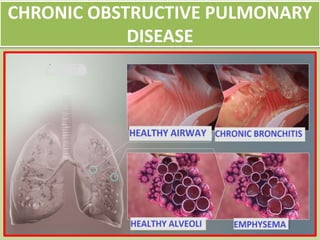
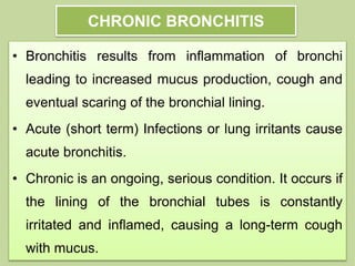
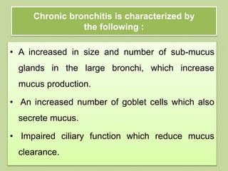
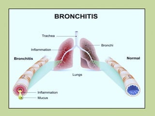
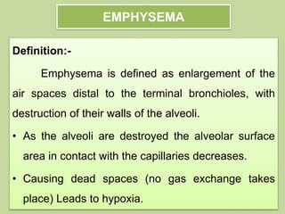
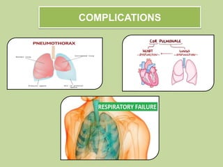
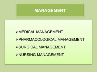
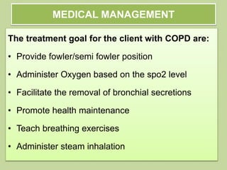
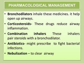
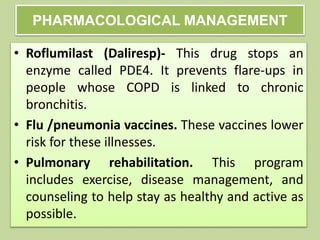
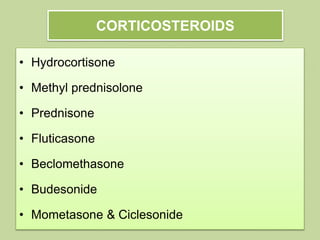
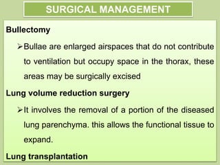
MR.GOPAL ,..MSC (N),
MEDICAL SURGICAL NURSING
ASSISTANT PROFESSOR
GANGA COLLEGE OF NURSING
COIMBATORE
CHRONIC OBSTRUCTIVE PULMONARY
DISEASE
CONTENT OVERVIEW
• Introduction
• Anatomy & physiology
• Definition
• Incidence
• Etiological factors
• Pathophysiology
• Signs & symptoms
• Diagnostic evaluation
• Complications
• Management
INTRODUCTION
Chronic obstructive pulmonary disease (COPD)
is a common lung disease. It is a preventable and
treatable lung disease. People with COPD must work
harder to breathe, which can lead to shortness of
breath and/or feeling tired. Early in the disease,
people with COPD may feel short of breath when they
exercise. As the disease progresses, it can be hard to
breathe out (exhale) or even breathe in (inhale)
ANATOMY & PHYSIOLOGY
DEFINITION
Chronic obstructive pulmonary disease
(COPD) is a progressive, irreversible chronic inflammatory
lung disease that causes obstruction of the airflow from the
lungs.
There are two main forms of COPD:
– Chronic bronchitis, which involves a long-term
cough with mucus
– Emphysema, which involves damage to the lungs
over time
INCIDENCE
• The Global Burden of Disease Study reports a
prevalence of 251 million cases of COPD globally
in 2016.
• Globally, it is estimated that 3.17 million deaths
were caused by the disease in 2015 (that is, 5% of
all deaths globally in that year).
• More than 90% of COPD deaths occur in low and
middleincome countries.
STAGES AND SYMPTOMS OF COPD ARE:
• Mild: Airflow is somewhat limited, but you don’t
notice it much. cough and have mucus every once
in a while.
• Moderate: Airflow is worse. Person often short of
breath after doing something active. This is the
point where most people notice symptoms and get
help.
• Severe: Airflow and shortness of breath are
worse. You can't do normal exercise anymore.
Your symptoms flare up frequently, also called an
exacerbation.
• Very severe: Airflow is limited, your flares are
more regular and intense, and your quality of life
is poor
STAGES AND SYMPTOMS OF COPD ARE:
CHRONIC BRONCHITIS
• Bronchitis results from inflammation of bronchi
leading to increased mucus production, cough and
eventual scaring of the bronchial lining.
• Acute (short term) Infections or lung irritants cause
acute bronchitis.
• Chronic is an ongoing, serious condition. It occurs if
the lining of the bronchial tubes is constantly
irritated and inflamed, causing a long-term cough
with mucus.
Chronic bronchitis is characterized by
the following :
• A increased in size and number of sub-mucus
glands in the large bronchi, which increase
mucus production.
• An increased number of goblet cells which also
secrete mucus.
• Impaired ciliary function which reduce mucus
clearance.
EMPHYSEMA
Definition:-
Emphysema is defined as enlargement of the
air spaces distal to the terminal bronchioles, with
destruction of their walls of the alveoli.
• As the alveoli are destroyed the alveolar surface
area in contact with the capillaries decreases.
• Causing dead spaces (no gas exchange takes
place) Leads to hypoxia.
TYPES
Pan
acinar
Centri
acinar
Para
septal
Centriacinar(centrilobular)
Centriacinar(centrilobular) emphysema the
most common type produce destruction in
bronchioles usually in the upper lung region.
Inflammation begins in the bronchioles and spread
peripherally but usually the alveolar sac remains
intact. This form of emphysema occurs most often in
smokers.
Panacinar
Panacinar emphysema destroys the entire
alveolus and most commonly involves the lower
portion of the lung. This form of disease is
generally observed in individuals with ATT
deficiency.
Paraseptal
Paraseptal or distal acinar emphysema
primarily involves the distal airway structures
alveolar ducts and alveolar sacs. The process is
localized around the septa of the lung or pleura. It is
believed to be the likely cause of spontaneous
pneumothorax
ETIOLOGICAL FACTORS
RISK FACTORS
• A history of childhood respiratory infection
• Exposure to tobacco smoke
• Exposure to fumes from burning fuel
• people at least 40 years of age
• Abnormal lung development
• Socio economic status
PATHOPHYSIOLOGY
EARLY SYMPTOMS INCLUDE:
• Occasional shortness of breath, especially after
exercise
• Mild but recurrent cough
• Needing to clear your throat often, especially first
thing in the morning
As the lungs become more damaged,
may experience:
• Shortness of breath, after even mild exercise such
as walking up a flight of stairs
• Wheezing, or noisy breathing
• Chest tightness &Lack of energy
• Chronic cough, with or without mucus
• Need to clear mucus from your lungs every day
• Frequent colds, flu, or other respiratory infections
In later stages of COPD, symptoms may:
• Fatigue
• Swelling of the feet, ankles, or legs
• Weight loss
• Bluish or gray fingernails or lips, as this indicates
low oxygen levels in your blood
• Trouble catching breath or cannot talk
• Confused, muddled, or faint
• Tachycardia
Signs: Pink puffers & blue bloaters (2
ends of a spectrum).
HISTORY COLLECTION
• A complete family history
• Environmental Factors
• occupational history is essential.
to establish the diagnosis
PHYSICAL EXAMINATION
• Hyper-expansion of the thorax
• Sounds of wheezing during normal breathing or a
prolonged phase of forced exhalation Increased
nasal secretions, mucosal swelling, sinusitis,
rhinitis, or nasal polyps
• Rales, Rhonchi,
• Tachypnea &Orthopnea
• Chest constriction
LABORATORY INVESTIGATIONS
• Arterial blood gas analysis
• Spo2 monitoring
• Elevated IgE level
• Complete blood count
• Blood levels of eosinophil
DIFFERENTIAL DIAGNOSIS
• Peak Expiratory Flow Rate
• Spirometry
• Chest X-ray
• Skin Prick Testing
• Measurement of Airway Hyper responsiveness
• Sputum Examination
• Pulmonary function test
PEFR is used to assess
the severity of wheezing in
those who have asthma.
PEFR measures how quickly
a person can exhale air from
the lungs
PEAK EXPIRATORY FLOW RATE
INCENTIVE SPIROMETRY
• It measures how much air you can exhale.
• FEV1(force expiratory volume) > 80% = normal
• Confirms the presence of airway obstruction and
measure the degree of lung function impairment.
• Monitor your response to asthma medications
PULMONARY FUNCTION TEST
S.No Paprameter
s
NORMAL VALUES ABNORMAL VALUES
1. FEV1/FVC – >75% Normal • 60%‐75% Mild obstruction
• 50‐59% Moderate obstruction
• <49% Severe obstruction
2. Forced
midexpiratory
flow
25‐75% (FEF25‐
75)
Interpretation of % predicted:
• >60% Normal
• 40‐60% Mild obstruction
• 20‐40% Moderate obstruction
• <10% Severe obstruction
3. Peak
expiratory
flow rates
Male : 450 ‐ 700 l/min
Females: 300 ‐ 500 l/min
<200/mins
PARAMETERS OBSTRUCTIVE RESTRICTIVE
Vital capacity Normal or decreased decreased
Total lung capacity Normal or increased decreased
Residual volume Increased decreased
FEV1/FVC decreased Normal or increased
Maximum mid
expiratory flow
decreased Normal
Maximum breathing
capacity
decreased Normal or decreased
PULMONARY FUNCTION TEST
CHEST X-RAY
COMPLICATIONS
MANAGEMENT
MEDICAL MANAGEMENT
PHARMACOLOGICAL MANAGEMENT
SURGICAL MANAGEMENT
NURSING MANAGEMENT
MEDICAL MANAGEMENT
The treatment goal for the client with COPD are:
• Provide fowler/semi fowler position
• Administer Oxygen based on the spo2 level
• Facilitate the removal of bronchial secretions
• Promote health maintenance
• Teach breathing exercises
• Administer steam inhalation
COLLABORATIVE THERAPY
• Cessation of cigarette smoking
• Treatment of exacerbation
• Drug therapies
• Breathing exercises
• Patient and caregiver teaching
• Influenza immunization yearly
• Pneumovax immunization
• Pulmonary rehabilitation program
PHARMACOLOGICAL MANAGEMENT
• Bronchodilators inhale these medicines. it help
open up airways.
• Corticosteroids- These drugs reduce airway
inflammation.
• Combination inhalers These inhalers
pair steroids with a bronchodilator.
• Antibiotics- might prescribe to fight bacterial
infections.
• Nebulization – to clear airway
• Roflumilast (Daliresp)- This drug stops an
enzyme called PDE4. It prevents flare-ups in
people whose COPD is linked to chronic
bronchitis.
• Flu /pneumonia vaccines. These vaccines lower
risk for these illnesses.
• Pulmonary rehabilitation. This program
includes exercise, disease management, and
counseling to help stay as healthy and active as
possible.
PHARMACOLOGICAL MANAGEMENT
BRONCHODILATORS
Three major classes of bronchodilators:
Β2 - agonists:
• Short acting: salbutamol & terbutaline
• Long acting :salmeterol & formoterol
Anticholinergic agents:
• ipratropium, tiotropium, Theophylline (a weak
bronchodilator, which may have some anti-
inflammatory properties)
CORTICOSTEROIDS
• Hydrocortisone
• Methyl prednisolone
• Prednisone
• Fluticasone
• Beclomethasone
• Budesonide
• Mometasone & Ciclesonide
SURGICAL MANAGEMENT
Bullectomy
Bullae are enlarged airspaces that do not contribute
to ventilation but occupy space in the thorax, these
areas may be surgically excised
Lung volume reduction surgery
It involves the removal of a portion of the diseased
lung parenchyma. this allows the functional tissue to
expand.
Lung transplantation
NURSING MANAGEMENT
• Ineffective airway clearance related to obstruction
of the airway, increased mucus secretion as
evidenced by secretion, decreased spo2 level,
tachypnea
• Ineffective breathing pattern related to
obstruction of airway, excessive mucus secretion
as evidenced by tachypnea and hypoxia.
• Acute pain on chest related to increased effort of breath
as evidenced by pain scale
• Impaired gas exchange related to decreased oxygen
level, bronchospasm as evidenced by decreased spo2
level
• Impaired tissue perfusion related to v/q mismatch,
hypoxia as evidenced by delayed capillary refills
• Disturbed sleeping pattern related to breathing
difficulty as evidenced by redness of eyes.
NURSING MANAGEMENT
REFERENCE
• Lewis & dirksen, (2015) textbook of medical –
surgical nursing, 2nd South asian edition, elsevier
publication.
• Brunner & suddarth’s, (2014) textbook of medical
– surgical nursing, 13th edition, wolters kluwer
publications. (620-630)
3. Al-Jahdali H, Alshimemeri A, Mobeireek A, Albanna
AS, Chang, Aliberti S. The Saudi Thoracic Society
guidelines for diagnosis and management of noncystic
fibrosis bronchiectasis. Ann Thorac Med 2017;12.
4. Margaret F Alexandra (2000) Nursing practice hospital
and homes, second edition
5. Merskey, H. (1964); International Study Of Pain: An
Unpleasant Experience That We Primarily Associate With
Tissue Damage Or Describe In Terms Of Tissue Damage.
COPD.pptx

COPD.pptx

  • 1.
    MR.GOPAL ,..MSC (N), MEDICALSURGICAL NURSING ASSISTANT PROFESSOR GANGA COLLEGE OF NURSING COIMBATORE
  • 2.
  • 3.
    CONTENT OVERVIEW • Introduction •Anatomy & physiology • Definition • Incidence • Etiological factors • Pathophysiology • Signs & symptoms • Diagnostic evaluation • Complications • Management
  • 4.
    INTRODUCTION Chronic obstructive pulmonarydisease (COPD) is a common lung disease. It is a preventable and treatable lung disease. People with COPD must work harder to breathe, which can lead to shortness of breath and/or feeling tired. Early in the disease, people with COPD may feel short of breath when they exercise. As the disease progresses, it can be hard to breathe out (exhale) or even breathe in (inhale)
  • 5.
  • 6.
    DEFINITION Chronic obstructive pulmonarydisease (COPD) is a progressive, irreversible chronic inflammatory lung disease that causes obstruction of the airflow from the lungs. There are two main forms of COPD: – Chronic bronchitis, which involves a long-term cough with mucus – Emphysema, which involves damage to the lungs over time
  • 7.
    INCIDENCE • The GlobalBurden of Disease Study reports a prevalence of 251 million cases of COPD globally in 2016. • Globally, it is estimated that 3.17 million deaths were caused by the disease in 2015 (that is, 5% of all deaths globally in that year). • More than 90% of COPD deaths occur in low and middleincome countries.
  • 8.
    STAGES AND SYMPTOMSOF COPD ARE: • Mild: Airflow is somewhat limited, but you don’t notice it much. cough and have mucus every once in a while. • Moderate: Airflow is worse. Person often short of breath after doing something active. This is the point where most people notice symptoms and get help.
  • 9.
    • Severe: Airflowand shortness of breath are worse. You can't do normal exercise anymore. Your symptoms flare up frequently, also called an exacerbation. • Very severe: Airflow is limited, your flares are more regular and intense, and your quality of life is poor STAGES AND SYMPTOMS OF COPD ARE:
  • 10.
    CHRONIC BRONCHITIS • Bronchitisresults from inflammation of bronchi leading to increased mucus production, cough and eventual scaring of the bronchial lining. • Acute (short term) Infections or lung irritants cause acute bronchitis. • Chronic is an ongoing, serious condition. It occurs if the lining of the bronchial tubes is constantly irritated and inflamed, causing a long-term cough with mucus.
  • 11.
    Chronic bronchitis ischaracterized by the following : • A increased in size and number of sub-mucus glands in the large bronchi, which increase mucus production. • An increased number of goblet cells which also secrete mucus. • Impaired ciliary function which reduce mucus clearance.
  • 13.
    EMPHYSEMA Definition:- Emphysema is definedas enlargement of the air spaces distal to the terminal bronchioles, with destruction of their walls of the alveoli. • As the alveoli are destroyed the alveolar surface area in contact with the capillaries decreases. • Causing dead spaces (no gas exchange takes place) Leads to hypoxia.
  • 14.
  • 16.
    Centriacinar(centrilobular) Centriacinar(centrilobular) emphysema the mostcommon type produce destruction in bronchioles usually in the upper lung region. Inflammation begins in the bronchioles and spread peripherally but usually the alveolar sac remains intact. This form of emphysema occurs most often in smokers.
  • 17.
    Panacinar Panacinar emphysema destroysthe entire alveolus and most commonly involves the lower portion of the lung. This form of disease is generally observed in individuals with ATT deficiency.
  • 18.
    Paraseptal Paraseptal or distalacinar emphysema primarily involves the distal airway structures alveolar ducts and alveolar sacs. The process is localized around the septa of the lung or pleura. It is believed to be the likely cause of spontaneous pneumothorax
  • 19.
  • 20.
    RISK FACTORS • Ahistory of childhood respiratory infection • Exposure to tobacco smoke • Exposure to fumes from burning fuel • people at least 40 years of age • Abnormal lung development • Socio economic status
  • 21.
  • 22.
    EARLY SYMPTOMS INCLUDE: •Occasional shortness of breath, especially after exercise • Mild but recurrent cough • Needing to clear your throat often, especially first thing in the morning
  • 23.
    As the lungsbecome more damaged, may experience: • Shortness of breath, after even mild exercise such as walking up a flight of stairs • Wheezing, or noisy breathing • Chest tightness &Lack of energy • Chronic cough, with or without mucus • Need to clear mucus from your lungs every day • Frequent colds, flu, or other respiratory infections
  • 24.
    In later stagesof COPD, symptoms may: • Fatigue • Swelling of the feet, ankles, or legs • Weight loss • Bluish or gray fingernails or lips, as this indicates low oxygen levels in your blood • Trouble catching breath or cannot talk • Confused, muddled, or faint • Tachycardia
  • 25.
    Signs: Pink puffers& blue bloaters (2 ends of a spectrum).
  • 26.
    HISTORY COLLECTION • Acomplete family history • Environmental Factors • occupational history is essential. to establish the diagnosis
  • 27.
    PHYSICAL EXAMINATION • Hyper-expansionof the thorax • Sounds of wheezing during normal breathing or a prolonged phase of forced exhalation Increased nasal secretions, mucosal swelling, sinusitis, rhinitis, or nasal polyps • Rales, Rhonchi, • Tachypnea &Orthopnea • Chest constriction
  • 28.
    LABORATORY INVESTIGATIONS • Arterialblood gas analysis • Spo2 monitoring • Elevated IgE level • Complete blood count • Blood levels of eosinophil
  • 29.
    DIFFERENTIAL DIAGNOSIS • PeakExpiratory Flow Rate • Spirometry • Chest X-ray • Skin Prick Testing • Measurement of Airway Hyper responsiveness • Sputum Examination • Pulmonary function test
  • 30.
    PEFR is usedto assess the severity of wheezing in those who have asthma. PEFR measures how quickly a person can exhale air from the lungs PEAK EXPIRATORY FLOW RATE
  • 31.
    INCENTIVE SPIROMETRY • Itmeasures how much air you can exhale. • FEV1(force expiratory volume) > 80% = normal • Confirms the presence of airway obstruction and measure the degree of lung function impairment. • Monitor your response to asthma medications
  • 32.
    PULMONARY FUNCTION TEST S.NoPaprameter s NORMAL VALUES ABNORMAL VALUES 1. FEV1/FVC – >75% Normal • 60%‐75% Mild obstruction • 50‐59% Moderate obstruction • <49% Severe obstruction 2. Forced midexpiratory flow 25‐75% (FEF25‐ 75) Interpretation of % predicted: • >60% Normal • 40‐60% Mild obstruction • 20‐40% Moderate obstruction • <10% Severe obstruction 3. Peak expiratory flow rates Male : 450 ‐ 700 l/min Females: 300 ‐ 500 l/min <200/mins
  • 33.
    PARAMETERS OBSTRUCTIVE RESTRICTIVE Vitalcapacity Normal or decreased decreased Total lung capacity Normal or increased decreased Residual volume Increased decreased FEV1/FVC decreased Normal or increased Maximum mid expiratory flow decreased Normal Maximum breathing capacity decreased Normal or decreased PULMONARY FUNCTION TEST
  • 34.
  • 35.
  • 36.
  • 37.
    MEDICAL MANAGEMENT The treatmentgoal for the client with COPD are: • Provide fowler/semi fowler position • Administer Oxygen based on the spo2 level • Facilitate the removal of bronchial secretions • Promote health maintenance • Teach breathing exercises • Administer steam inhalation
  • 38.
    COLLABORATIVE THERAPY • Cessationof cigarette smoking • Treatment of exacerbation • Drug therapies • Breathing exercises • Patient and caregiver teaching • Influenza immunization yearly • Pneumovax immunization • Pulmonary rehabilitation program
  • 39.
    PHARMACOLOGICAL MANAGEMENT • Bronchodilatorsinhale these medicines. it help open up airways. • Corticosteroids- These drugs reduce airway inflammation. • Combination inhalers These inhalers pair steroids with a bronchodilator. • Antibiotics- might prescribe to fight bacterial infections. • Nebulization – to clear airway
  • 40.
    • Roflumilast (Daliresp)-This drug stops an enzyme called PDE4. It prevents flare-ups in people whose COPD is linked to chronic bronchitis. • Flu /pneumonia vaccines. These vaccines lower risk for these illnesses. • Pulmonary rehabilitation. This program includes exercise, disease management, and counseling to help stay as healthy and active as possible. PHARMACOLOGICAL MANAGEMENT
  • 41.
    BRONCHODILATORS Three major classesof bronchodilators: Β2 - agonists: • Short acting: salbutamol & terbutaline • Long acting :salmeterol & formoterol Anticholinergic agents: • ipratropium, tiotropium, Theophylline (a weak bronchodilator, which may have some anti- inflammatory properties)
  • 42.
    CORTICOSTEROIDS • Hydrocortisone • Methylprednisolone • Prednisone • Fluticasone • Beclomethasone • Budesonide • Mometasone & Ciclesonide
  • 43.
    SURGICAL MANAGEMENT Bullectomy Bullae areenlarged airspaces that do not contribute to ventilation but occupy space in the thorax, these areas may be surgically excised Lung volume reduction surgery It involves the removal of a portion of the diseased lung parenchyma. this allows the functional tissue to expand. Lung transplantation
  • 44.
    NURSING MANAGEMENT • Ineffectiveairway clearance related to obstruction of the airway, increased mucus secretion as evidenced by secretion, decreased spo2 level, tachypnea • Ineffective breathing pattern related to obstruction of airway, excessive mucus secretion as evidenced by tachypnea and hypoxia.
  • 45.
    • Acute painon chest related to increased effort of breath as evidenced by pain scale • Impaired gas exchange related to decreased oxygen level, bronchospasm as evidenced by decreased spo2 level • Impaired tissue perfusion related to v/q mismatch, hypoxia as evidenced by delayed capillary refills • Disturbed sleeping pattern related to breathing difficulty as evidenced by redness of eyes. NURSING MANAGEMENT
  • 46.
    REFERENCE • Lewis &dirksen, (2015) textbook of medical – surgical nursing, 2nd South asian edition, elsevier publication. • Brunner & suddarth’s, (2014) textbook of medical – surgical nursing, 13th edition, wolters kluwer publications. (620-630)
  • 47.
    3. Al-Jahdali H,Alshimemeri A, Mobeireek A, Albanna AS, Chang, Aliberti S. The Saudi Thoracic Society guidelines for diagnosis and management of noncystic fibrosis bronchiectasis. Ann Thorac Med 2017;12. 4. Margaret F Alexandra (2000) Nursing practice hospital and homes, second edition 5. Merskey, H. (1964); International Study Of Pain: An Unpleasant Experience That We Primarily Associate With Tissue Damage Or Describe In Terms Of Tissue Damage.