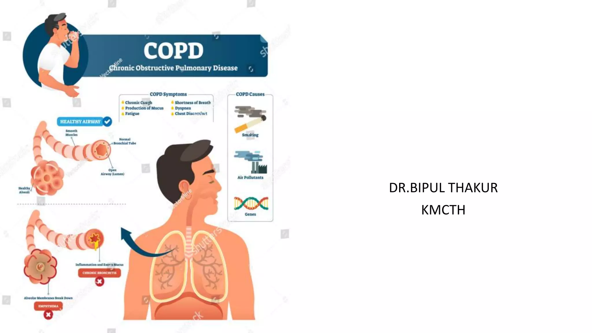
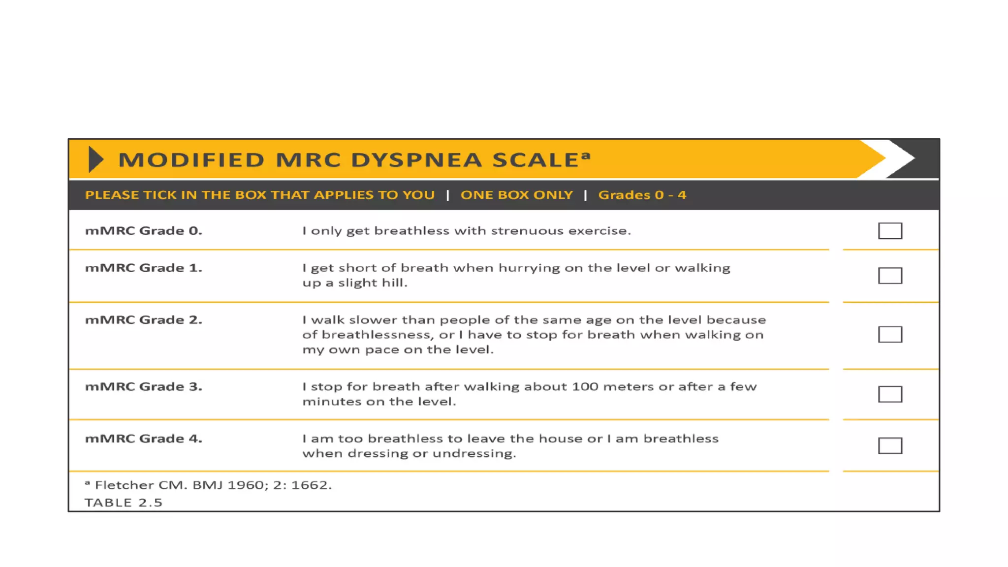
DR.BIPUL THAKUR
KMCTH
Definition
• COPD is a common, preventable, and treatable disease that is
characterized by persistent respiratory symptoms and airflow
limitation that is due to airway and/or alveolar abnormalities
usually caused by significant exposure to noxious particles or
gases.
COPD includes
• 1. Chronic bronchitis:
is defines as chronic productive cough onmost days for at least 3
consecutive months in 2 successive years.
• 2.Emphysema:
is defined as dilatation and destruction of air spaces distal to
terminal bronchioles without obvious fibrosis.
EPIDEMIOLOGY
• Prevalence directly related to tobacco smoking and use of biomass fuel in
low and middle income countries.
• Current estimates suggest that 80 million people worldwide
suffer from moderate to severe disease.
• A/c to BOLD and other large scale epidemiological studies, it is estimated
that no. of COPD cases was 384M in 2010 with global prevalence of 11.7%
• Globally around 3 million deaths occur annually
• With increasing prevalence and ageing , COPD prevalence expected to rise
over next 40 years and by 2060 there may be 5.4 million deaths annually.
RISK FACTORS
Clinical features:
• Dyspnea:
- Progressive over time
- Characteristically worse with exercise
- Persistent
• Cough:
- May be Intermittent or may be unproductive
- Recurrent Wheeze
• Chronic Sputum production:
- May be dry or productive
- Usually mucoid in nature
- Mucopurulent during acute exacerbation
Physical signs:
• Inspection:
- Barrel-shaped chest ,
- Accessory respiratory muscle participate ,Prolonged
expiration during quiet breathing. Expiration through
pursed lips
- Paradoxical retraction of the lower interspaces during
inspiration (ie, hoover's sign)
- Tripod Position
Tripod Position
• Patients with end-stage COPD
may adopt positions that relieve
dyspnea, such as leaning forward
with arms outstretched and
weight supported on the palms or
elbows.
• Palpation:
- Decreased vocal fremitus
• Percussion :
- Hyper resonant
- Depressed diaphragm,
- Diminution of the area of absolute cardiac dullness.
• Auscultation:
- Vesicular with Prolonged expiration
- Reduced breath sounds
- The presence of wheezing during quiet breathing
- Crackle can be heard if infection exist.
• The presence of a post-
bronchodilator FEV1/FVC <
0.70 confirms the presence
of persistent airflow
limitation and thus of
COPD.
• ABG: pH<7.3- sign of acute respiratory compromise
Type II RF: Chronic Bronchitis
Type I RF: Emphysema
• CBC: Polycythemia
Hematocrit>50
• Sputum examination:
Streptococcus pneumonia
Hemophilus influenzae
Moraxella catarrhalis
Klebsiella pneumonia
Chest x-ray-Chronic Bronchitis
• No apparent
abnormality
• Or thickened and
increased lung
markings are noted.
No apparent
abnormality
Or thickened and
increased lung
markings are noted.
Chest X-Ray -Emphysema
• Marked over inflation is noted
with flattened and low diaphragm
• Intercostal space becomes widen
• A horizontal
pattern of ribs
• A long thin
heart shadow
• Decreased markings of lung
peripheral vessels
CT(Computed tomography)
• Greater sensitivity and
specificity for emphysema
for evaluation of bullous
disease
© 2020 Global Initiative for Chronic Obstructive Lung Disease
• General: Exercise and management of Nutritional status
Weight loss if obese
• Reduce exposure to noxious agents:
1.Smoking cessation: aided by Bupropion(Noradrenergic
antidepressant), varenicline(Nicotinic receptor agonist antagonist)
2.Reduce Indoor and outdoor air pollution
3.Avoid dusty and smoke laden environment
Surgical Intervention
• Bulla: Bullectomy
• Lung volume reduction surgery
• Lung Transplantation
Acute exacerbation of COPD
• Is defined as any event in natural course of COPD characterized by a
change in patient’s baseline dyspnoea, cough and/or sputum that is
beyond normal day to day variations.
• Causes:
1.Infection
2.Air pollution
3.Cold
Management
• Initial treatment: 1. Position the patient up in bed
2. O2 therapy
3. If condition is not improving, intubation may be
required
• Bronchodilator: 1. SABA: Nebulized Salbutamol 2.5mg every 20 min
for initial 1-2 hr &/or
2. Short anticholinergic: Nebulized Ipratropium
bromide 0.5mg &/or
3. IV Aminophylline: Failure of above treatment
Loading dose:250mg IV in 20 min
Maintenance dose: 0.5-0.7mg/kg/hr
in 1 ltr of saline at 2.4ml/kg/hr
• Antibiotics:
1. Outpatient:
a. Doxycycline,cotrimoxazole or amoxiclav
b. Hospitalized pt >65 yrs: give one of the newer
FQs(Levoflox,Gemiflox,Moxiflox)
2. Hosptalized: IV anibiotics: Azithro or FQs or 3rd gen
Cephalosporins
3. Severe exacerbations: 3rd gen Cephalosporin + FQs or an
aminoglycoside
• Antibiotics should cover S. pneumoniae, H. influenza, Legionella sp.
• Steroids: Shortens recovery time, improve Lung function and hypoxia.
Hydrocortisone 200mg IV repea 6-8 hrly
or
Methyl prednisolone1-2mg/kg IV 6hrly not to exceed 125mg
: F/U with oral steroid: Presdnisolone 40-60mg/day in
tapering dose
• Monitoring
• Mgso4 IV single dose : 1.2-2gm infused over 20 min
• Diuretics: In pts with gross Rt. Ventricular failure
• NIPPV: 1.CPAP
2.BiPAP
MOA: 1. Prevents airways to collapse and air trapping
2. Reduces need for ET intubation
Copd
Copd
Copd
Copd

Copd

  • 1.
  • 2.
    Definition • COPD isa common, preventable, and treatable disease that is characterized by persistent respiratory symptoms and airflow limitation that is due to airway and/or alveolar abnormalities usually caused by significant exposure to noxious particles or gases.
  • 3.
    COPD includes • 1.Chronic bronchitis: is defines as chronic productive cough onmost days for at least 3 consecutive months in 2 successive years. • 2.Emphysema: is defined as dilatation and destruction of air spaces distal to terminal bronchioles without obvious fibrosis.
  • 4.
    EPIDEMIOLOGY • Prevalence directlyrelated to tobacco smoking and use of biomass fuel in low and middle income countries. • Current estimates suggest that 80 million people worldwide suffer from moderate to severe disease. • A/c to BOLD and other large scale epidemiological studies, it is estimated that no. of COPD cases was 384M in 2010 with global prevalence of 11.7% • Globally around 3 million deaths occur annually • With increasing prevalence and ageing , COPD prevalence expected to rise over next 40 years and by 2060 there may be 5.4 million deaths annually.
  • 5.
  • 8.
    Clinical features: • Dyspnea: -Progressive over time - Characteristically worse with exercise - Persistent • Cough: - May be Intermittent or may be unproductive - Recurrent Wheeze • Chronic Sputum production: - May be dry or productive - Usually mucoid in nature - Mucopurulent during acute exacerbation
  • 9.
    Physical signs: • Inspection: -Barrel-shaped chest , - Accessory respiratory muscle participate ,Prolonged expiration during quiet breathing. Expiration through pursed lips - Paradoxical retraction of the lower interspaces during inspiration (ie, hoover's sign) - Tripod Position
  • 11.
    Tripod Position • Patientswith end-stage COPD may adopt positions that relieve dyspnea, such as leaning forward with arms outstretched and weight supported on the palms or elbows.
  • 12.
    • Palpation: - Decreasedvocal fremitus • Percussion : - Hyper resonant - Depressed diaphragm, - Diminution of the area of absolute cardiac dullness.
  • 13.
    • Auscultation: - Vesicularwith Prolonged expiration - Reduced breath sounds - The presence of wheezing during quiet breathing - Crackle can be heard if infection exist.
  • 17.
    • The presenceof a post- bronchodilator FEV1/FVC < 0.70 confirms the presence of persistent airflow limitation and thus of COPD.
  • 22.
    • ABG: pH<7.3-sign of acute respiratory compromise Type II RF: Chronic Bronchitis Type I RF: Emphysema • CBC: Polycythemia Hematocrit>50 • Sputum examination: Streptococcus pneumonia Hemophilus influenzae Moraxella catarrhalis Klebsiella pneumonia
  • 23.
    Chest x-ray-Chronic Bronchitis •No apparent abnormality • Or thickened and increased lung markings are noted. No apparent abnormality Or thickened and increased lung markings are noted.
  • 24.
    Chest X-Ray -Emphysema •Marked over inflation is noted with flattened and low diaphragm • Intercostal space becomes widen • A horizontal pattern of ribs • A long thin heart shadow • Decreased markings of lung peripheral vessels
  • 25.
    CT(Computed tomography) • Greatersensitivity and specificity for emphysema for evaluation of bullous disease
  • 27.
    © 2020 GlobalInitiative for Chronic Obstructive Lung Disease
  • 28.
    • General: Exerciseand management of Nutritional status Weight loss if obese • Reduce exposure to noxious agents: 1.Smoking cessation: aided by Bupropion(Noradrenergic antidepressant), varenicline(Nicotinic receptor agonist antagonist) 2.Reduce Indoor and outdoor air pollution 3.Avoid dusty and smoke laden environment
  • 37.
    Surgical Intervention • Bulla:Bullectomy • Lung volume reduction surgery • Lung Transplantation
  • 39.
    Acute exacerbation ofCOPD • Is defined as any event in natural course of COPD characterized by a change in patient’s baseline dyspnoea, cough and/or sputum that is beyond normal day to day variations. • Causes: 1.Infection 2.Air pollution 3.Cold
  • 40.
    Management • Initial treatment:1. Position the patient up in bed 2. O2 therapy 3. If condition is not improving, intubation may be required • Bronchodilator: 1. SABA: Nebulized Salbutamol 2.5mg every 20 min for initial 1-2 hr &/or 2. Short anticholinergic: Nebulized Ipratropium bromide 0.5mg &/or 3. IV Aminophylline: Failure of above treatment Loading dose:250mg IV in 20 min Maintenance dose: 0.5-0.7mg/kg/hr in 1 ltr of saline at 2.4ml/kg/hr
  • 41.
    • Antibiotics: 1. Outpatient: a.Doxycycline,cotrimoxazole or amoxiclav b. Hospitalized pt >65 yrs: give one of the newer FQs(Levoflox,Gemiflox,Moxiflox) 2. Hosptalized: IV anibiotics: Azithro or FQs or 3rd gen Cephalosporins 3. Severe exacerbations: 3rd gen Cephalosporin + FQs or an aminoglycoside • Antibiotics should cover S. pneumoniae, H. influenza, Legionella sp.
  • 42.
    • Steroids: Shortensrecovery time, improve Lung function and hypoxia. Hydrocortisone 200mg IV repea 6-8 hrly or Methyl prednisolone1-2mg/kg IV 6hrly not to exceed 125mg : F/U with oral steroid: Presdnisolone 40-60mg/day in tapering dose • Monitoring • Mgso4 IV single dose : 1.2-2gm infused over 20 min • Diuretics: In pts with gross Rt. Ventricular failure
  • 44.
    • NIPPV: 1.CPAP 2.BiPAP MOA:1. Prevents airways to collapse and air trapping 2. Reduces need for ET intubation