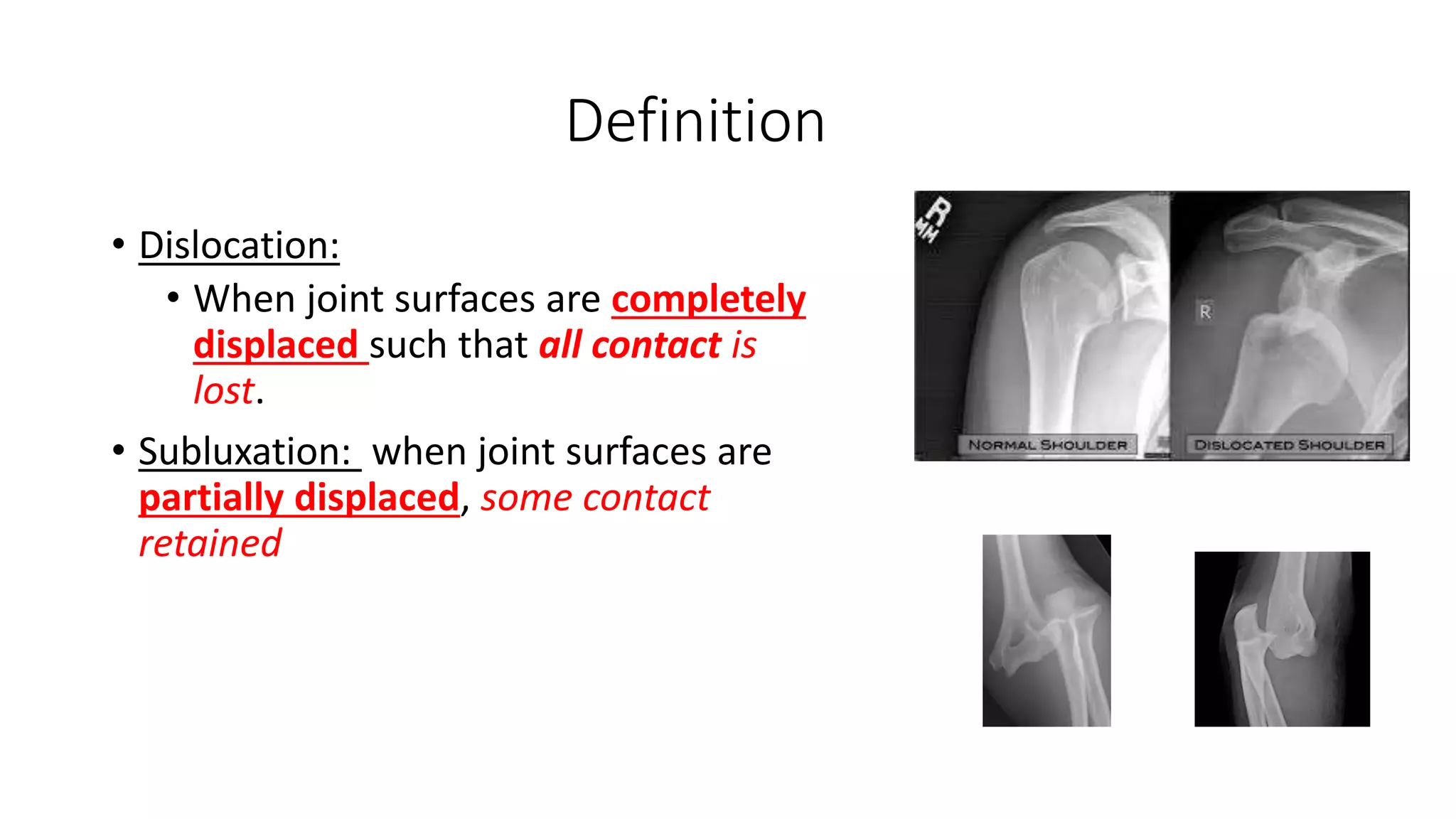
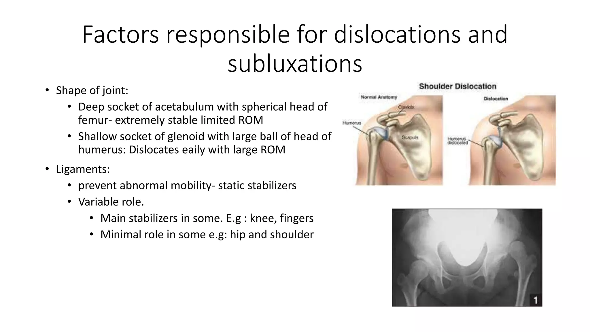
Dislocations and
Subluxations
Dr. Siddhartha Sinha
Assistant Professor, Department of Orthopaedics
Hamdard Institute of Medical sciences and Research, New Delhi,India
Definition
• Dislocation:
• When joint surfaces are completely
displaced such that all contact is
lost.
• Subluxation: when joint surfaces are
partially displaced, some contact
retained
Factors responsible for dislocations and
subluxations
• Shape of joint:
• Deep socket of acetabulum with spherical head of
femur- extremely stable limited ROM
• Shallow socket of glenoid with large ball of head of
humerus: Dislocates eaily with large ROM
• Ligaments:
• prevent abnormal mobility- static stabilizers
• Variable role.
• Main stabilizers in some. E.g : knee, fingers
• Minimal role in some e.g: hip and shoulder
• Muscles:
• Dynamic stabilizers- contract by
contracting when ligaments come
under stress
• Cover joints to give stability
Classification
•Aetiology:
•Congenital / Acquired
•Traumatic/ Pathological
Traumatic dislocations
• MOST COMMON CAUSE
• Force required to dislocate the joint is variable for each joint.
• Types
• Acute Traumatic Dislocation: force of injury is main contributing
factor (shoulder)
• Old unreduced dislocation: e.g hip, shoulder
• Recurrent dislocation: improper healing after 1st dislocation-
weakness of supporting structures- easy dislocation (e.g: Shoulder
and Patella)
• Fracture dislocation: dislocations associated with fracture of one or
both articular bones ( hip and acetacbulum)
Pathological Dislocation
• Articular surfaces destroyed by
• Infection
• Neoplasia
• Ligament damage secondary to
disease
Diagnosis
• Pain
• Deformity: specific for each joint
• Swelling
• Loss of movement
• Shortening of limb
• Telescopy: produce to and fro movement of all
dislocated joints
Typical deformities in dislocation
• Shoulder- abduction deformities
• Elbow- flexion deformities
• Hip:
Anterior- flexion abduction and internal rotation deformities.
Posterior-flexion, adduction and internal rotation deformity
• Knee-flexion deformity
• Ankle-varus deformity
X ray
• 2 planes of each other
• Opposite limb for comparison
• For fractures or osteochondral
fragment
Principles of management
• Acute dislocation should be reduced as soon as possible.
• Open reduction is rarely necessary for acute dislocation.
• Close reduction with intravenous analgesia and sedation or under GA
should be attempted first for most uncomplicated dislocation.
Treatment
• Acute traumatic dislocation:
• Conservative methods: closed reduction (specific techniques for
each )- instant pain relief
• Operative methods:
• Failure of reduction (late detection of dislocation)
• Fracture dislocation
• Incongruity in joint
• Loose piece in joint
• Dislocation difficult to maintain in closed reduction
(unstable)
• Old dislocation –ORIF
• Recurrent dislocations: Reconstruction Procedures
Complications
• Immediate Complications:
• Neurovascular deficit
• Early complications
• Recurrence
• Myositis ossificans
• Persistent instability
• Joint stiffness
• Late complications
• Recurrence
• Osteoarthritis
• Avascular necrosis (head of femur)
Glenohumeral Dislocations
• Most common dislocated joint
• Recurrence rate in all ages is 50% but rises to
almost 89% in the 14 to 20 year age group
• Shoulder instability:
• broad term when head of humerus is not stable in
the glenoid
• Pain d/t stretching of capsule caused by abnormal
movement of head of humerus
• Unidirectional, bidirectional or multi directional
• Mechanism of injury: fall on outstretched
hand with shoulder abducted and externally
rotated (anterior dislocation)
• Convulsive mechanisms and electrical shock
(posterior dislocation)
Glenohumeral stability
• Passive:
• Joint conformity
• Vacuum effect of limited joint
volume
• Adhesion and cohesion owing to the
presence of synovial fluid.
• Scapular inclination:
• Ligamentous and capsular restraints
(capsule of joint, superior inferior
and middle glenohumeral ligament,
coracohumeral ligament.
• Glenoid labrum
• Bony restraints: acromion, coracoid,
glenoid fossa.
■ Active:
Biceps, long-head
Rotator cuff
Scapular stabilizing muscles
Classification
• Anterior dislocation
• Preglenoid
• Subcoracoid sublcavivluar
• Subclavicular
• Posterior dislocation
• Luxatio erecta: head in subglenoid
position
Pathological changes
• Bankart lesion:
• Stripping of the glenoidal labrum
with periosteum from anterior
surface of glenoid and scapular
neck
• Head lies in front of scapular neck
• Avulsion of bone from anterior-
inferior glenoidal rin: BONY
BANKART LESION
• Hill Sachs Lesion:
• Depression of humeral head in
posterolateral quadrant due to
impingement by anterior edge
of glenoid on head as it
dislocates
• Rounding off anterior
glenoidal rim in chronic cases-
easy dislocation
• Other associated # : greater
tuberosity, rotator cuff
injuries, chondral damage
Presentation
• Pain in shoulder with
shoulder abducted &
supported by elbow (anterior)
• Internal rotation and
adduction. (posterior
dislocation)
• h/o trauma,similar episodes
in the past
• h/o seizure disorder
Examination for shoulder dislocation
• Arm abducted
• Round contour of joint lost flattened
• Fullness below clavicle
• Signs:
• Dugas test: inability to touch opposite shoulder
• Hamilton ruler test: d/t flattening of shoulder it
becomes possible to place a ruler from the
lateral side of arm touching the acromion and
lateral condyle simultaneously ( normally it is
not possible).
• Regiment Badge sign: loss of sensations over
deltoid s/o axillary nerve involvement.
Radiography
• Chest x-rays showing B/L shoulder joints for
comparison of the 2 joints
• Scapular Y view
• Axillary views
• West Point axillary: tangential view of the
anteroinferior glenoid rim
• Hill-Sachs view: visualize a posterolateral defect
• Stryker notch view: can visualize 90% of
posterolateral humeral head defects
Techniques of reduction
• Traction counter traction:
• Apply traction – counter
traction for a period of time
• Stimson technique:
• After administration of analgesics
and/or sedatives
• the patient is placed prone on the
stretcher with the affected upper
extremity hanging free.
• Gentle, manual traction or 5 lb of
weight is applied to the wrist, with
reduction effected over 15 to 20
minutes
• Hippocratic technique:
• Only one person performing reduction,
• One foot placed across the axillary folds
and onto the chest wall
• gentle internal and external rotation
with axial traction on the affected upper
extremity
• Milch technique:patient supine and
the upper extremity abducted and
externally rotated, thumb pressure is
applied by the physician to push the
humeral head into place.
• Kocher maneuver: The humeral
head levered on the anterior glenoid
to effect reduction; this is not
recommended because of increased
risk of fracture.
Post reduction care
• Immobilization for 2 to 5 weeks in shoulder
detorator immobilizer of appropriate size.
• use shorter duration for older patients to
prevent stiffness
• Physiotherapy:
• increasing degrees of shoulder external rotation,
flexion, and abduction as time progresses,
accompanied by full, active range of motion to the
hand, wrist, and elbow
Operative management
• Indications for surgery include:
• First-time dislocation in young active men
• Soft tissue interposition
• Displaced greater tuberosity fracture that remains >5 mm superiorly
displaced following joint reduction
• Glenoid rim fracture >5 mm in size
• arthroscopic ligamentous repair
Operative options
• Putti- Platt operation:
• Bankart operation
• Bristows operation
• Arthroscopic bankart repair
Complications
• Early:
• Axillary nerve: deltoid muscle paralysis;absence of contraction of deltoid
• Late complications:
• Recurrent anterior dislocation (most common complication)
• Hill-Sachs lesion
• Glenoid lip fracture (“bony Bankart lesion”)
• Greater tuberosity fracture
• Fracture of acromion or coracoid
• Posttraumatic degenerative changes
• Rotator cuff tear (older patients)
• Capsular or subscapularis tendon tears
Elbow dislocations
• 11% to 28% of elbow injuries.
• Posterior dislocation is most common
• Simple dislocations are purely ligamentous
• eComplex dislocations are those that occur
with an associated fracture
• Highest incidence occurs in the 10- to 20-year-
old age group and is associated with sports
injuriesx dislocations are those that occur with
an associated fracture
• recurrent dislocation is uncommon.
MECHANISM OF INJURY
• Fall onto an outstretched hand or elbow,
resulting in a levering force to unlock the
olecranon from the trochlea combined with
translation of the articular surfaces to
produce the dislocation.
CLASSIFICATION
• Simple versus complex (associated with
fracture)
• Direction of displacement of the ulna
relative to the humerus:
• Posterior
• Posterolateral
• Posteromedial
• Lateral
• Medial
• Anterior
• Fracture-Dislocations:
• Associated radial head fracture:
• Associated medial or lateral
epicondyle
• Associated coronoid process fracture
• Posterior dislocation with fractures
of the radial head and coronoid
process—”terrible triad” injury
CLINICAL EVALUATION
• Pain in elbow guard the injured upper
extremity, gross instability and
swelling.
• Prominent triceps tendon ( triceps
bow stringing)
• 3 point bony relationship of elbow
reversed
• Acute neurovascular injuries
uncommon; the ulnar nerve and
anterior interosseous branches of the
median nerve are most commonly
involved.
• ■ The brachial artery may be injured,
particularly with an open dislocation
Radiographically
• X ray elbow AP and lateral views
Treatment
• Nonoperative:
• closed reduction with the patient under
sedation and adequate analgesia.
• Correction of medial or lateral
displacement followed by longitudinal
traction and flexion is usually successful
for posterior dislocations
• A/E slab for 3 weeks
• Always assess neurovascular status after
reduction
• Operative: if closed methods fail
Hip dislocations
• 16- to 40-year-old males involved in motor vehicle accidents.
• Almost all posterior hip dislocations result from motor vehicle
accidents more common.
• femoral head osteonecrosis is between 2% and 17% of patients,
• 16% of patients develop posttraumatic arthritis
• Sciatic nerve injury is present in 10% to 20% of posterior dislocations
Anatomy
• Ball and socket articulation
• Bony and ligamentous restraints give
stability
• 40% of femoral head covered by
acetabulum in any position of hip
• iliofemoral, pubofemoral, and
ischiofemoral ligaments run in a spiral
fashion preventing excess hip
extension.
MECHANISM OF INJURY
• High-energy trauma: motor vehicle accident, fall from a height, or an
industrial accident.
• Force transmission to the hip joint results from one of three common
sources:
• The anterior surface of the flexed knee striking an object
• The sole of the foot, with the ipsilateral knee extended
• The greater trochanter
• Classified as:
• Posterior dislocation
• Anterior dislocation
• Central fracture dislocation
• Anterior dislocations:
• from external rotation and abduction of the hip
• degree of hip flexion determines whether a superior or inferior type of
anterior hip dislocation
• Inferior (obturator) dislocation :simultaneous abduction, external rotation,
and hip flexion.
• Superior (iliac or pubic) dislocation s:imultaneous abduction, external
rotation,and hip extension
• Posterior Dislocations:
• trauma to the flexed knee (e.g., dashboard injury), with the hip in varying
degrees of flexion.
• Central fracture dislocation:
• femoral head is driven through the medial wall of acetabulum
CLINICAL EVALUATION
• full trauma survey is essential- high energy of trauma
• unable to move the lower extremity and are in severe discomfort.
• posterior hip dislocation:
• flexion, internal rotation, and adduction.
• Anterior dislocation :
• external rotation with mild flexion and abduction.
• injury to the sciatic nerve or femoral neurovascular structures
• Ipsilateral knee, patella, and femur fractures
• Pelvic fractures and spine injuries may also be seen.
Radiography
• (AP) radiograph of the pelvis is essential
• femoral heads should appear similar in size
• joint spaces should be symmetric
• posterior dislocations, the affected femoral head
will appear smaller than the normal femoral head
• anterior dislocation, the femoral head will appear
slightly larger.
• Shenton line should be smooth and continuous
• relative appearance of the greater and lesser
trochanters may indicate pathologic internal or
external rotation of the hip.
• The adducted or abducted position of the femoral
shaft should also be noted.
TREATMENT
• reduce the hip on an urgent basis to minimize the risk of
osteonecrosis
• long-term prognosis worsens if reduction is delayed more than 12
hours
• Most recommend an immediate attempt at a closed reduction (closed
or open).
Closed Reduction
• Allis Method:
• traction applied in line with the deformity
• patient is placed supine with the surgeon
standing above the patient on the
stretcher or table
• in-line traction while the assistant applies
countertraction by stabilizing the patient’s
pelvis.
• slowly increase the degree of flexion to
approximately 70 degrees.
• Gentle rotational motions of the hip as
well as slight adduction will often help the
femoral head to clear the lip of the
acetabulum.
• lateral force to the proximal thigh may
assist in reduction
• audible “clunk” is a sign of a successful
closed reduction
• Stimson Gravity Technique.
• Patient prone on the
stretcher with the affected
leg hanging off the side of the
stretcher.
• Brings the extremity into a
position of hip flexion and
knee flexion of 90 degrees
each.
• The assistant immobilizes the
pelvis, and the surgeon
applies an anteriorly directed
force on the proximal calf.
• Gentle rotation of the limb
may assist in reduction
• Bigelow and Reverse Bigelow Maneuvers
• associated with iatrogenic femoral neck fractures- not as frequently
used
• Bigelow maneuver: the patient is supine, and the surgeon applies
longitudinal traction on the limb. The adducted and internally rotated
thigh is then flexed at least 90 degrees. The femoral head is then
levered into the acetabulum by abduction, external rotation, and
extension of the hip.
• Reverse Bigelow:for anterior dislocations, traction is again applied in
the line of the deformity. The hip is then adducted, sharply internally
rotated, and extended.
• AP pelvis radiographs should be obtained to confirm the adequacy of
reduction.
• Open Reduction:
• Dislocation irreducible by closed means
• Nonconcentric reduction
• Fracture of the acetabulum or femoral head requiring excision or open
reduction and internal fixation
• Ipsilateral femoral neck fracture
Complications of hip dislocation
• Osteonecrosis (AVN)
• Posttraumatic osteoarthritis
• Recurrent dislocation
• Neurovascular injury :Sciatic nerve
• Femoral head fractures
• Heterotopic ossification
• Thromboembolism
Knee dislocations
• Uncommon injury that may be limb
threatening- orthopaedic emergency.
• Result of high-energy injuries, such as motor
vehicle or industrial accidents.
• The dynamic and static stability of the knee is
conferred mainly by soft tissues (ligaments,
muscles, tendons, menisci) in addition to the
bony articulations.
• Significant soft tissue injury is necessary for
knee dislocation.( rupture of
cruciate,collateral,capsular elements & menisci.
MECHANISM OF INJURY
• High-energy: A motor vehicle accident with a “dashboard” injury
• Low-energy: athletic injuries , falls in an obese patient.
• Hyperextension with or without varus/valgus leads to anterior
dislocation.
• Flexion plus posterior force leads to posterior dislocation (dashboard
injury).
• Associated injuries include fractures of the femur, acetabulum, and
tibial plateau
Classification
CLINICAL EVALUATION
• Gross knee distortion unless the knee
underwent spontaneous reduction
• Immediate reduction should be undertaken
without waiting for radiograph
• careful neurovascular examination is critical:
• Vascular injury—popliteal artery disruption
(20% to 60%): owing to the bowstring effect
across the popliteal fossa secondary to
proximal and distal tethering( usually intimal
tear due to traction injury)
Disruption of flow in right
popliteal artery after
dislocation
Treatment
• IMMEDIATE REDUCTION (WITHOUT WAITING FOR RADIOGRAPHS).
• Immobilization in extension for 6 weeks
• External fixation
• Arthroscopy
• Operative
• Unsuccessful closed reduction
• Residual soft issue interposition
• Open injuries
• Vascular injuries
• Assess distal pulses pre and post reduction
• If any doubt do angiogram
• Revasularization should be done within 8 hours
COMPLICATIONS
• Limited range of motion:
• Ligamentous laxity and instability:
• Vascular compromise: popliteal artery injury is of paramount
importance, particularly 24 to 72 hours after the initial injury, when
late thrombosis related to intimal injury may be overlooked.
• Nerve traction injury:
References
• Essential Orthopaedics by J. Maheshwari
• Rockwood and Green’s Fracture in adults (5th Edition).
• www.google.com ( Image search)
Common Dislocations and subluxations

Common Dislocations and subluxations

  • 1.
    Dislocations and Subluxations Dr. SiddharthaSinha Assistant Professor, Department of Orthopaedics Hamdard Institute of Medical sciences and Research, New Delhi,India
  • 2.
    Definition • Dislocation: • Whenjoint surfaces are completely displaced such that all contact is lost. • Subluxation: when joint surfaces are partially displaced, some contact retained
  • 3.
    Factors responsible fordislocations and subluxations • Shape of joint: • Deep socket of acetabulum with spherical head of femur- extremely stable limited ROM • Shallow socket of glenoid with large ball of head of humerus: Dislocates eaily with large ROM • Ligaments: • prevent abnormal mobility- static stabilizers • Variable role. • Main stabilizers in some. E.g : knee, fingers • Minimal role in some e.g: hip and shoulder
  • 4.
    • Muscles: • Dynamicstabilizers- contract by contracting when ligaments come under stress • Cover joints to give stability
  • 5.
  • 6.
    Traumatic dislocations • MOSTCOMMON CAUSE • Force required to dislocate the joint is variable for each joint. • Types • Acute Traumatic Dislocation: force of injury is main contributing factor (shoulder) • Old unreduced dislocation: e.g hip, shoulder • Recurrent dislocation: improper healing after 1st dislocation- weakness of supporting structures- easy dislocation (e.g: Shoulder and Patella) • Fracture dislocation: dislocations associated with fracture of one or both articular bones ( hip and acetacbulum)
  • 7.
    Pathological Dislocation • Articularsurfaces destroyed by • Infection • Neoplasia • Ligament damage secondary to disease
  • 8.
    Diagnosis • Pain • Deformity:specific for each joint • Swelling • Loss of movement • Shortening of limb • Telescopy: produce to and fro movement of all dislocated joints
  • 9.
    Typical deformities indislocation • Shoulder- abduction deformities • Elbow- flexion deformities • Hip: Anterior- flexion abduction and internal rotation deformities. Posterior-flexion, adduction and internal rotation deformity • Knee-flexion deformity • Ankle-varus deformity
  • 10.
    X ray • 2planes of each other • Opposite limb for comparison • For fractures or osteochondral fragment
  • 11.
    Principles of management •Acute dislocation should be reduced as soon as possible. • Open reduction is rarely necessary for acute dislocation. • Close reduction with intravenous analgesia and sedation or under GA should be attempted first for most uncomplicated dislocation.
  • 12.
    Treatment • Acute traumaticdislocation: • Conservative methods: closed reduction (specific techniques for each )- instant pain relief • Operative methods: • Failure of reduction (late detection of dislocation) • Fracture dislocation • Incongruity in joint • Loose piece in joint • Dislocation difficult to maintain in closed reduction (unstable)
  • 13.
    • Old dislocation–ORIF • Recurrent dislocations: Reconstruction Procedures
  • 14.
    Complications • Immediate Complications: •Neurovascular deficit • Early complications • Recurrence • Myositis ossificans • Persistent instability • Joint stiffness • Late complications • Recurrence • Osteoarthritis • Avascular necrosis (head of femur)
  • 15.
    Glenohumeral Dislocations • Mostcommon dislocated joint • Recurrence rate in all ages is 50% but rises to almost 89% in the 14 to 20 year age group • Shoulder instability: • broad term when head of humerus is not stable in the glenoid • Pain d/t stretching of capsule caused by abnormal movement of head of humerus • Unidirectional, bidirectional or multi directional • Mechanism of injury: fall on outstretched hand with shoulder abducted and externally rotated (anterior dislocation) • Convulsive mechanisms and electrical shock (posterior dislocation)
  • 16.
    Glenohumeral stability • Passive: •Joint conformity • Vacuum effect of limited joint volume • Adhesion and cohesion owing to the presence of synovial fluid. • Scapular inclination: • Ligamentous and capsular restraints (capsule of joint, superior inferior and middle glenohumeral ligament, coracohumeral ligament. • Glenoid labrum • Bony restraints: acromion, coracoid, glenoid fossa. ■ Active: Biceps, long-head Rotator cuff Scapular stabilizing muscles
  • 17.
    Classification • Anterior dislocation •Preglenoid • Subcoracoid sublcavivluar • Subclavicular • Posterior dislocation • Luxatio erecta: head in subglenoid position
  • 18.
    Pathological changes • Bankartlesion: • Stripping of the glenoidal labrum with periosteum from anterior surface of glenoid and scapular neck • Head lies in front of scapular neck • Avulsion of bone from anterior- inferior glenoidal rin: BONY BANKART LESION
  • 19.
    • Hill SachsLesion: • Depression of humeral head in posterolateral quadrant due to impingement by anterior edge of glenoid on head as it dislocates • Rounding off anterior glenoidal rim in chronic cases- easy dislocation • Other associated # : greater tuberosity, rotator cuff injuries, chondral damage
  • 20.
    Presentation • Pain inshoulder with shoulder abducted & supported by elbow (anterior) • Internal rotation and adduction. (posterior dislocation) • h/o trauma,similar episodes in the past • h/o seizure disorder
  • 21.
    Examination for shoulderdislocation • Arm abducted • Round contour of joint lost flattened • Fullness below clavicle • Signs: • Dugas test: inability to touch opposite shoulder • Hamilton ruler test: d/t flattening of shoulder it becomes possible to place a ruler from the lateral side of arm touching the acromion and lateral condyle simultaneously ( normally it is not possible). • Regiment Badge sign: loss of sensations over deltoid s/o axillary nerve involvement.
  • 22.
    Radiography • Chest x-raysshowing B/L shoulder joints for comparison of the 2 joints • Scapular Y view • Axillary views • West Point axillary: tangential view of the anteroinferior glenoid rim • Hill-Sachs view: visualize a posterolateral defect • Stryker notch view: can visualize 90% of posterolateral humeral head defects
  • 23.
    Techniques of reduction •Traction counter traction: • Apply traction – counter traction for a period of time
  • 24.
    • Stimson technique: •After administration of analgesics and/or sedatives • the patient is placed prone on the stretcher with the affected upper extremity hanging free. • Gentle, manual traction or 5 lb of weight is applied to the wrist, with reduction effected over 15 to 20 minutes • Hippocratic technique: • Only one person performing reduction, • One foot placed across the axillary folds and onto the chest wall • gentle internal and external rotation with axial traction on the affected upper extremity
  • 25.
    • Milch technique:patientsupine and the upper extremity abducted and externally rotated, thumb pressure is applied by the physician to push the humeral head into place. • Kocher maneuver: The humeral head levered on the anterior glenoid to effect reduction; this is not recommended because of increased risk of fracture.
  • 26.
    Post reduction care •Immobilization for 2 to 5 weeks in shoulder detorator immobilizer of appropriate size. • use shorter duration for older patients to prevent stiffness • Physiotherapy: • increasing degrees of shoulder external rotation, flexion, and abduction as time progresses, accompanied by full, active range of motion to the hand, wrist, and elbow
  • 27.
    Operative management • Indicationsfor surgery include: • First-time dislocation in young active men • Soft tissue interposition • Displaced greater tuberosity fracture that remains >5 mm superiorly displaced following joint reduction • Glenoid rim fracture >5 mm in size • arthroscopic ligamentous repair
  • 28.
    Operative options • Putti-Platt operation: • Bankart operation • Bristows operation • Arthroscopic bankart repair
  • 29.
    Complications • Early: • Axillarynerve: deltoid muscle paralysis;absence of contraction of deltoid • Late complications: • Recurrent anterior dislocation (most common complication) • Hill-Sachs lesion • Glenoid lip fracture (“bony Bankart lesion”) • Greater tuberosity fracture • Fracture of acromion or coracoid • Posttraumatic degenerative changes • Rotator cuff tear (older patients) • Capsular or subscapularis tendon tears
  • 30.
    Elbow dislocations • 11%to 28% of elbow injuries. • Posterior dislocation is most common • Simple dislocations are purely ligamentous • eComplex dislocations are those that occur with an associated fracture • Highest incidence occurs in the 10- to 20-year- old age group and is associated with sports injuriesx dislocations are those that occur with an associated fracture • recurrent dislocation is uncommon.
  • 31.
    MECHANISM OF INJURY •Fall onto an outstretched hand or elbow, resulting in a levering force to unlock the olecranon from the trochlea combined with translation of the articular surfaces to produce the dislocation.
  • 32.
    CLASSIFICATION • Simple versuscomplex (associated with fracture) • Direction of displacement of the ulna relative to the humerus: • Posterior • Posterolateral • Posteromedial • Lateral • Medial • Anterior
  • 33.
    • Fracture-Dislocations: • Associatedradial head fracture: • Associated medial or lateral epicondyle • Associated coronoid process fracture • Posterior dislocation with fractures of the radial head and coronoid process—”terrible triad” injury
  • 34.
    CLINICAL EVALUATION • Painin elbow guard the injured upper extremity, gross instability and swelling. • Prominent triceps tendon ( triceps bow stringing) • 3 point bony relationship of elbow reversed • Acute neurovascular injuries uncommon; the ulnar nerve and anterior interosseous branches of the median nerve are most commonly involved. • ■ The brachial artery may be injured, particularly with an open dislocation
  • 35.
    Radiographically • X rayelbow AP and lateral views
  • 36.
    Treatment • Nonoperative: • closedreduction with the patient under sedation and adequate analgesia. • Correction of medial or lateral displacement followed by longitudinal traction and flexion is usually successful for posterior dislocations • A/E slab for 3 weeks • Always assess neurovascular status after reduction • Operative: if closed methods fail
  • 37.
    Hip dislocations • 16-to 40-year-old males involved in motor vehicle accidents. • Almost all posterior hip dislocations result from motor vehicle accidents more common. • femoral head osteonecrosis is between 2% and 17% of patients, • 16% of patients develop posttraumatic arthritis • Sciatic nerve injury is present in 10% to 20% of posterior dislocations
  • 38.
    Anatomy • Ball andsocket articulation • Bony and ligamentous restraints give stability • 40% of femoral head covered by acetabulum in any position of hip • iliofemoral, pubofemoral, and ischiofemoral ligaments run in a spiral fashion preventing excess hip extension.
  • 39.
    MECHANISM OF INJURY •High-energy trauma: motor vehicle accident, fall from a height, or an industrial accident. • Force transmission to the hip joint results from one of three common sources: • The anterior surface of the flexed knee striking an object • The sole of the foot, with the ipsilateral knee extended • The greater trochanter
  • 40.
    • Classified as: •Posterior dislocation • Anterior dislocation • Central fracture dislocation • Anterior dislocations: • from external rotation and abduction of the hip • degree of hip flexion determines whether a superior or inferior type of anterior hip dislocation • Inferior (obturator) dislocation :simultaneous abduction, external rotation, and hip flexion. • Superior (iliac or pubic) dislocation s:imultaneous abduction, external rotation,and hip extension
  • 41.
    • Posterior Dislocations: •trauma to the flexed knee (e.g., dashboard injury), with the hip in varying degrees of flexion. • Central fracture dislocation: • femoral head is driven through the medial wall of acetabulum
  • 42.
    CLINICAL EVALUATION • fulltrauma survey is essential- high energy of trauma • unable to move the lower extremity and are in severe discomfort. • posterior hip dislocation: • flexion, internal rotation, and adduction. • Anterior dislocation : • external rotation with mild flexion and abduction. • injury to the sciatic nerve or femoral neurovascular structures • Ipsilateral knee, patella, and femur fractures • Pelvic fractures and spine injuries may also be seen.
  • 43.
    Radiography • (AP) radiographof the pelvis is essential • femoral heads should appear similar in size • joint spaces should be symmetric • posterior dislocations, the affected femoral head will appear smaller than the normal femoral head • anterior dislocation, the femoral head will appear slightly larger. • Shenton line should be smooth and continuous • relative appearance of the greater and lesser trochanters may indicate pathologic internal or external rotation of the hip. • The adducted or abducted position of the femoral shaft should also be noted.
  • 44.
    TREATMENT • reduce thehip on an urgent basis to minimize the risk of osteonecrosis • long-term prognosis worsens if reduction is delayed more than 12 hours • Most recommend an immediate attempt at a closed reduction (closed or open).
  • 45.
    Closed Reduction • AllisMethod: • traction applied in line with the deformity • patient is placed supine with the surgeon standing above the patient on the stretcher or table • in-line traction while the assistant applies countertraction by stabilizing the patient’s pelvis. • slowly increase the degree of flexion to approximately 70 degrees. • Gentle rotational motions of the hip as well as slight adduction will often help the femoral head to clear the lip of the acetabulum. • lateral force to the proximal thigh may assist in reduction • audible “clunk” is a sign of a successful closed reduction
  • 46.
    • Stimson GravityTechnique. • Patient prone on the stretcher with the affected leg hanging off the side of the stretcher. • Brings the extremity into a position of hip flexion and knee flexion of 90 degrees each. • The assistant immobilizes the pelvis, and the surgeon applies an anteriorly directed force on the proximal calf. • Gentle rotation of the limb may assist in reduction
  • 47.
    • Bigelow andReverse Bigelow Maneuvers • associated with iatrogenic femoral neck fractures- not as frequently used • Bigelow maneuver: the patient is supine, and the surgeon applies longitudinal traction on the limb. The adducted and internally rotated thigh is then flexed at least 90 degrees. The femoral head is then levered into the acetabulum by abduction, external rotation, and extension of the hip. • Reverse Bigelow:for anterior dislocations, traction is again applied in the line of the deformity. The hip is then adducted, sharply internally rotated, and extended.
  • 48.
    • AP pelvisradiographs should be obtained to confirm the adequacy of reduction. • Open Reduction: • Dislocation irreducible by closed means • Nonconcentric reduction • Fracture of the acetabulum or femoral head requiring excision or open reduction and internal fixation • Ipsilateral femoral neck fracture
  • 49.
    Complications of hipdislocation • Osteonecrosis (AVN) • Posttraumatic osteoarthritis • Recurrent dislocation • Neurovascular injury :Sciatic nerve • Femoral head fractures • Heterotopic ossification • Thromboembolism
  • 50.
    Knee dislocations • Uncommoninjury that may be limb threatening- orthopaedic emergency. • Result of high-energy injuries, such as motor vehicle or industrial accidents. • The dynamic and static stability of the knee is conferred mainly by soft tissues (ligaments, muscles, tendons, menisci) in addition to the bony articulations. • Significant soft tissue injury is necessary for knee dislocation.( rupture of cruciate,collateral,capsular elements & menisci.
  • 51.
    MECHANISM OF INJURY •High-energy: A motor vehicle accident with a “dashboard” injury • Low-energy: athletic injuries , falls in an obese patient. • Hyperextension with or without varus/valgus leads to anterior dislocation. • Flexion plus posterior force leads to posterior dislocation (dashboard injury). • Associated injuries include fractures of the femur, acetabulum, and tibial plateau
  • 52.
  • 53.
    CLINICAL EVALUATION • Grossknee distortion unless the knee underwent spontaneous reduction • Immediate reduction should be undertaken without waiting for radiograph • careful neurovascular examination is critical: • Vascular injury—popliteal artery disruption (20% to 60%): owing to the bowstring effect across the popliteal fossa secondary to proximal and distal tethering( usually intimal tear due to traction injury) Disruption of flow in right popliteal artery after dislocation
  • 55.
    Treatment • IMMEDIATE REDUCTION(WITHOUT WAITING FOR RADIOGRAPHS). • Immobilization in extension for 6 weeks • External fixation • Arthroscopy • Operative • Unsuccessful closed reduction • Residual soft issue interposition • Open injuries • Vascular injuries • Assess distal pulses pre and post reduction • If any doubt do angiogram • Revasularization should be done within 8 hours
  • 56.
    COMPLICATIONS • Limited rangeof motion: • Ligamentous laxity and instability: • Vascular compromise: popliteal artery injury is of paramount importance, particularly 24 to 72 hours after the initial injury, when late thrombosis related to intimal injury may be overlooked. • Nerve traction injury:
  • 57.
    References • Essential Orthopaedicsby J. Maheshwari • Rockwood and Green’s Fracture in adults (5th Edition). • www.google.com ( Image search)