
Recommended
More Related Content
Similar to Cardiovascular System Lecture on CHF and Hypertension
Similar to Cardiovascular System Lecture on CHF and Hypertension (20)
Recently uploaded
Recently uploaded (20)
Cardiovascular System Lecture on CHF and Hypertension
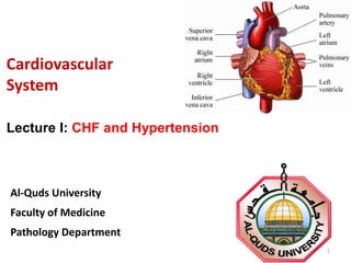
- 1. Cardiovascular System Al-Quds University Faculty of Medicine Pathology Department 1 Lecture I: CHF and Hypertension
- 2. Congestive Heart Failure (CHF) • Definition: diminished functional capacity of the heart which is unable to maintain an output sufficient for metabolic needs. • Can involve the heart's left side, right side, or both sides • Called congestive because of pulmonary congestion and peripheral edema 2
- 3. CHF: Causes • Congenital heart disease (valvular problems) • Ischemic heart disease • Rheumatic heart disease and other immune disease : – E.g., SLE, RA, Scleroderma • Hypertensive heart disease • Inflammatory disease of the heart • Nutritional, endocrine, and metabolic diseases including: – Thyrotoxicosis – Myxedema – Beriberi – Carcinoid syndrome – Storage disease – Amyloidosis • Cardiomyopathy 3
- 4. Types of heart failure • High output failure: inability of the heart to meet the increased demand for blood by tissues – Thyrotoxicosis, anemias, arteriovenous shunts • Low output failure: cardiac output is insufficient at rest or at exertion: – MI, cardiomyopathy, valvular disease • Forward failure: Decreased left ventricular output – Cardiac tamponade, aortic stenosis • Backward failure: increased congestion of the venous circulation, because the failing ventricle is unable to eject the normal volume of venous blood delivered to it during diastole 4
- 5. • Compensated heart failure: If the dilated ventricle is able to maintain cardiac output at a level that meets the needs of the body • Decompensated heart failure: With time, the failing myocardium is no longer able to propel sufficient blood to meet the needs of the body, even at rest. • Right-sided cardiac failure • Left-sided cardiac failure Types of heart failure 5
- 6. • Systolic heart failure: is the result of impaired contractile function – E.g., MI, Dilated cardiomyopathy • Diastolic heart failure: reduced ventricular compliance in the setting of normal or near normal systolic function – E.g., HTN, aortic stenosis Types of heart failure 6
- 7. CHF: Symptoms • Dyspnea on ordinary exertion • Exercise intolerance • Fatigue • Nocturia • Cough (nonproductive) • Orthopnea • Paroxysmal nocturnal dyspnea 7
- 8. CHF: Signs • Acute pulmonary edema • Cardiomegaly • Laterally displaced apical impulse • Increased central venous pressure • Hepatojugular reflux • Hepatomegaly • Lower extremity edema • Neck vein distention • Pleural effusion • Rales (abnormal lung sounds). • S3 gallop (third heart sound, aka “ventricular gallop) • Tachycardia • Unexplained weight gain 8
- 9. The most common causes: • Left sided heart failure (the most common cause) • Intrinsic diseases of the lung: (in the absence of LHF) – pulmonary HTN (cor pulmonale) – COPD – Pulmonary fibrosis • Pulmonic or tricuspid valve disease • Congenital heart diseases (left-to-right shunt) Right-sided cardiac failure 9
- 10. Clinical Features: Right heart failure Systemic venous congestion: • distended neck veins • passive congestion of the liver • generalized venous congestion including jugular veins • enlarged, sometimes tender liver • increased frequency of deep venous thrombi & pulmonary embolism Edema: • subcutaneous edema • weight gain • ascites, pleural effusions • dependent edema (the feet and lower legs) 10
- 11. Left-sided cardiac failure The most common causes: • systemic hypertension • mitral or aortic valve disease • ischemic heart disease • primary diseases of the myocardium. 11
- 12. • Dyspnea due to pulmonary congestion and edema: – Exertional dyspnea: during physical activity – Orthopnea: when the person is lying down – Paroxysmal nocturnal dyspnea: sudden, severe dyspnea, with coughing, choking sensation, & wheezing • muscle fatigue • Enlarged heart, Tachycardia • Third heart sound (S3) • Rales at the lung bases • Mitral regurgitation with systolic murmur • Atrial fibrillation: "irregularly irregular" heartbeat due to chronic dilation of the left atrium Clinical Features: Left heart failure 12
- 13. Compensated heart failure: adaptive responses • Neurohumoral reactions: release of catecholamines in response to reduced cardiac output, this leads to: – Increased force of contraction (inotropic effect) – Increased heart rate (Tachycardia) • Hypertrophy: 1) Concentric, 2) Eccentric • Dilatation • Activation of the renin-angiotensin system 13
- 14. Cardiac hypertrophy: • Concentric hypertrophy: the thickness of the ventricular wall increases without increase in the size of the chamber • Pure pressure load • increase the diameter of individual muscle fibers • e.g., HTN, valvular stenosis Compensated heart failure: adaptive responses 14
- 15. Cardiac hypertrophy: • Eccentric hypertrophy: • an increase in heart size as well as an increase in ventricular wall thickness • pure volume load • increase in cardiac fiber length • E.g., valvular regurgitation, abnormal shunts Compensated heart failure: adaptive responses 15
- 16. 16 Hypertension (HTN) - Introduction “Sustained increase in blood pressure” Silent Killer– asymptomatic until late Symptoms: dizziness, headache, and visual difficulties Risk factor for – MI, DM, Stroke, heart & renal failure 25% of population, <35% aware Chronic, end organ & vascular damage Complications: ATH, IHD, Renal damage, Stroke 95% of HTN is idiopathic, 5% is secondary HTN
- 17. 17
- 18. 18 Malignant (Accelerated) Hypertension Occurs in 5% of hypertensive persons Rapidly rising blood pressure that if untreated leads to death within 1 or 2 years. May complicate any type of HTN. Severe hypertension (diastolic > 120 mm Hg) Characterized by: • renal failure • retinal hemorrhages and exudates, with or without papilledema
- 19. 19 Causes of Hypertension Essential Hypertension (90%-95% of cases) Secondary Hypertension (5%-10%) Renal: Acute glomerulonephritis Chronic renal disease Polycystic disease Renal artery stenosis Renal vasculitis Renin-producing tumors
- 20. 20 Renal Causes: Polycystic Disease
- 21. 21 Endocrine: Adrenocortical hyperfunction (Cushing syndrome, primary aldosteronism, congenital adrenal hyperplasia) Exogenous hormones (glucocorticoids, estrogen [including pregnancy-induced and oral contraceptives], sympathomimetics and tyramine-containing foods, monoamine oxidase inhibitors) Pheochromocytoma Acromegaly Hypothyroidism (myxedema) Hyperthyroidism (thyrotoxicosis) Pregnancy-induced Causes of Hypertension
- 22. 22 Cardiovascular: Coarctation of aorta Polyarteritis nodosa Increased intravascular volume Increased cardiac output Rigidity of the aorta Neurologic: Psychogenic Increased intracranial pressure Sleep apnea Acute stress, including surgery Causes of Hypertension
- 24. 24 Pathogenesis of HTN both increased blood volume and increased peripheral resistance contribute to the increased pressure Cardiac output is influenced by blood volume, which is greatly dependent on body sodium Total peripheral resistance is determined at the level of arterioles, and depends on the effects of the neural and hormonal influences Normal vascular tone depends on the competition between vasoconstricting influences and vasodilators
- 25. 25 Through renin angiotensin system, the kidney influences both peripheral resistance and sodium homeostasis Natriuretic factor is believed to be a neurohormone that is secreted by atria and left ventricle in response to volume expansion. It inhibits Na reabsorption in distal tubules and cause vasodilation Mutations in proteins that affect sodium reabsorption, for example mutations in an epithelial sodium channel protein lead to increased distal tubular reabsorption of Na induced by aldosterone, and decreased renal Na excretion. This results in a moderate to severe form of salt sensitive hypertension called Liddle’s syndrome
- 26. 26 Pathogenesis of Renovascular HTN GFR Renin by JGA Angiotensin II Vasoconstriction Peripheral Vascular Resestance Sodium Retention Blood Volume Aldosterone Hypertension
- 27. 27 Pathogenesis of Essential Hypertension Essential hypertension is a complex multifactorial disorder Genetic factors: • Polygenic mutations are more likely • Gene defects in some enzymes like: • Aldosterone synthase, 11 hydroxylase, 17 hydroxylase • Lead to increase in secretion of aldosterone with the result of increased Na and water reabsorption, plasma volume expansion, and HTN Environmental factors • Stress • Obesity • Physical inactivity • Heavy consumption of salt
- 28. 28 Complications of HTN Large Blood Vessels – Macroangiopathy • Atherosclerosis and its complications Small Blood Vessels – Microangiopathy • hyaline and hyperplastic arteriolosclerosis Heart: • LVH Kidney: • Benign nephrosclerosis Eyes: • Hypertensive retinopathy Brain: • Hemorrhage, infarction
- 29. 29 Benign Nephrosclerosis Leathery Granularity due to minute scarring
- 30. 30 LV Hypertrophy secondary to HTN
- 31. 31 Retinopathy secondary to HTN Normal retina
- 32. Hypertensive Heart Disease • Left ventricular hypertrophy with a history of hypertension • other causes of ventricular hypertrophy have been excluded. – (e.g., aortic stenosis or primary hypertrophic cardiomyopathy) • Cause: sustained pressure load on LV • with increasing degrees of hypertrophy, the metabolic requirements continue to increase but the ability of the heart to meet these demands decreases. • hypertrophy decreases diastolic filling and stroke volume • Complication: CHF, MI, arrhythmias. 32
- 33. Morphology • LV concentric hypertrophy • Heart weight > 450 g. • wall thicknesses > 2.0 cm. • When LV fails, RV undergoes hypertrophy, and dilation may also develop • Histology: cardiac myocytes enlarge, and contain hyperchromatic , rectangular box-car shaped nuclei 33
- 34. LV hypertrophy: Clinical features: • Initially: no symptoms • Later: LV may decompensate (fail) • Diagnosis: CXR, echo, ECG • Complications: – heart failure is a poor prognosis – progressive renal damage – cerebrovascular accidents – sudden cardiac death 34
- 35. Cor Pulmonale (Pulmonary Heart Disease) • Describe disease of right side chambers of the heart. • This results from pulmonary hypertension, occurred from pulmonary parenchymal or vascular disease • Characterized by hypertrophy and/or dilation Morphology: • In acute cor pulmonale: RV is usually dilated. • In chronic cor pulmonale: – RV & right atrium hypertrophy. – Thickness of the RV may exceed that of the LV – Later: RV failure with RV & right atrium dilation 35
- 36. DISORDERS THAT PREDISPOSE TO COR PULMONALE Diseases of the Lungs: Chronic obstructive lung disease (the most common chronic cause) Diffuse pulmonary interstitial fibrosis Extensive, persistent atelectasis Cystic fibrosis Diseases of Pulmonary Vessels: Pulmonary embolism (the most common acute cause) Primary pulmonary vascular sclerosis Pulmonary arteritis (e.g., Wegener granulomatosis) Drug-, toxin-, or radiation-induced vascular sclerosis Disorders Affecting Chest Movement: Kyphoscoliosis Marked obesity Neuromuscular diseases Disorders Inducing Pulmonary Arteriolar Constriction: Metabolic acidosis Hypoxemia 36
Editor's Notes
- 1
- 2
- 3
- 4
- 5
- 6
- 7
- 8
- 9
- 10
- 11
- 12
- 13
- 14
- 15
- 16
- 17
- 18
- 19
- 20
- 21
- 22
- 23
- 24
- 25
- 26
- 27
- 28
- 29
- 30
- 32
- 33
- 34
- 35
- 36