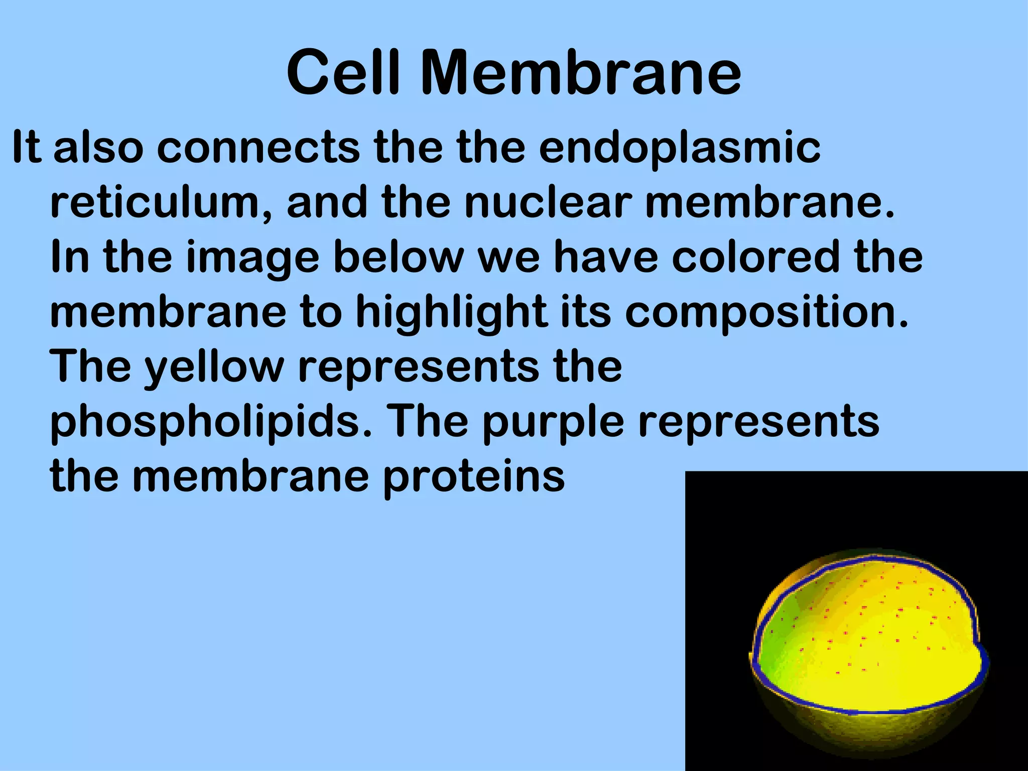
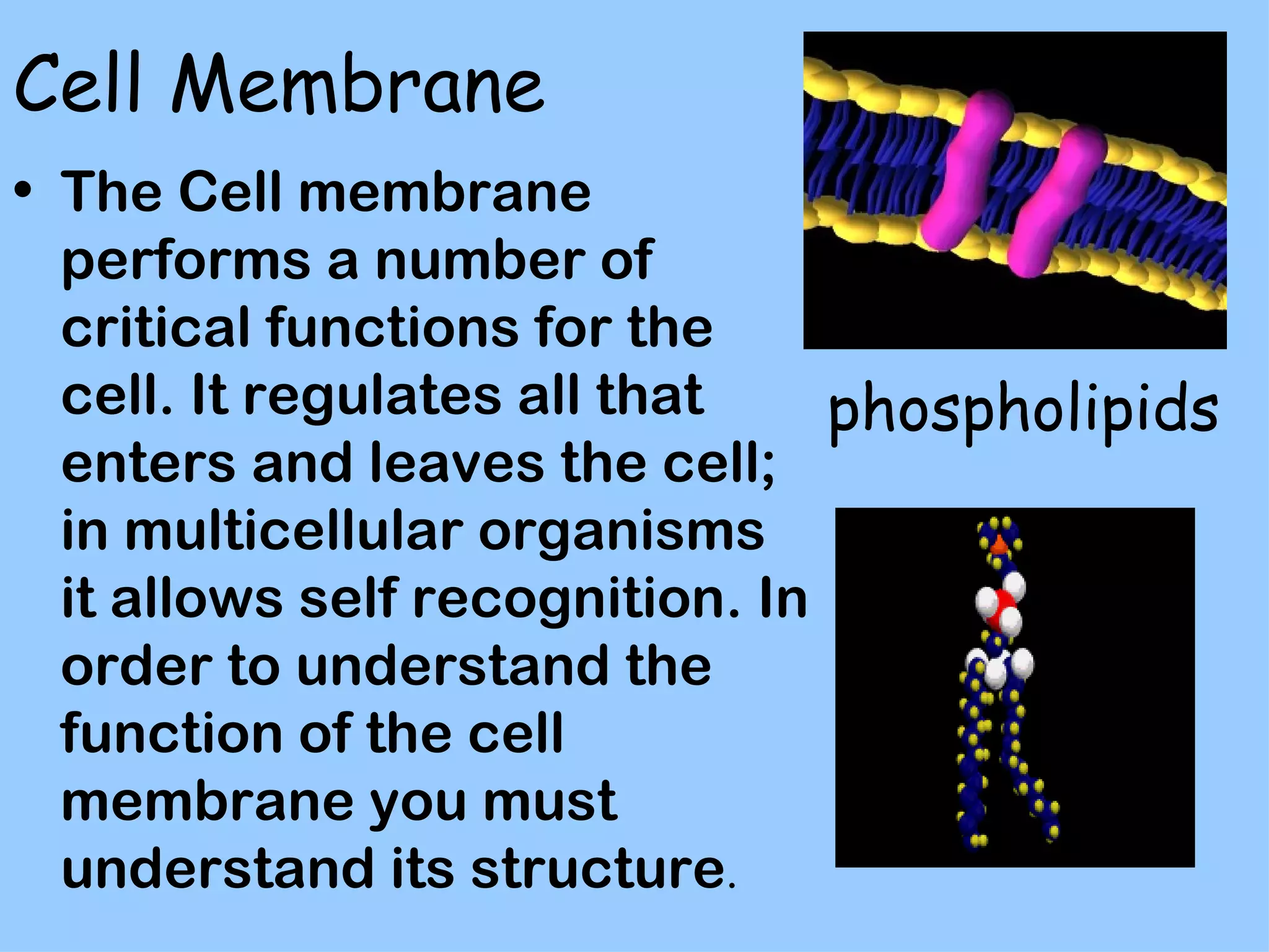
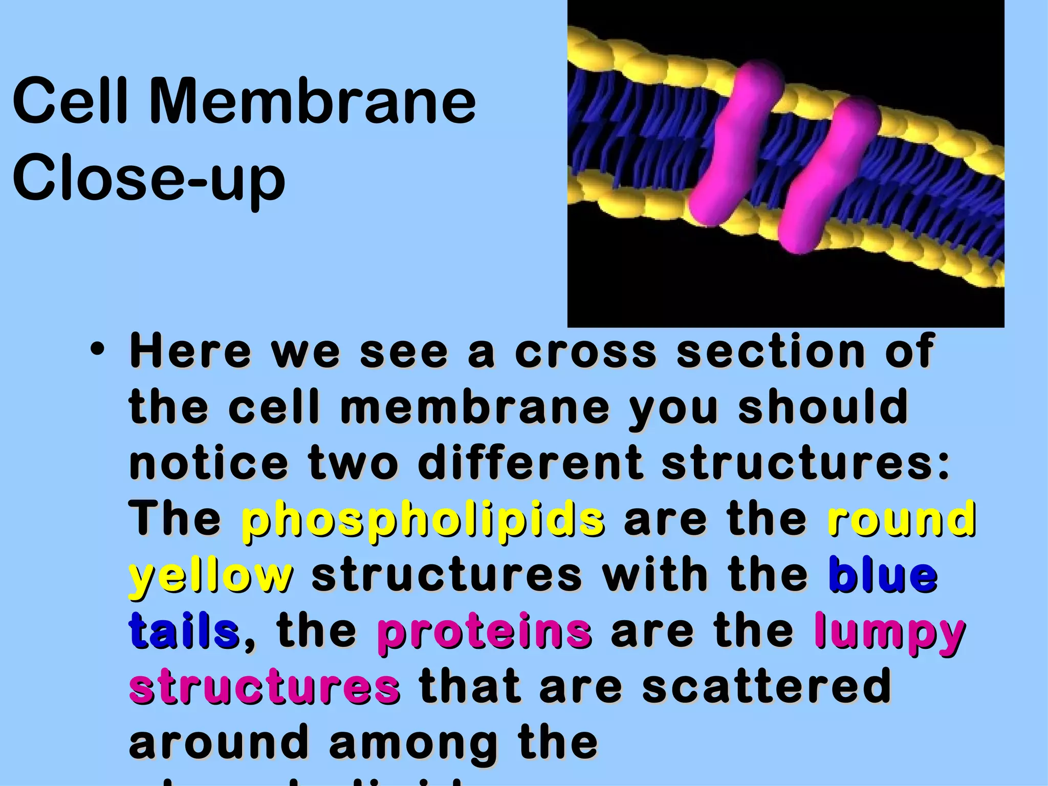
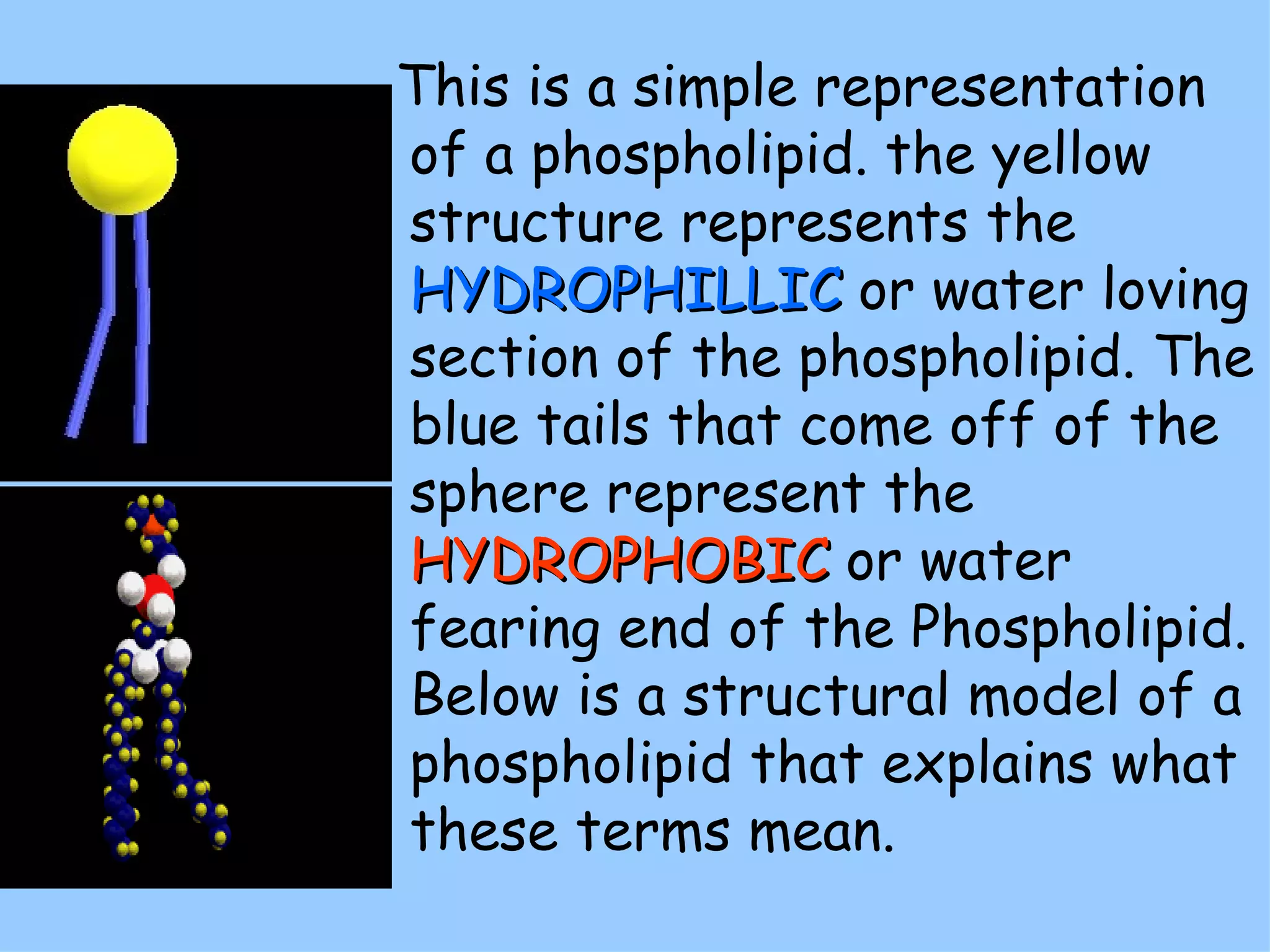
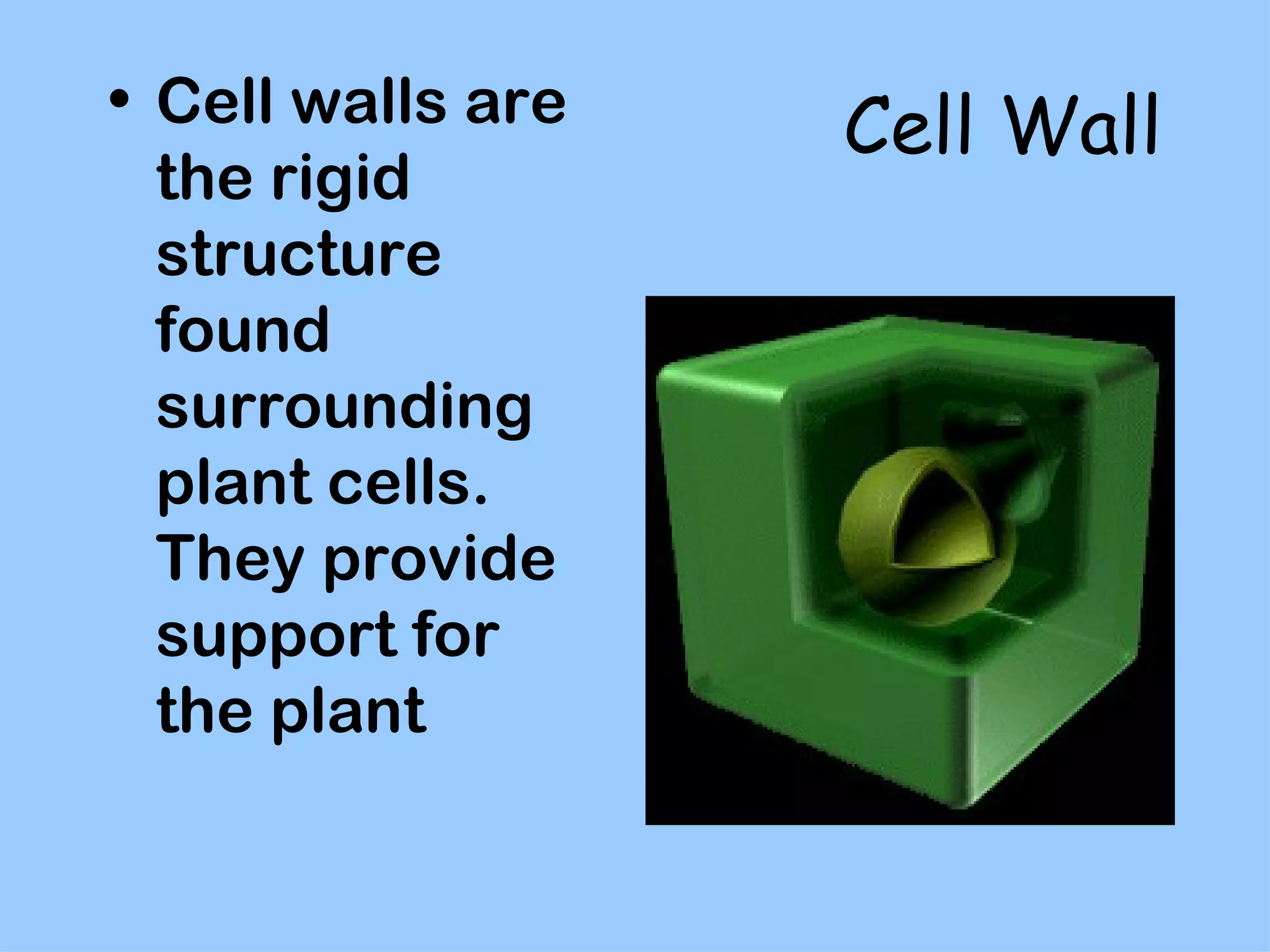
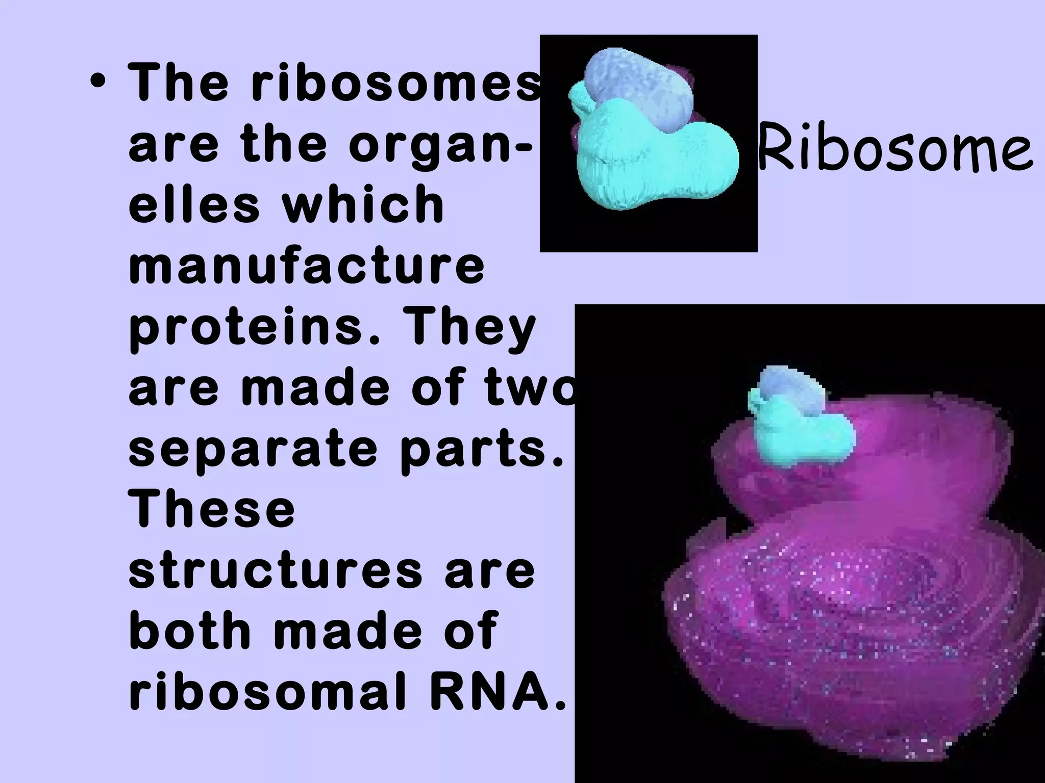
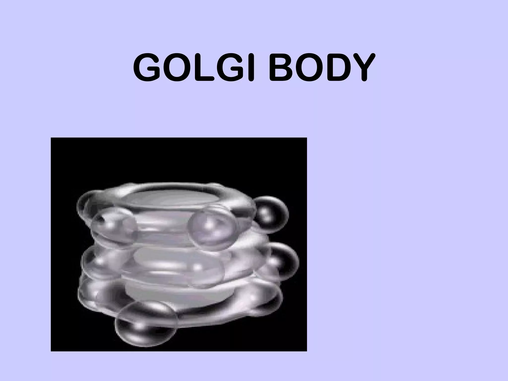
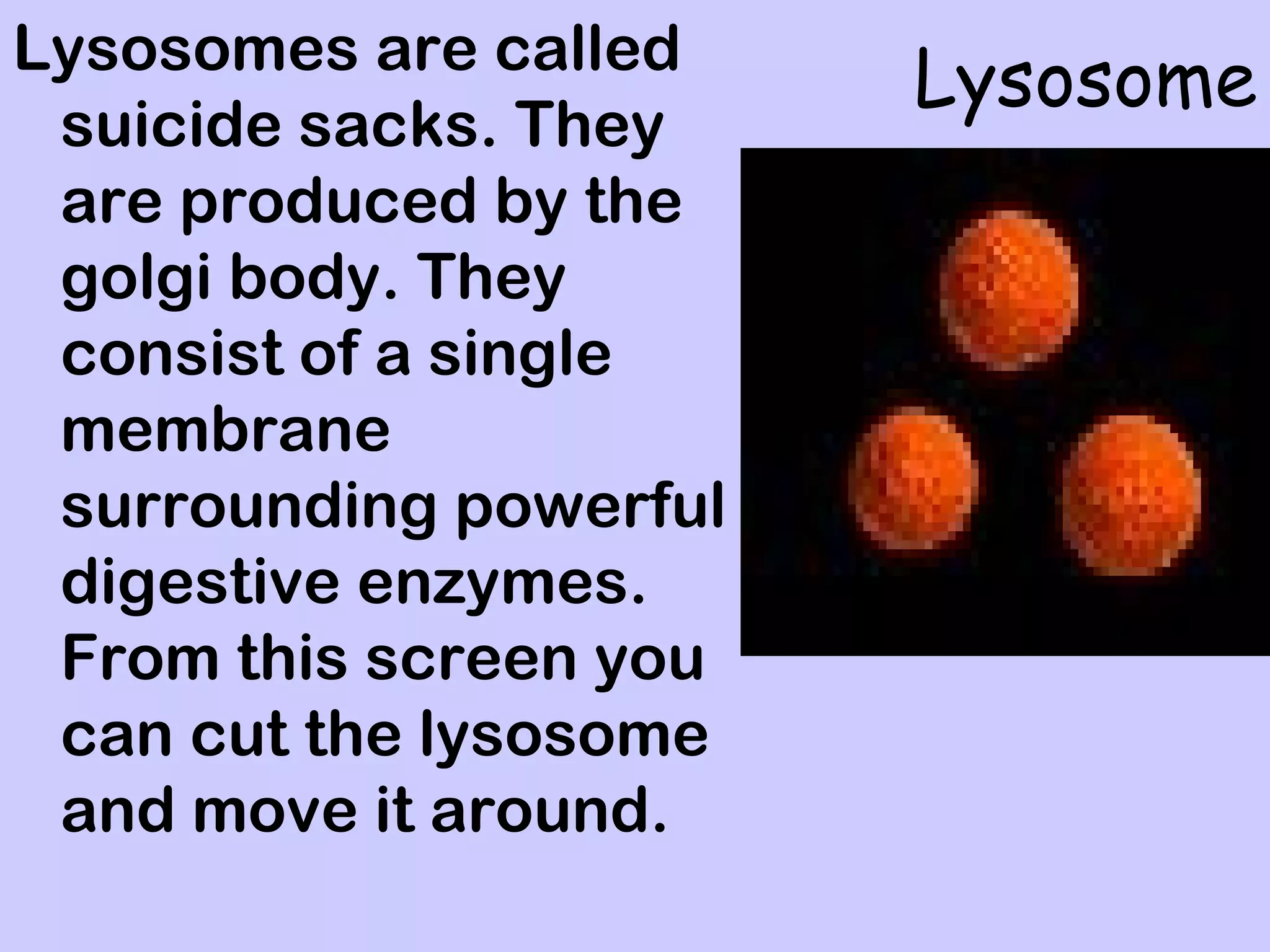
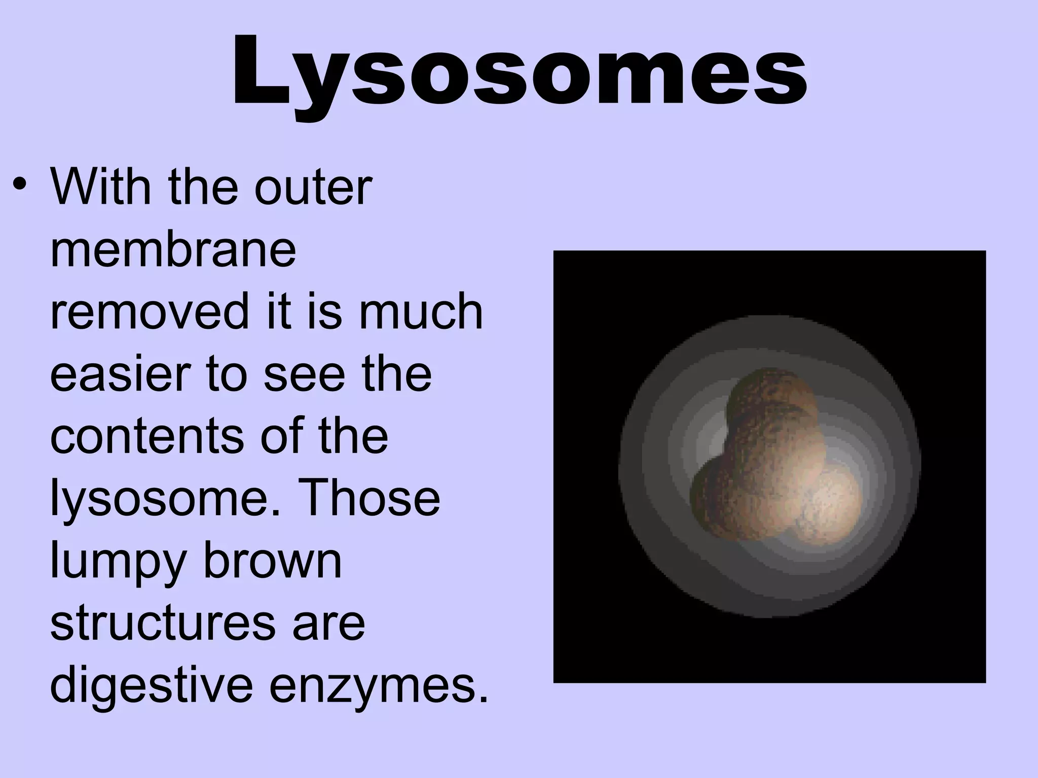
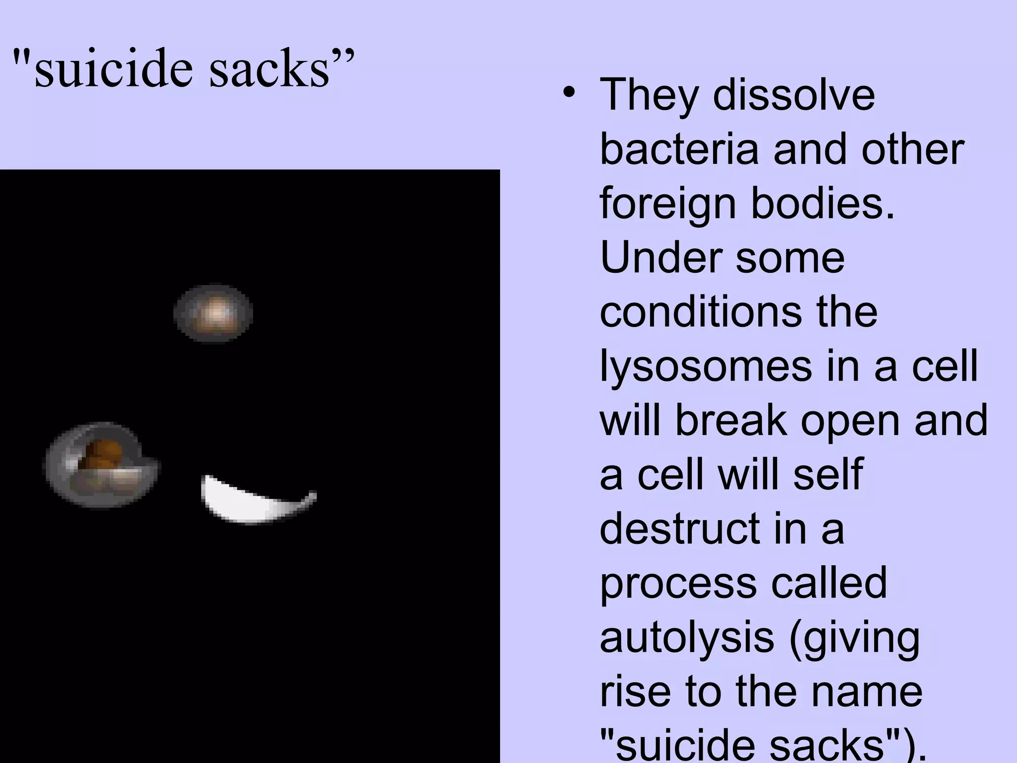
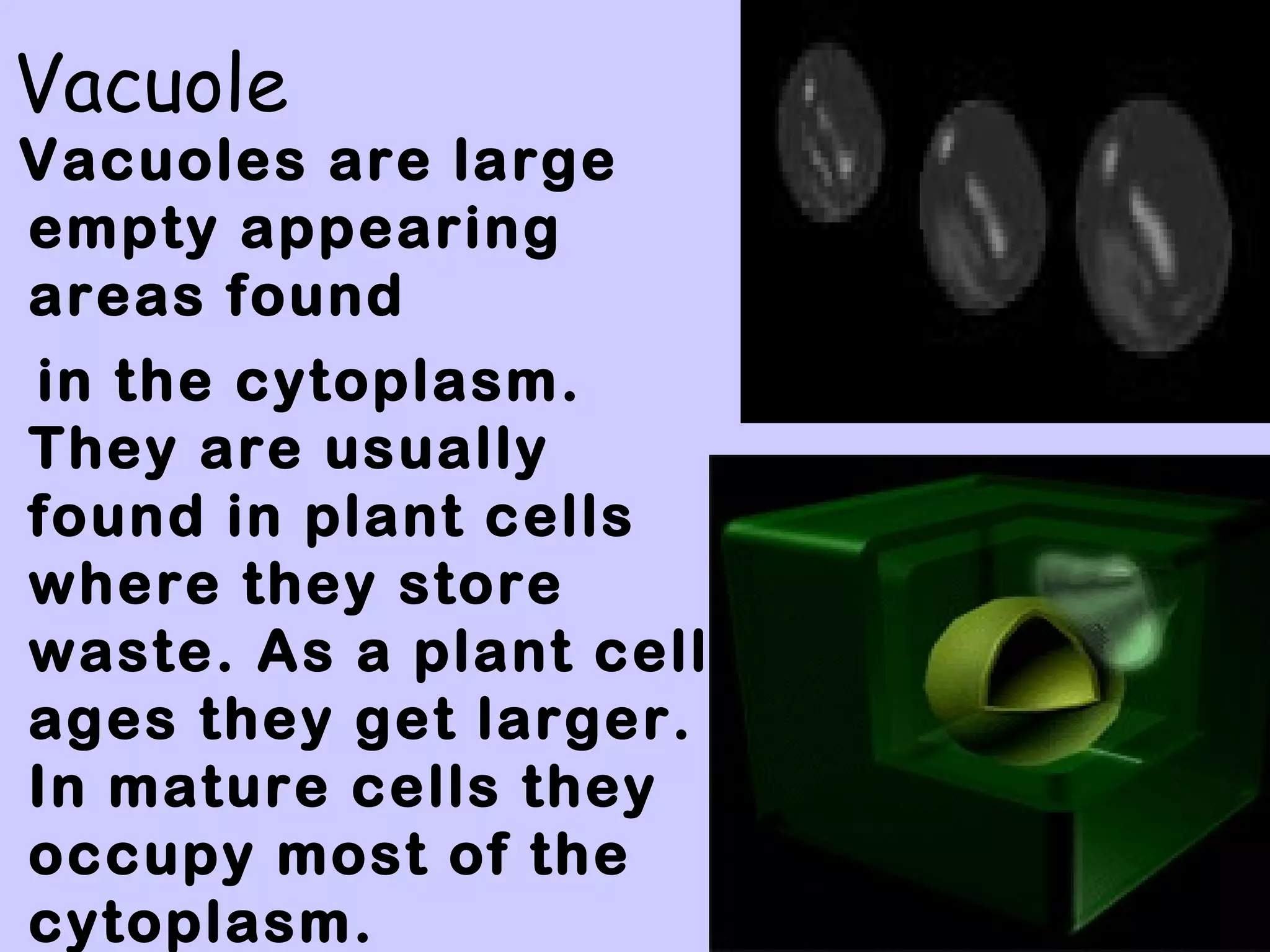
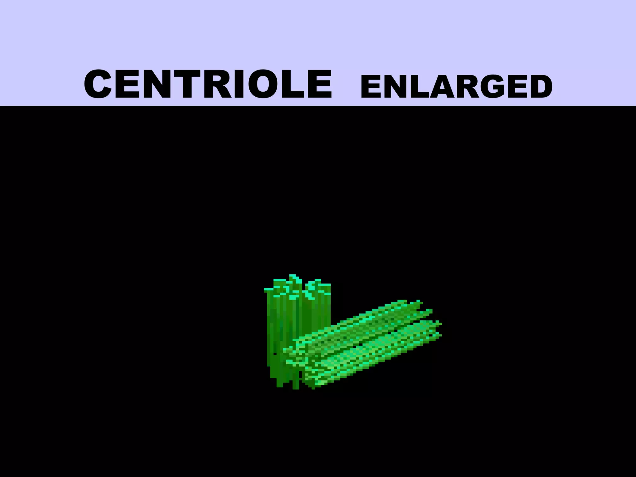
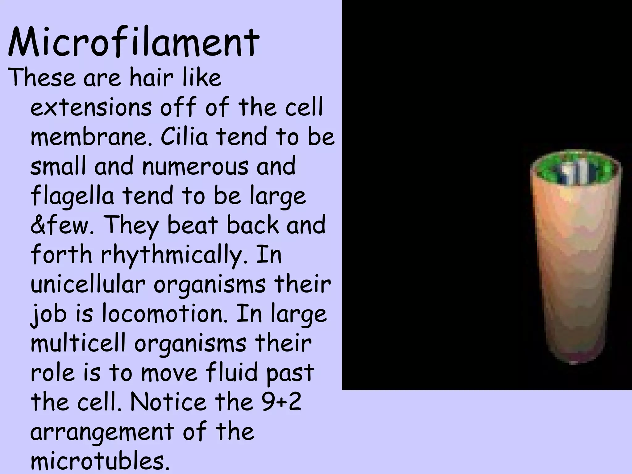
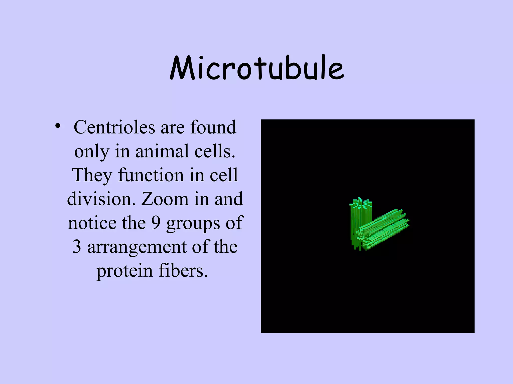
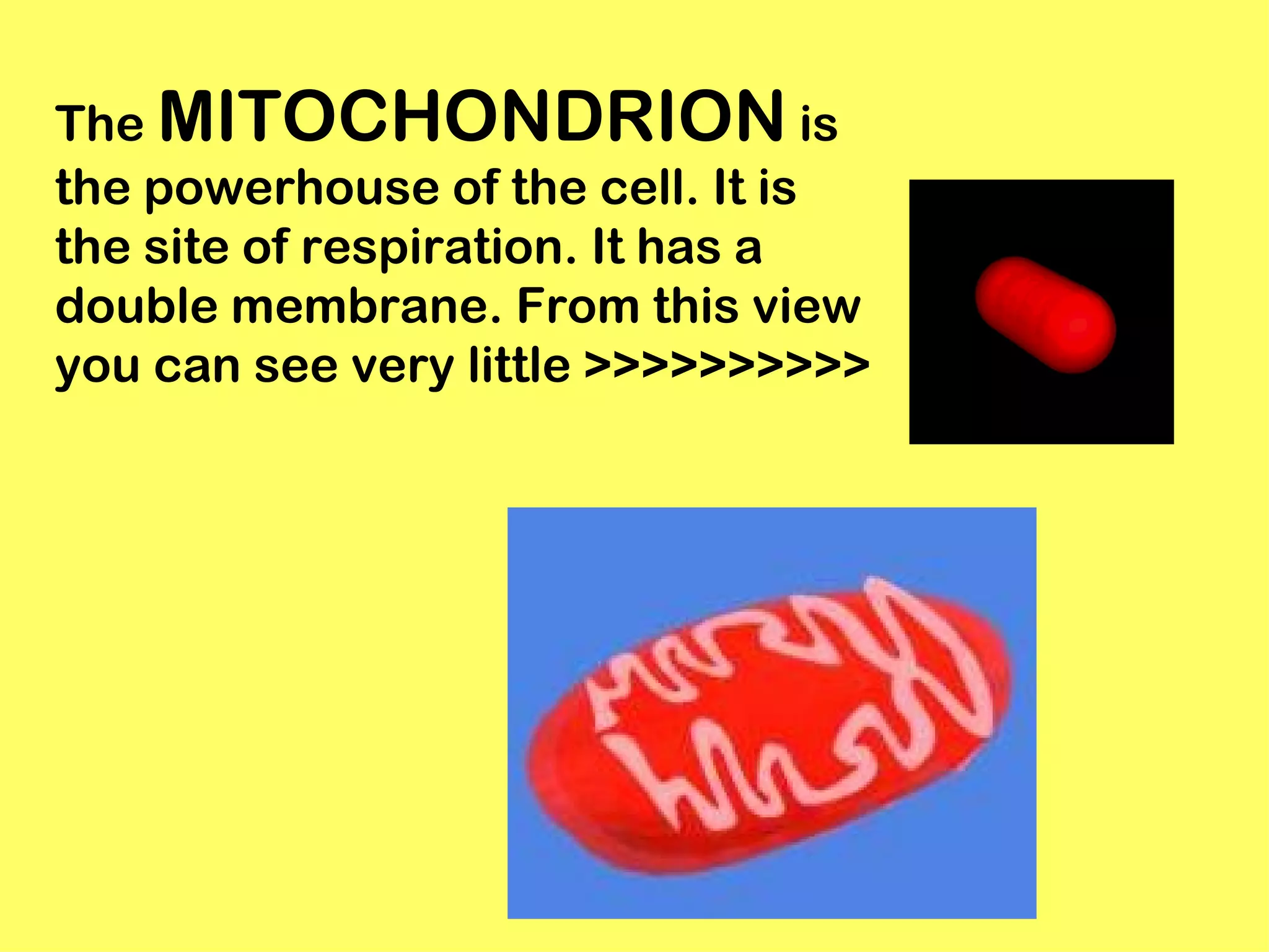
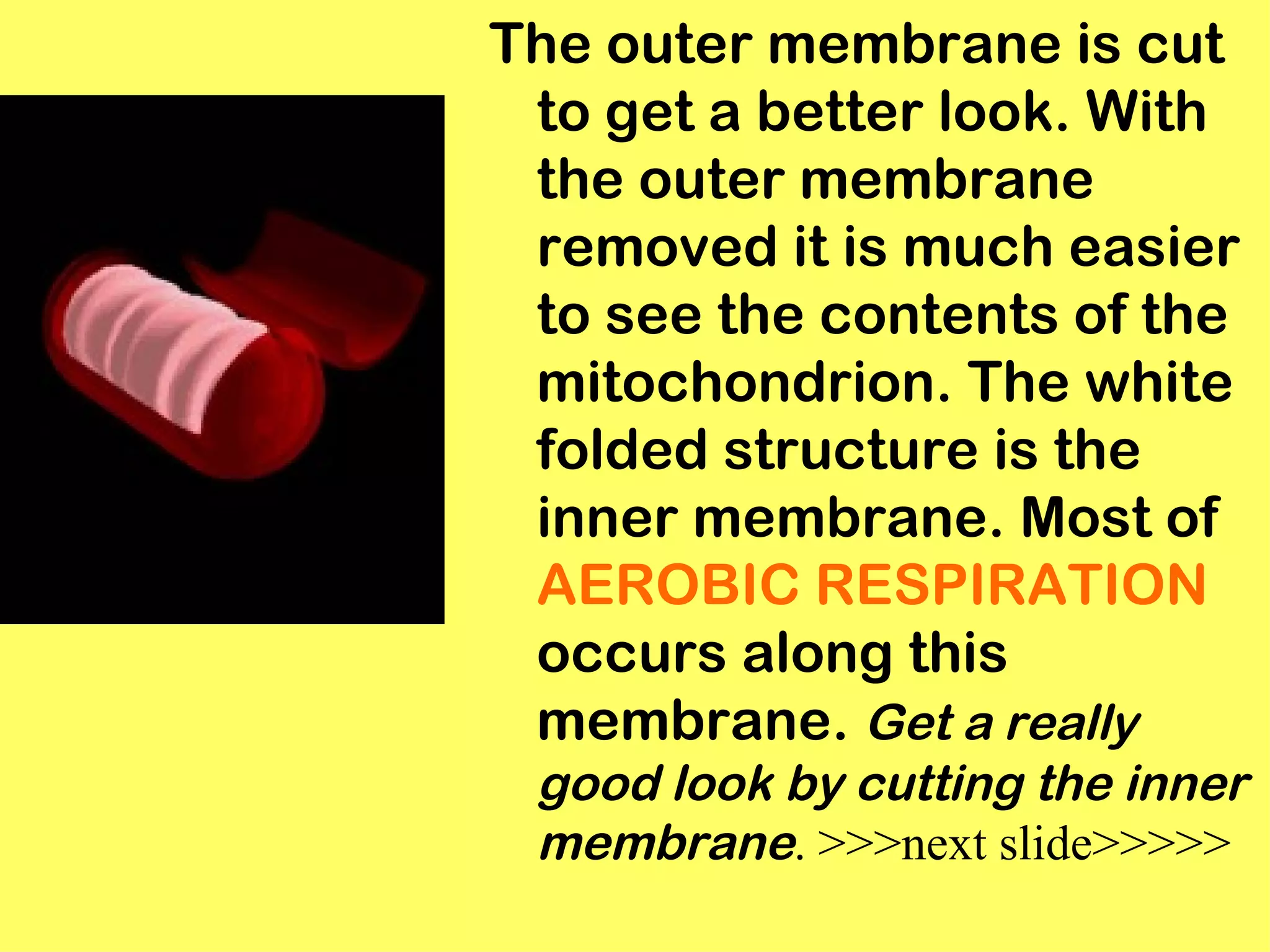
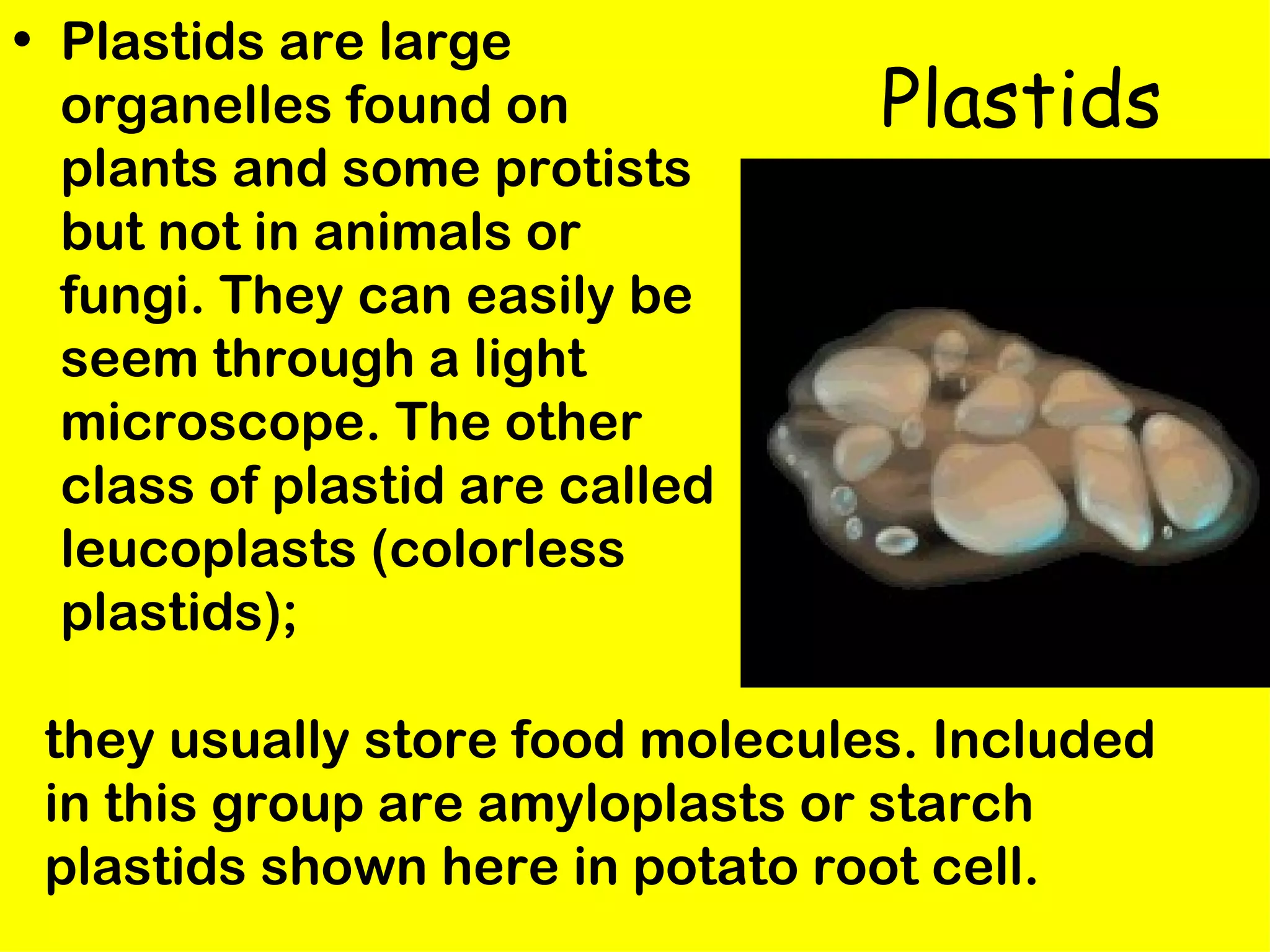
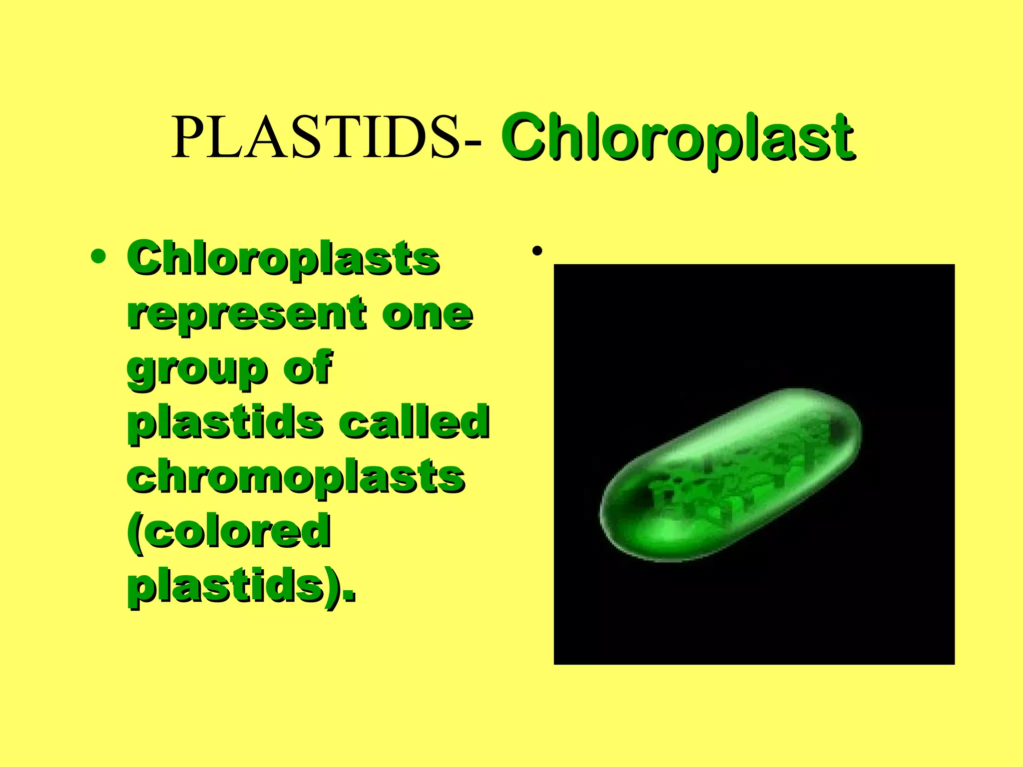
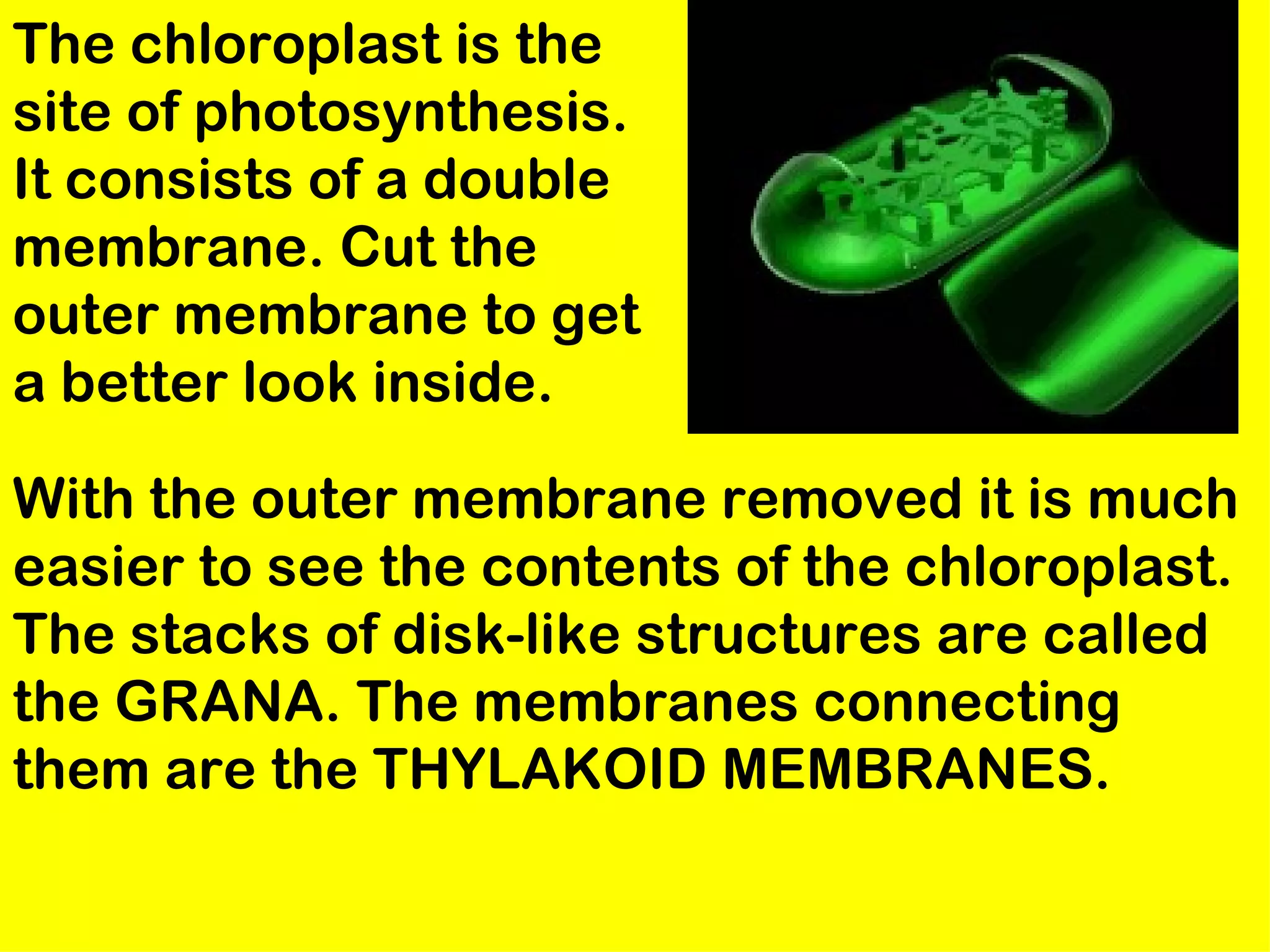
The Cell
Cell Parts
• Cell Membrane, Cell Wall, Cytoplasm
• Protein Production- Nucleus,
  Nucleolus, Endoplasmic Reticulum,
  Ribosomes, Golgi Bodies,
  Lysosomes
• Energy- Mitochondrion, Chloroplasts
  (Energy- Mitochondrion, Chloroplasts)
• Miscellaneous- Microtubules,
  Microfilaments, Plastids
Cell Membrane
It also connects the the endoplasmic
   reticulum, and the nuclear membrane.
   In the image below we have colored the
   membrane to highlight its composition.
   The yellow represents the
   phospholipids. The purple represents
   the membrane proteins
Cell Membrane
• The Cell membrane
  performs a number of
  critical functions for the
  cell. It regulates all that    phospholipids
  enters and leaves the cell;
  in multicellular organisms
  it allows self recognition. In
  order to understand the
  function of the cell
  membrane you must
  understand its structure.
Cell Membrane
Close-up

  • Here we see a cross section of
    the cell membrane you should
    notice two different structures:
    The phospholipids are the round
    yellow structures with the blue
    tails , the proteins are the lumpy
    structures that are scattered
    around among the
This is a simple representation
of a phospholipid. the yellow
structure represents the
HYDROPHILLIC or water loving
section of the phospholipid. The
blue tails that come off of the
sphere represent the
HYDROPHOBIC or water
fearing end of the Phospholipid.
Below is a structural model of a
phospholipid that explains what
these terms mean.
• Cell walls are
                   Cell Wall
  the rigid
  structure
  found
  surrounding
  plant cells.
  They provide
  support for
  the plant
Cytoplasm
• The term cytoplasm refers to
  everything between the cell
  membrane and the nuclear
  envelope. It consists of primarily
  of water. It also contains various
  organelles as well as salts,
  dissolved gasses and nutrients.
Cytoplasm
• Protein Producing
  Organelles: Endoplasmic
  Reticulum, Ribosomes, Golgi
  Apparatus, Lysosomes
• Energy Producing Organelles
  -Mitochondrion, Chloroplasts
• Specialty Structures
  -centrioles, vacuoles, cell
  walls, cilia, flagella,
  plastids
NUCLEUS-
   nuclear envelope


The nuclear envelope is a double
 membrane. Is has 4 phospholipid
 layers. It is also has large pores
 through which materials pass
 back and forth.
Nucleus


The headquarters of the cell. It is a
 large dark spot in EUKARYOTIC
 cells. It controls all cell activity.
 Close up you will see that the
 nuclear membrane has many
 pores. The nuclear membrane is
 continuous with the E.R.
With the outer membrane removed it is
much easier to see the contents of the
nucleus.
                  The thick ropy
                   strands are the
                   CHROMATIN . The
                   large solid spot is
                   the NUCLEOLUS .
                   The nucleolus is a
                   knot of chromatin.
                   It manufactures
                   ribosomes.
Chromatin


Within the nucleus are found
 chromatin and a structure called
 the nucleolus. Chromatin is DNA in
 its active form. It consists of DNA
 looped around histone proteins.
 The nucleolus is a knot of
 chromatin. It is the nucleolus that
Endoplasmic Reticulum

  • Smooth, no
    ribosomes
  • rough, has
    ribosomes
Smooth E.R.
• The endoplasmic reticulum is a series of
  double membranes that loop back and forth
  between the cell membrane and the
  nucleus.
• These membranes fill the cytoplasm but
  you cannot see them because they are very
  transparent. There are two distinct types of
  E.R.: The rough E.R. has ribosomes and is
  the site of protein synthesis; the smooth
  E.R. has no ribosomes
•
The endoplasmic
R.E.R.    reticulum is a series
          of double membranes
          that loop back and
          forth between the cell
          membrane and the
          nucleus. These
          membranes fill the
          cytoplasm but you
          cannot see them
          because they are very
          transparent.
• The ribosomes
  are the organ-    Ribosome
  elles which
  manufacture
  proteins. They
  are made of two
  separate parts.
  These
  structures are
  both made of
  ribosomal RNA.
GOLGI BODY
The golgi body is      Golgi Apparatus
 responsible for
 packaging proteins
 for the cell. Once
 the proteins are
 produced by the
 rough E.R. they
 pass into the sack-
 like cisternae that
 are the main part of
 the golgi body.
  These proteins are then squeezed off into the
  little blebs which drift off into the cytoplasm.
Lysosomes are called
                        Lysosome
 suicide sacks. They
 are produced by the
 golgi body. They
 consist of a single
 membrane
 surrounding powerful
 digestive enzymes.
 From this screen you
 can cut the lysosome
 and move it around.
Lysosomes
• With the outer
  membrane
  removed it is much
  easier to see the
  contents of the
  lysosome. Those
  lumpy brown
  structures are
  digestive enzymes.
"suicide sacks”   • They dissolve
                    bacteria and other
                    foreign bodies.
                    Under some
                    conditions the
                    lysosomes in a cell
                    will break open and
                    a cell will self
                    destruct in a
                    process called
                    autolysis (giving
                    rise to the name
                    "suicide sacks").
Vacuole
Vacuoles are large
empty appearing
areas found
in the cytoplasm.
They are usually
found in plant cells
where they store
waste. As a plant cell
ages they get larger.
In mature cells they
occupy most of the
cytoplasm.
CENTRIOLE ENLARGED
Microfilament
These are hair like
 extensions off of the cell
 membrane. Cilia tend to be
 small and numerous and
 flagella tend to be large
 &few. They beat back and
 forth rhythmically. In
 unicellular organisms their
 job is locomotion. In large
 multicell organisms their
 role is to move fluid past
 the cell. Notice the 9+2
 arrangement of the
 microtubles.
Microtubule
• Centrioles are found
   only in animal cells.
  They function in cell
 division. Zoom in and
  notice the 9 groups of
  3 arrangement of the
      protein fibers.
The MITOCHONDRION is
the powerhouse of the cell. It is
the site of respiration. It has a
double membrane. From this view
you can see very little >>>>>>>>>>
The outer membrane is cut
 to get a better look. With
 the outer membrane
 removed it is much easier
 to see the contents of the
 mitochondrion. The white
 folded structure is the
 inner membrane. Most of
 AEROBIC RESPIRATION
 occurs along this
 membrane. Get a really
 good look by cutting the inner
 membrane. >>>next slide>>>>>
The inner membranes is ruffled. It has
a very large surface area. These ruffles are
called cristae. Mitochondria have their
own DNA and manufacture some their
own proteins. It is thought that the
mitochondrion evolved from symbiotic
bacteria that took up residence inside
the first eukaryotic cells.
INSIDE THE INNER
 MITOCHONDRION
• Plastids are large
  organelles found on            Plastids
  plants and some protists
  but not in animals or
  fungi. They can easily be
  seem through a light
  microscope. The other
  class of plastid are called
  leucoplasts (colorless
  plastids);

 they usually store food molecules. Included
 in this group are amyloplasts or starch
 plastids shown here in potato root cell.
PLASTIDS- Chloroplast
• Chloroplasts    •
  represent one
  group of
  plastids called
  chromoplasts
  (colored
  plastids).
The chloroplast is the
site of photosynthesis.
It consists of a double
membrane. Cut the
outer membrane to get
a better look inside.

With the outer membrane removed it is much
easier to see the contents of the chloroplast.
The stacks of disk-like structures are called
the GRANA. The membranes connecting
them are the THYLAKOID MEMBRANES.
Grana and Thylakoid
           Membranes
                         • The membranes
                           that you see
Dissolve the Remaining
 membrane and zoom in      here are the
to get a better look.      site of
                           photosynthesis.
                           It is here that
                           the energy
                           harnessing
                           process of
                           photosynthesis
                           occurs.

Ch.1 Cell Structures and Functions

  • 1.
  • 2.
    Cell Parts • CellMembrane, Cell Wall, Cytoplasm • Protein Production- Nucleus, Nucleolus, Endoplasmic Reticulum, Ribosomes, Golgi Bodies, Lysosomes • Energy- Mitochondrion, Chloroplasts (Energy- Mitochondrion, Chloroplasts) • Miscellaneous- Microtubules, Microfilaments, Plastids
  • 3.
    Cell Membrane It alsoconnects the the endoplasmic reticulum, and the nuclear membrane. In the image below we have colored the membrane to highlight its composition. The yellow represents the phospholipids. The purple represents the membrane proteins
  • 4.
    Cell Membrane • TheCell membrane performs a number of critical functions for the cell. It regulates all that phospholipids enters and leaves the cell; in multicellular organisms it allows self recognition. In order to understand the function of the cell membrane you must understand its structure.
  • 5.
    Cell Membrane Close-up • Here we see a cross section of the cell membrane you should notice two different structures: The phospholipids are the round yellow structures with the blue tails , the proteins are the lumpy structures that are scattered around among the
  • 6.
    This is asimple representation of a phospholipid. the yellow structure represents the HYDROPHILLIC or water loving section of the phospholipid. The blue tails that come off of the sphere represent the HYDROPHOBIC or water fearing end of the Phospholipid. Below is a structural model of a phospholipid that explains what these terms mean.
  • 7.
    • Cell wallsare Cell Wall the rigid structure found surrounding plant cells. They provide support for the plant
  • 8.
    Cytoplasm • The termcytoplasm refers to everything between the cell membrane and the nuclear envelope. It consists of primarily of water. It also contains various organelles as well as salts, dissolved gasses and nutrients.
  • 9.
    Cytoplasm • Protein Producing Organelles: Endoplasmic Reticulum, Ribosomes, Golgi Apparatus, Lysosomes • Energy Producing Organelles -Mitochondrion, Chloroplasts • Specialty Structures -centrioles, vacuoles, cell walls, cilia, flagella, plastids
  • 10.
    NUCLEUS- nuclear envelope The nuclear envelope is a double membrane. Is has 4 phospholipid layers. It is also has large pores through which materials pass back and forth.
  • 11.
    Nucleus The headquarters ofthe cell. It is a large dark spot in EUKARYOTIC cells. It controls all cell activity. Close up you will see that the nuclear membrane has many pores. The nuclear membrane is continuous with the E.R.
  • 12.
    With the outermembrane removed it is much easier to see the contents of the nucleus. The thick ropy strands are the CHROMATIN . The large solid spot is the NUCLEOLUS . The nucleolus is a knot of chromatin. It manufactures ribosomes.
  • 13.
    Chromatin Within the nucleusare found chromatin and a structure called the nucleolus. Chromatin is DNA in its active form. It consists of DNA looped around histone proteins. The nucleolus is a knot of chromatin. It is the nucleolus that
  • 14.
    Endoplasmic Reticulum • Smooth, no ribosomes • rough, has ribosomes
  • 15.
    Smooth E.R. • Theendoplasmic reticulum is a series of double membranes that loop back and forth between the cell membrane and the nucleus. • These membranes fill the cytoplasm but you cannot see them because they are very transparent. There are two distinct types of E.R.: The rough E.R. has ribosomes and is the site of protein synthesis; the smooth E.R. has no ribosomes •
  • 16.
    The endoplasmic R.E.R. reticulum is a series of double membranes that loop back and forth between the cell membrane and the nucleus. These membranes fill the cytoplasm but you cannot see them because they are very transparent.
  • 17.
    • The ribosomes are the organ- Ribosome elles which manufacture proteins. They are made of two separate parts. These structures are both made of ribosomal RNA.
  • 18.
  • 19.
    The golgi bodyis Golgi Apparatus responsible for packaging proteins for the cell. Once the proteins are produced by the rough E.R. they pass into the sack- like cisternae that are the main part of the golgi body. These proteins are then squeezed off into the little blebs which drift off into the cytoplasm.
  • 20.
    Lysosomes are called Lysosome suicide sacks. They are produced by the golgi body. They consist of a single membrane surrounding powerful digestive enzymes. From this screen you can cut the lysosome and move it around.
  • 21.
    Lysosomes • With theouter membrane removed it is much easier to see the contents of the lysosome. Those lumpy brown structures are digestive enzymes.
  • 22.
    "suicide sacks” • They dissolve bacteria and other foreign bodies. Under some conditions the lysosomes in a cell will break open and a cell will self destruct in a process called autolysis (giving rise to the name "suicide sacks").
  • 23.
    Vacuole Vacuoles are large emptyappearing areas found in the cytoplasm. They are usually found in plant cells where they store waste. As a plant cell ages they get larger. In mature cells they occupy most of the cytoplasm.
  • 24.
  • 25.
    Microfilament These are hairlike extensions off of the cell membrane. Cilia tend to be small and numerous and flagella tend to be large &few. They beat back and forth rhythmically. In unicellular organisms their job is locomotion. In large multicell organisms their role is to move fluid past the cell. Notice the 9+2 arrangement of the microtubles.
  • 26.
    Microtubule • Centrioles arefound only in animal cells. They function in cell division. Zoom in and notice the 9 groups of 3 arrangement of the protein fibers.
  • 27.
    The MITOCHONDRION is thepowerhouse of the cell. It is the site of respiration. It has a double membrane. From this view you can see very little >>>>>>>>>>
  • 28.
    The outer membraneis cut to get a better look. With the outer membrane removed it is much easier to see the contents of the mitochondrion. The white folded structure is the inner membrane. Most of AEROBIC RESPIRATION occurs along this membrane. Get a really good look by cutting the inner membrane. >>>next slide>>>>>
  • 29.
    The inner membranesis ruffled. It has a very large surface area. These ruffles are called cristae. Mitochondria have their own DNA and manufacture some their own proteins. It is thought that the mitochondrion evolved from symbiotic bacteria that took up residence inside the first eukaryotic cells.
  • 30.
    INSIDE THE INNER MITOCHONDRION
  • 31.
    • Plastids arelarge organelles found on Plastids plants and some protists but not in animals or fungi. They can easily be seem through a light microscope. The other class of plastid are called leucoplasts (colorless plastids); they usually store food molecules. Included in this group are amyloplasts or starch plastids shown here in potato root cell.
  • 32.
    PLASTIDS- Chloroplast • Chloroplasts • represent one group of plastids called chromoplasts (colored plastids).
  • 33.
    The chloroplast isthe site of photosynthesis. It consists of a double membrane. Cut the outer membrane to get a better look inside. With the outer membrane removed it is much easier to see the contents of the chloroplast. The stacks of disk-like structures are called the GRANA. The membranes connecting them are the THYLAKOID MEMBRANES.
  • 34.
    Grana and Thylakoid Membranes • The membranes that you see Dissolve the Remaining membrane and zoom in here are the to get a better look. site of photosynthesis. It is here that the energy harnessing process of photosynthesis occurs.