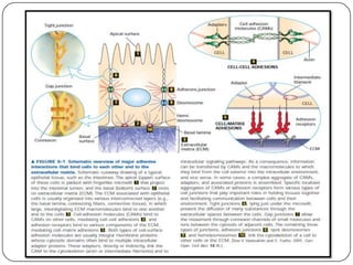
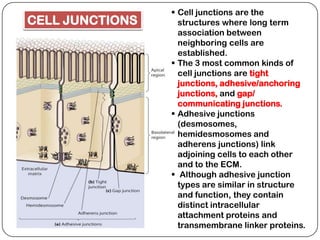
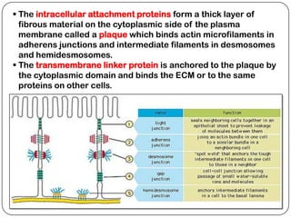
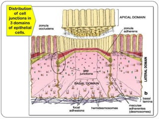
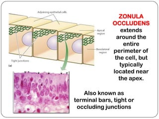
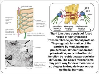
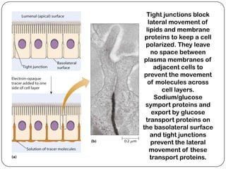
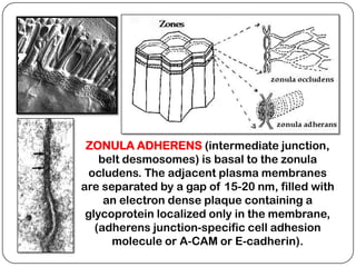
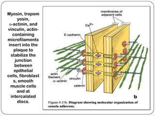
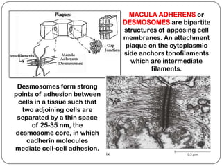
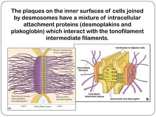
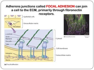
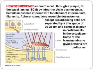
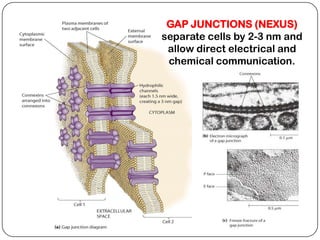
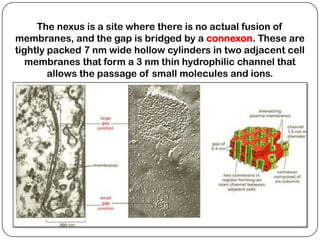
CELL JUNCTIONS


Department of Natural Sciences
   University of St. La Salle
        Bacolod City
 Cell junctions are the
CELL JUNCTIONS     structures where long term
                   association between
                   neighboring cells are
                   established.
                  The 3 most common kinds of
                   cell junctions are tight
                   junctions, adhesive/anchoring
                   junctions, and gap/
                   communicating junctions.
                  Adhesive junctions
                   (desmosomes,
                   hemidesmosomes and
                   adherens junctions) link
                   adjoining cells to each other
                   and to the ECM.
                  Although adhesive junction
                   types are similar in structure
                   and function, they contain
                   distinct intracellular
                   attachment proteins and
                   transmembrane linker proteins.
 The intracellular attachment proteins form a thick layer of
  fibrous material on the cytoplasmic side of the plasma
  membrane called a plaque which binds actin microfilaments in
  adherens junctions and intermediate filaments in desmosomes
  and hemidesmosomes.
 The transmembrane linker protein is anchored to the plaque by
  the cytoplasmic domain and binds the ECM or to the same
  proteins on other cells.
Distribution
   of cell
junctions in
 3 domains
of epithelial
    cells.
ZONULA
              OCCLUDENS
                  extends
                around the
                   entire
              perimeter of
               the cell, but
                 typically
              located near
                 the apex.

   Also known as
terminal bars, tight or
 occluding junctions
Tight junctions consist of fused
       ridges of tightly packed
transmembrane junctional proteins.
    They regulate formation of the
      barriers by modulating cell
   proliferation, differentiation and
   polarization, and control barrier
 function by restricting paracellular
  diffusion. The above mechanisms
  may pave way for new therapeutic
  strategies in drug delivery across
          epithelial barriers.
Tight junctions block
   lateral movement of
  lipids and membrane
 proteins to keep a cell
 polarized. They leave
    no space between
 plasma membranes of
      adjacent cells to
 prevent the movement
   of molecules across
         cell layers.
      Sodium/glucose
 symport proteins and
     export by glucose
 transport proteins on
the basolateral surface
    and tight junctions
    prevent the lateral
    movement of these
    transport proteins.
ZONULA ADHERENS (intermediate junction,
    belt desmosomes) is basal to the zonula
 ocludens. The adjacent plasma membranes
are separated by a gap of 15-20 nm, filled with
     an electron dense plaque containing a
 glycoprotein localized only in the membrane,
   (adherens junction-specific cell adhesion
      molecule or A-CAM or E-cadherin).
Myosin, tropom
      yosin,
 α-actinin, and
vinculin, actin-
   containing
microfilaments
 insert into the
    plaque to
  stabilize the
     junction
    between
    epithelial
cells, fibroblast
    s, smooth
  muscle cells
      and at
  intercalated
      discs.
MACULA ADHERENS or
                               DESMOSOMES are bipartite
                               structures of apposing cell
                               membranes. An attachment
                                plaque on the cytoplasmic
                               side anchors tonofilaments
                                  which are intermediate
                                        filaments.

 Desmosomes form strong
points of adhesion between
 cells in a tissue such that
  two adjoining cells are
 separated by a thin space
      of 25-35 nm, the
desmosome core, in which
    cadherin molecules
mediate cell-cell adhesion.
The plaques on the inner surfaces of cells joined
 by desmosomes have a mixture of intracellular
     attachment proteins (desmoplakins and
plakoglobin) which interact with the tonofilament
             intermediate filaments.
Adherens junctions called FOCAL ADHESION can join
   a cell to the ECM, primarily through fibronectin
                      receptors.
HEMIDESMOSOMES connect a cell, through a plaque, to
the basal lamina (ECM) by integrins. As in desmosomes,
hemidesmosomes interact with tonofilament intermediate
filaments. Adherens junctions resemble desmosomes
                            except two adjoining cells are
                            separated by a thin space of
                            20-25 nm and connect to actin
                                          microfilaments
                                          in the cytoplasm.
                                          Some of the
                                          transmembrane
                                          glycoproteins are
                                                 cadherins.
Hemidesmosomes
                                  occur at most basal
                                 surface of stratified
                                  squamous epithelia
                                      where the
                                 superficial layer lack
                                      junctional
                                  complexes, and the
                                    basal cells are
                                    exposed to the
                                    underlying CT.


 They serve mainly as sites of
attachment for the actin-based
   contractile system of the
         cytoplasm.
GAP JUNCTIONS (NEXUS)
separate cells by 2-3 nm and
 allow direct electrical and
 chemical communication.
The nexus is a site where there is no actual fusion of
membranes, and the gap is bridged by a connexon. These are
tightly packed 7 nm wide hollow cylinders in two adjacent cell
   membranes that form a 3 nm thin hydrophilic channel that
        allows the passage of small molecules and ions.
 The connexons of each membrane link to form
  continuous pores that bridge the intercellular gap,
  allowing passage of ions, cyclic AMP, amino acids
  and other small molecules.
 As sites of electronic coupling (reduced resistance to
  ion flow), it is the only type of junction mediating flow
  of current between cells important in intercellular
  communication and coordination.
 An influx of Ca+2 ions results in the closure of their
  channels, preventing spread of damage to other cells.
 Also found between osteocytes, astrocytes, cardiac
  muscle cells, smooth muscle cells, & endocrine cells.
 Cancer cells generally do not have gap junctions, so
  that cells fail to communicate their mitotic activity to
  each other, which may explain their uncontrolled
  growth.
 When they were originally discovered cell
  junctions were considered to be relatively static
  structures. This was likely because they appeared
  to have a consistent, unchanging structure when
  viewed with the electron microscope.
 New techniques have revealed that proteins can
  move in and out of these junctions allowing the
  cell to sense the status of its intercellular
  adhesions.
 For example, occludin and ZO1, two proteins from
  adherens junctions have been shown to move into
  the nucleus to regulate gene activity.
 The interaction of junctional adhesion molecules
  with the cytoskeleton has also been shown to be a
  dynamic process that is still being elucidated.
(A) In a single cell, a subset of E-cadherin is found in a complex with Nectin-2α
     and components of the Exocyst complex. (B) Upon cell-cell adhesion, E-
   cadherin and Nectin-2α homodimers form trans-interactions with E-cadherin
and Nectin-2α homodimers from the opposing cell, respectively. This interaction
  initiates the recruitment of microtubules, the Exocyst complex and the basal-
  lateral SNARE complex to the forming cell-cell contact. (C) Following cell-cell
     contact formation, microtubules extend into the contact, and post-Golgi
    carriers carrying basal-lateral cargo travel via microtubules to the forming
   contact. At the forming contact, the Exocyst and SNARE complexes are fully
 functional in mediating docking and fusion of basal-lateral post-Golgi carriers.
http://www.mhhe.com/biosci/gen
bio/biolink/j_explorations/explor
ations.html

Cell junctions

  • 1.
    CELL JUNCTIONS Department ofNatural Sciences University of St. La Salle Bacolod City
  • 3.
     Cell junctionsare the CELL JUNCTIONS structures where long term association between neighboring cells are established.  The 3 most common kinds of cell junctions are tight junctions, adhesive/anchoring junctions, and gap/ communicating junctions.  Adhesive junctions (desmosomes, hemidesmosomes and adherens junctions) link adjoining cells to each other and to the ECM.  Although adhesive junction types are similar in structure and function, they contain distinct intracellular attachment proteins and transmembrane linker proteins.
  • 4.
     The intracellularattachment proteins form a thick layer of fibrous material on the cytoplasmic side of the plasma membrane called a plaque which binds actin microfilaments in adherens junctions and intermediate filaments in desmosomes and hemidesmosomes.  The transmembrane linker protein is anchored to the plaque by the cytoplasmic domain and binds the ECM or to the same proteins on other cells.
  • 5.
    Distribution of cell junctions in 3 domains of epithelial cells.
  • 6.
    ZONULA OCCLUDENS extends around the entire perimeter of the cell, but typically located near the apex. Also known as terminal bars, tight or occluding junctions
  • 7.
    Tight junctions consistof fused ridges of tightly packed transmembrane junctional proteins. They regulate formation of the barriers by modulating cell proliferation, differentiation and polarization, and control barrier function by restricting paracellular diffusion. The above mechanisms may pave way for new therapeutic strategies in drug delivery across epithelial barriers.
  • 8.
    Tight junctions block lateral movement of lipids and membrane proteins to keep a cell polarized. They leave no space between plasma membranes of adjacent cells to prevent the movement of molecules across cell layers. Sodium/glucose symport proteins and export by glucose transport proteins on the basolateral surface and tight junctions prevent the lateral movement of these transport proteins.
  • 9.
    ZONULA ADHERENS (intermediatejunction, belt desmosomes) is basal to the zonula ocludens. The adjacent plasma membranes are separated by a gap of 15-20 nm, filled with an electron dense plaque containing a glycoprotein localized only in the membrane, (adherens junction-specific cell adhesion molecule or A-CAM or E-cadherin).
  • 10.
    Myosin, tropom yosin, α-actinin, and vinculin, actin- containing microfilaments insert into the plaque to stabilize the junction between epithelial cells, fibroblast s, smooth muscle cells and at intercalated discs.
  • 11.
    MACULA ADHERENS or DESMOSOMES are bipartite structures of apposing cell membranes. An attachment plaque on the cytoplasmic side anchors tonofilaments which are intermediate filaments. Desmosomes form strong points of adhesion between cells in a tissue such that two adjoining cells are separated by a thin space of 25-35 nm, the desmosome core, in which cadherin molecules mediate cell-cell adhesion.
  • 12.
    The plaques onthe inner surfaces of cells joined by desmosomes have a mixture of intracellular attachment proteins (desmoplakins and plakoglobin) which interact with the tonofilament intermediate filaments.
  • 13.
    Adherens junctions calledFOCAL ADHESION can join a cell to the ECM, primarily through fibronectin receptors.
  • 14.
    HEMIDESMOSOMES connect acell, through a plaque, to the basal lamina (ECM) by integrins. As in desmosomes, hemidesmosomes interact with tonofilament intermediate filaments. Adherens junctions resemble desmosomes except two adjoining cells are separated by a thin space of 20-25 nm and connect to actin microfilaments in the cytoplasm. Some of the transmembrane glycoproteins are cadherins.
  • 15.
    Hemidesmosomes occur at most basal surface of stratified squamous epithelia where the superficial layer lack junctional complexes, and the basal cells are exposed to the underlying CT. They serve mainly as sites of attachment for the actin-based contractile system of the cytoplasm.
  • 16.
    GAP JUNCTIONS (NEXUS) separatecells by 2-3 nm and allow direct electrical and chemical communication.
  • 17.
    The nexus isa site where there is no actual fusion of membranes, and the gap is bridged by a connexon. These are tightly packed 7 nm wide hollow cylinders in two adjacent cell membranes that form a 3 nm thin hydrophilic channel that allows the passage of small molecules and ions.
  • 18.
     The connexonsof each membrane link to form continuous pores that bridge the intercellular gap, allowing passage of ions, cyclic AMP, amino acids and other small molecules.  As sites of electronic coupling (reduced resistance to ion flow), it is the only type of junction mediating flow of current between cells important in intercellular communication and coordination.  An influx of Ca+2 ions results in the closure of their channels, preventing spread of damage to other cells.  Also found between osteocytes, astrocytes, cardiac muscle cells, smooth muscle cells, & endocrine cells.  Cancer cells generally do not have gap junctions, so that cells fail to communicate their mitotic activity to each other, which may explain their uncontrolled growth.
  • 19.
     When theywere originally discovered cell junctions were considered to be relatively static structures. This was likely because they appeared to have a consistent, unchanging structure when viewed with the electron microscope.  New techniques have revealed that proteins can move in and out of these junctions allowing the cell to sense the status of its intercellular adhesions.  For example, occludin and ZO1, two proteins from adherens junctions have been shown to move into the nucleus to regulate gene activity.  The interaction of junctional adhesion molecules with the cytoskeleton has also been shown to be a dynamic process that is still being elucidated.
  • 20.
    (A) In asingle cell, a subset of E-cadherin is found in a complex with Nectin-2α and components of the Exocyst complex. (B) Upon cell-cell adhesion, E- cadherin and Nectin-2α homodimers form trans-interactions with E-cadherin and Nectin-2α homodimers from the opposing cell, respectively. This interaction initiates the recruitment of microtubules, the Exocyst complex and the basal- lateral SNARE complex to the forming cell-cell contact. (C) Following cell-cell contact formation, microtubules extend into the contact, and post-Golgi carriers carrying basal-lateral cargo travel via microtubules to the forming contact. At the forming contact, the Exocyst and SNARE complexes are fully functional in mediating docking and fusion of basal-lateral post-Golgi carriers.
  • 21.