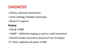
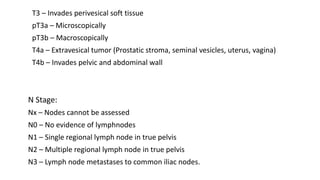
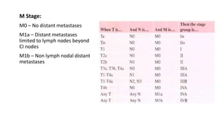
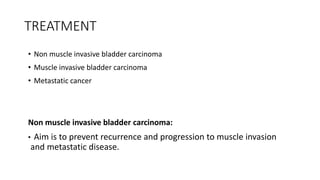
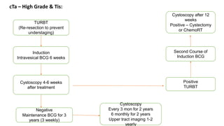
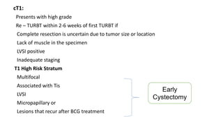
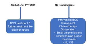
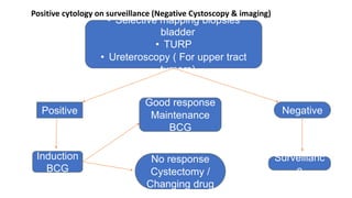
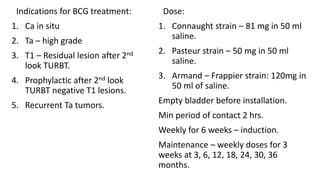
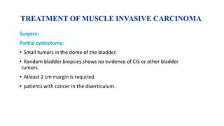
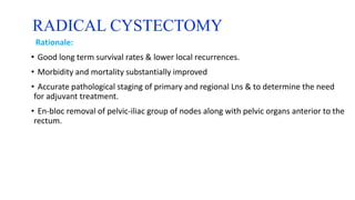
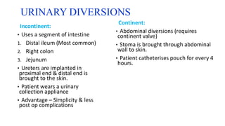
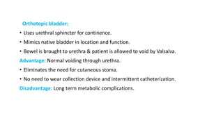
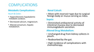
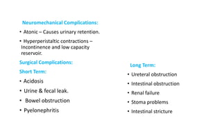
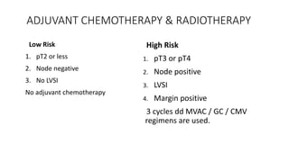
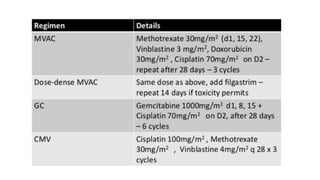
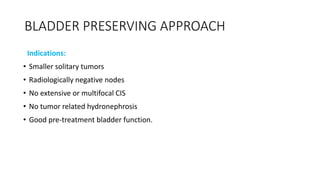
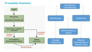
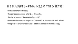
MANAGEMENT OF CARCINOMA
BLADDER
DR KIRAN KUMAR
DIAGNOSIS
• History, physical examination
• Urine cytology, flexible Cystoscopy
• Renal CT urogram
Staging:
• EUA & TURBT
• TURBT – Definitive staging as well as initial treatment.
• Should include muscularis propria (if not re-biopsy)
• CT chest, abdomen & pelvis or MRI
STAGING
T Stage
Tx – primary tumor cannot be assessed.
T0 – No evidence of primary tumor
Ta – Non-invasive papillary carcinoma
Tis – Urothelial carcinoma in situ
T1 – Tumor invades lamina propria
T2 – Tumor invades muscularis propria
pT2a – Superficial (Inner half)
pT2b – Deep muscularis propria (Outer half)
T3 – Invades perivesical soft tissue
pT3a – Microscopically
pT3b – Macroscopically
T4a – Extravesical tumor (Prostatic stroma, seminal vesicles, uterus, vagina)
T4b – Invades pelvic and abdominal wall
N Stage:
Nx – Nodes cannot be assessed
N0 – No evidence of lymphnodes
N1 – Single regional lymph node in true pelvis
N2 – Multiple regional lymph node in true pelvis
N3 – Lymph node metastases to common iliac nodes.
M Stage:
M0 – No distant metastases
M1a – Distant metastases
limited to lymph nodes beyond
CI nodes
M1b – Non lymph nodal distant
metastases
TREATMENT
• Non muscle invasive bladder carcinoma
• Muscle invasive bladder carcinoma
• Metastatic cancer
Non muscle invasive bladder carcinoma:
• Aim is to prevent recurrence and progression to muscle invasion
and metastatic disease.
EORT
C
Low risk
• Grade 1 or 2
• Ta without Tis
• < 3 cm tumor
• Unifocal
• Recurrence – 50%
• Progression – 5%
High Risk
• Grade -3
• Multifocal Tis
• Ta or T1 with Tis
• 70 % recurrence
• 30 – 50 % progression
Treatment for Ta – Low grade:
TURBT
Intravesical
Gemcitabine / MM-C
within 24 hrs
Cystoscopy within 4
months of TURBT
Negative
Cystoscopy at 6-9 mon
& Yearly for 5 years
Persistent / Recurrence
TURBT & Intravesical
treatment
Cystoscopy after 4
months
Positive
Cystectomy /
Chemoradiation
cTa – High Grade & Tis:
TURBT
(Re-resection to prevent
understaging)
Induction
Intravesical BCG 6 weeks
Cystoscopy 4-6 weeks
after treatment
Negative
Maintenance BCG for 3
years (3 weekly)
Cystoscopy
Every 3 mon for 2 years
6 monthly for 2 years
Upper tract imaging 1-2
yearly
Positive
TURBT
Second Course of
Induction BCG
Cystoscopy after 12
weeks
Positive – Cystectomy
or ChemoRT
cT1:
Presents with high grade
Re – TURBT within 2-6 weeks of first TURBT if
Complete resection is uncertain due to tumor size or location
Lack of muscle in the specimen
LVSI positive
Inadequate staging
T1 High Risk Stratum
Multifocal
Associated with Tis
LVSI
Micropapillary or
Lesions that recur after BCG treatment
Early
Cystectomy
Residual after 2nd TURBT. No residual disease
BCG treatment &
further treatment like
cTa high grade
Intravesical BCG
Intravesical
Chemotherapy
Observation
• Small volume lesions
• Limited lamina propria
involvement
• No CIS
Positive cytology on surveillance (Negative Cystoscopy & imaging)
• Selective mapping biopsies
bladder
• TURP
• Ureteroscopy ( For upper tract
tumors)
Positive Negative
Surveillanc
e
Induction
BCG
Good response
Maintenance
BCG
No response
Cystectomy /
Changing drug
Indications for BCG treatment:
1. Ca in situ
2. Ta – high grade
3. T1 – Residual lesion after 2nd
look TURBT.
4. Prophylactic after 2nd look
TURBT negative T1 lesions.
5. Recurrent Ta tumors.
Dose:
1. Connaught strain – 81 mg in 50 ml
saline.
2. Pasteur strain – 50 mg in 50 ml
saline.
3. Armand – Frappier strain: 120mg in
50 ml of saline.
Empty bladder before installation.
Min period of contact 2 hrs.
Weekly for 6 weeks – induction.
Maintenance – weekly doses for 3
weeks at 3, 6, 12, 18, 24, 30, 36
months.
TREATMENT OF MUSCLE INVASIVE CARCINOMA
Surgery:
Partial cystectomy:
• Small tumors in the dome of the bladder.
• Random bladder biopsies shows no evidence of CIS or other bladder
tumors.
• Atleast 2 cm margin is required.
• patients with cancer in the diverticulum.
RADICAL CYSTECTOMY
Rationale:
• Good long term survival rates & lower local recurrences.
• Morbidity and mortality substantially improved
• Accurate pathological staging of primary and regional Lns & to determine the need
for adjuvant treatment.
• En-bloc removal of pelvic-iliac group of nodes along with pelvic organs anterior to the
rectum.
URINARY DIVERSIONS
Incontinent:
• Uses a segment of intestine
1. Distal ileum (Most common)
2. Right colon
3. Jejunum
• Ureters are implanted in
proximal end & distal end is
brought to the skin.
• Patient wears a urinary
collection appliance
• Advantage – Simplicity & less
post op complications
Continent:
• Abdominal diversions (requires
continent valve)
• Stoma is brought through abdominal
wall to skin.
• Patient catheterises pouch for every 4
hours.
Orthotopic bladder:
• Uses urethral sphincter for continence.
• Mimics native bladder in location and function.
• Bowel is brought to urethra & patient is allowed to void by Valsalva.
Advantage: Normal voiding through urethra.
• Eliminates the need for cutaneous stoma.
• No need to wear collection device and intermittent catheterization.
Disadvantage: Long term metabolic complications.
COMPLICATIONS
Metabolic Complications:
Ileum & Colon:
• Hyperchloremic hypokalemic
metabolic acidosis.
• Decreased calcium, magnesium.
• Altered sensorium, hepatic
metabolism.
Renal Calculi:
• More with reservoir type due to surgical
foreign body or mucus serving as nidus.
Sepsis:
• Diminished antibacterial activity of
intestinal mucosa due to decreased
immunoglobulin secretion.
Altered Drug Metabolism:
• Unchanged drug from kidney collects in
pouch.
• Reabsorbed by the gut.
• High incidence of complications with
chemotherapy.
Neuromechanical Complications:
• Atonic – Causes urinary retention.
• Hyperperistaltic contractions –
Incontinence and low capacity
reservoir.
Surgical Complications:
Short Term:
• Acidosis
• Urine & fecal leak.
• Bowel obstruction
• Pyelonephritis
Long Term:
• Ureteral obstruction
• Intestinal obstruction
• Renal failure
• Stoma problems
• Intestinal stricture
ADJUVANT CHEMOTHERAPY & RADIOTHERAPY
Low Risk
1. pT2 or less
2. Node negative
3. No LVSI
No adjuvant chemotherapy
High Risk
1. pT3 or pT4
2. Node positive
3. LVSI
4. Margin positive
3 cycles dd MVAC / GC / CMV
regimens are used.
BLADDER PRESERVING APPROACH
Indications:
• Smaller solitary tumors
• Radiologically negative nodes
• No extensive or multifocal CIS
• No tumor related hydronephrosis
• Good pre-treatment bladder function.
Tri modality Treatment:
Neoadjuvant
Chemotherapy
Radiotherapy Cystectomy
Salvage
Cystectomy
Chemotherapy on
relapse
Adjuvant
chemotherapy in
selected cases
IIIB & IVA(PT1 – PT4A, N2,3 & T4B DISEASE)
• Induction chemotherapy.
• Response assessment after 2 or 3 months.
• Partial response – Surgery or Chemo-RT
• Complete response – Surgery or Chemo-RT or observation until relapse
• Progression or Distant disease – additional lines of chemotherapy.
IV B
Systemic therapy
• Metastatectomy – if good response to systemic therapy & solitary mets or Lns
involvement.
• GC or dd MVAC regimens
• Cisplatin can be replaced with carboplatin if GFR is low or cisplatin is CI.
Targeted therapies:
• Bladder cancer is the third highest mutated cancer.
• Commonly identified clinically relevant genetic alterations are CDKN2A, FGFR3,
PI3KCA & ERBB2.
• PD-1 inhibitors – Nivolumab, Pembrolizumab
• PD-L1 inhibitor – Atezolizumab, Durvalumab, Avelumab
• FGFR inhibitor – Erdafitinib
• As second line after cisplatin based chemotherapy.
• Atezolizumab & Pembrolizumab – first line if not eligible for cisplatin
containing chemotherapy & shows PD-L 1 expression or
• Indicated in patients who are not eligible for platinum containing
agents regardless of levels of PD-L1 expression in locally advanced or
metastatic setting.
IMMUNOTHERAPY
RADIOTHERAPY
Conventional:
Dose:
• Radical – 45 to 50.4 Gy to the pelvis
Boost – 64 – 66 Gy to the tumor
• Adjuvant – Cystectomy bed & Pelvic Lymphnodes (45 – 50.4 Gy)
Involved resection margins & areas of ENE – 54 – 60 Gy.
• Palliative – 21 Gy in 3 fractions on alternate days.
• Thank you

Carcinoma Bladder.pptx

  • 1.
  • 2.
    DIAGNOSIS • History, physicalexamination • Urine cytology, flexible Cystoscopy • Renal CT urogram Staging: • EUA & TURBT • TURBT – Definitive staging as well as initial treatment. • Should include muscularis propria (if not re-biopsy) • CT chest, abdomen & pelvis or MRI
  • 3.
    STAGING T Stage Tx –primary tumor cannot be assessed. T0 – No evidence of primary tumor Ta – Non-invasive papillary carcinoma Tis – Urothelial carcinoma in situ T1 – Tumor invades lamina propria T2 – Tumor invades muscularis propria pT2a – Superficial (Inner half) pT2b – Deep muscularis propria (Outer half)
  • 4.
    T3 – Invadesperivesical soft tissue pT3a – Microscopically pT3b – Macroscopically T4a – Extravesical tumor (Prostatic stroma, seminal vesicles, uterus, vagina) T4b – Invades pelvic and abdominal wall N Stage: Nx – Nodes cannot be assessed N0 – No evidence of lymphnodes N1 – Single regional lymph node in true pelvis N2 – Multiple regional lymph node in true pelvis N3 – Lymph node metastases to common iliac nodes.
  • 5.
    M Stage: M0 –No distant metastases M1a – Distant metastases limited to lymph nodes beyond CI nodes M1b – Non lymph nodal distant metastases
  • 6.
    TREATMENT • Non muscleinvasive bladder carcinoma • Muscle invasive bladder carcinoma • Metastatic cancer Non muscle invasive bladder carcinoma: • Aim is to prevent recurrence and progression to muscle invasion and metastatic disease.
  • 7.
  • 8.
    Low risk • Grade1 or 2 • Ta without Tis • < 3 cm tumor • Unifocal • Recurrence – 50% • Progression – 5% High Risk • Grade -3 • Multifocal Tis • Ta or T1 with Tis • 70 % recurrence • 30 – 50 % progression
  • 9.
    Treatment for Ta– Low grade: TURBT Intravesical Gemcitabine / MM-C within 24 hrs Cystoscopy within 4 months of TURBT Negative Cystoscopy at 6-9 mon & Yearly for 5 years Persistent / Recurrence TURBT & Intravesical treatment Cystoscopy after 4 months Positive Cystectomy / Chemoradiation
  • 10.
    cTa – HighGrade & Tis: TURBT (Re-resection to prevent understaging) Induction Intravesical BCG 6 weeks Cystoscopy 4-6 weeks after treatment Negative Maintenance BCG for 3 years (3 weekly) Cystoscopy Every 3 mon for 2 years 6 monthly for 2 years Upper tract imaging 1-2 yearly Positive TURBT Second Course of Induction BCG Cystoscopy after 12 weeks Positive – Cystectomy or ChemoRT
  • 11.
    cT1: Presents with highgrade Re – TURBT within 2-6 weeks of first TURBT if Complete resection is uncertain due to tumor size or location Lack of muscle in the specimen LVSI positive Inadequate staging T1 High Risk Stratum Multifocal Associated with Tis LVSI Micropapillary or Lesions that recur after BCG treatment Early Cystectomy
  • 12.
    Residual after 2ndTURBT. No residual disease BCG treatment & further treatment like cTa high grade Intravesical BCG Intravesical Chemotherapy Observation • Small volume lesions • Limited lamina propria involvement • No CIS
  • 13.
    Positive cytology onsurveillance (Negative Cystoscopy & imaging) • Selective mapping biopsies bladder • TURP • Ureteroscopy ( For upper tract tumors) Positive Negative Surveillanc e Induction BCG Good response Maintenance BCG No response Cystectomy / Changing drug
  • 14.
    Indications for BCGtreatment: 1. Ca in situ 2. Ta – high grade 3. T1 – Residual lesion after 2nd look TURBT. 4. Prophylactic after 2nd look TURBT negative T1 lesions. 5. Recurrent Ta tumors. Dose: 1. Connaught strain – 81 mg in 50 ml saline. 2. Pasteur strain – 50 mg in 50 ml saline. 3. Armand – Frappier strain: 120mg in 50 ml of saline. Empty bladder before installation. Min period of contact 2 hrs. Weekly for 6 weeks – induction. Maintenance – weekly doses for 3 weeks at 3, 6, 12, 18, 24, 30, 36 months.
  • 15.
    TREATMENT OF MUSCLEINVASIVE CARCINOMA Surgery: Partial cystectomy: • Small tumors in the dome of the bladder. • Random bladder biopsies shows no evidence of CIS or other bladder tumors. • Atleast 2 cm margin is required. • patients with cancer in the diverticulum.
  • 16.
    RADICAL CYSTECTOMY Rationale: • Goodlong term survival rates & lower local recurrences. • Morbidity and mortality substantially improved • Accurate pathological staging of primary and regional Lns & to determine the need for adjuvant treatment. • En-bloc removal of pelvic-iliac group of nodes along with pelvic organs anterior to the rectum.
  • 17.
    URINARY DIVERSIONS Incontinent: • Usesa segment of intestine 1. Distal ileum (Most common) 2. Right colon 3. Jejunum • Ureters are implanted in proximal end & distal end is brought to the skin. • Patient wears a urinary collection appliance • Advantage – Simplicity & less post op complications Continent: • Abdominal diversions (requires continent valve) • Stoma is brought through abdominal wall to skin. • Patient catheterises pouch for every 4 hours.
  • 18.
    Orthotopic bladder: • Usesurethral sphincter for continence. • Mimics native bladder in location and function. • Bowel is brought to urethra & patient is allowed to void by Valsalva. Advantage: Normal voiding through urethra. • Eliminates the need for cutaneous stoma. • No need to wear collection device and intermittent catheterization. Disadvantage: Long term metabolic complications.
  • 19.
    COMPLICATIONS Metabolic Complications: Ileum &Colon: • Hyperchloremic hypokalemic metabolic acidosis. • Decreased calcium, magnesium. • Altered sensorium, hepatic metabolism. Renal Calculi: • More with reservoir type due to surgical foreign body or mucus serving as nidus. Sepsis: • Diminished antibacterial activity of intestinal mucosa due to decreased immunoglobulin secretion. Altered Drug Metabolism: • Unchanged drug from kidney collects in pouch. • Reabsorbed by the gut. • High incidence of complications with chemotherapy.
  • 20.
    Neuromechanical Complications: • Atonic– Causes urinary retention. • Hyperperistaltic contractions – Incontinence and low capacity reservoir. Surgical Complications: Short Term: • Acidosis • Urine & fecal leak. • Bowel obstruction • Pyelonephritis Long Term: • Ureteral obstruction • Intestinal obstruction • Renal failure • Stoma problems • Intestinal stricture
  • 21.
    ADJUVANT CHEMOTHERAPY &RADIOTHERAPY Low Risk 1. pT2 or less 2. Node negative 3. No LVSI No adjuvant chemotherapy High Risk 1. pT3 or pT4 2. Node positive 3. LVSI 4. Margin positive 3 cycles dd MVAC / GC / CMV regimens are used.
  • 23.
    BLADDER PRESERVING APPROACH Indications: •Smaller solitary tumors • Radiologically negative nodes • No extensive or multifocal CIS • No tumor related hydronephrosis • Good pre-treatment bladder function.
  • 24.
    Tri modality Treatment: Neoadjuvant Chemotherapy RadiotherapyCystectomy Salvage Cystectomy Chemotherapy on relapse Adjuvant chemotherapy in selected cases
  • 25.
    IIIB & IVA(PT1– PT4A, N2,3 & T4B DISEASE) • Induction chemotherapy. • Response assessment after 2 or 3 months. • Partial response – Surgery or Chemo-RT • Complete response – Surgery or Chemo-RT or observation until relapse • Progression or Distant disease – additional lines of chemotherapy.
  • 26.
    IV B Systemic therapy •Metastatectomy – if good response to systemic therapy & solitary mets or Lns involvement. • GC or dd MVAC regimens • Cisplatin can be replaced with carboplatin if GFR is low or cisplatin is CI. Targeted therapies: • Bladder cancer is the third highest mutated cancer. • Commonly identified clinically relevant genetic alterations are CDKN2A, FGFR3, PI3KCA & ERBB2.
  • 27.
    • PD-1 inhibitors– Nivolumab, Pembrolizumab • PD-L1 inhibitor – Atezolizumab, Durvalumab, Avelumab • FGFR inhibitor – Erdafitinib • As second line after cisplatin based chemotherapy. • Atezolizumab & Pembrolizumab – first line if not eligible for cisplatin containing chemotherapy & shows PD-L 1 expression or • Indicated in patients who are not eligible for platinum containing agents regardless of levels of PD-L1 expression in locally advanced or metastatic setting. IMMUNOTHERAPY
  • 28.
  • 29.
    Dose: • Radical –45 to 50.4 Gy to the pelvis Boost – 64 – 66 Gy to the tumor • Adjuvant – Cystectomy bed & Pelvic Lymphnodes (45 – 50.4 Gy) Involved resection margins & areas of ENE – 54 – 60 Gy. • Palliative – 21 Gy in 3 fractions on alternate days.
  • 30.