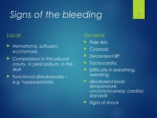
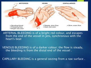
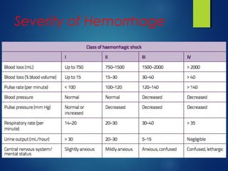
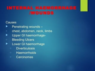
BLEEDING
CONTROL
SEQUENCE
IMPORTANCE
Physiology/homeostasis
Integrity of circulatory system
TYPES/CAUSES
CONTROL METHODS
BLOOD TRANSFUSION
Subject’s importance
 Hemorrhage is one of the basic problems and
considerations in surgery.
 From-trivial trauma or major abdominal organ
injuries-to- congenital and acquired coagulation
disorders.
 A wide spectrum of problems involves
hemorrhage.
Transfusion of blood is the main remedy
Clinical Situation-
Bleeding Trauma /accidents
 General operative interventions
 Gynecological procedures
 Common surgical conditions that presents with bleeding-
Intracranial hemorrhages/CVA
Upper GIT bleed/ hematemesis and melena
Bleeding hemorrhoids
Chronic wounds
Aneurysms
 Coagulation disorders
 Congenital- Hemophilia, vWF deficiency
 Acquired
DIC
Anticoagulants
Fulminant sepsis
What Prevents
Hemorrhage
NATURAL BARRIERS AGAINST
HAEMORRHAGE
 Integrity of vascular wall
 Coagulation system
Body’s response to
hemorrhage/injury
Attempts to repair the loss & restore normality
There are several interrelated stages
Local response / Generalized response
Aims at:
 Wall repair
 Restoration of volume loss
 Correction of coagulation abnormalities
Signs of the bleeding
Local
 Hematoma, suffusion,
ecchymosis
 Compression in the pleural
cavity, in pericardium, in the
skull
 Functional disturbancies –
e.g. hyperperistalsis
General
 Pale skin,
 Cyanosis,
 Decreased BP,
 Tachycardia,
 Difficulty in breathing,
sweating,
 decreased body
temperature,
unconsciousness, cardiac
standstill
 Signs of shock
7
Body’s response to
hemorrhage/injury
 Local
 Vasoconstriction
 Platelet aggregation and plug formation
 Coagulation leading to Fibrin formation –Intrinsic
& Extrinsic Pathways
 General
 Cardiac stimulation
 Compartmental Volume
movement
TYPES OF HAEMORRHAGE
 AMOUNT OF LOSS -MINOR/MAJOR
 ACUTE/CHRONIC
 ARTERIAL/VENOUS/CAPILLARY/MIXED
 LOCALIZED/DIFFUSE
 EXTERNAL/ INTERNAL
 OVERT/OCCULT
TYPES OF HAEMORRHAGE
ARTERIAL BLEEDING is of a bright red colour, and escapes
from the end of the vessel in jets, synchronous with the
heart's beat
VENOUS BLEEDING is of a darker colour; the flow is steady,
the bleeding is from the distal end of the vessel .
CAPILLARY BLEEDING is a general oozing from a raw surface .
Hemorrhage and Shock
What happens when you start to
bleed? – it depends on how much
blood you lose
Normal Adult Blood
Volume is about
5 Litres
Severity of Hemorrhage
The Direction Of
Hemorrage
 External
 Internal
 In a luminar organ (hematuria, hemoptysis, melena)
 In body cavities (intracranial, hemothorax, hemoperitoneum,
hemopericardium, hemarthros)
 Among the tissues (hematoma, suffusion)
13
Internal Hemorrhage
INTERNAL HAEMORRHAGE
/WOUNDS
Causes
 Penetrating wounds –
o chest, abdomen, neck, limbs
 Upper GI haemorrhage-
o Bleeding Ulcers
 Lower GI haemorrhage
o Diverticulosis
o Haemorrhoids
o Carcinomas
External Hemorrhage
Bleeding
PREOPERATIVE HEMORRHAGE
Prehospital care! – maintenance of the airways, ventillation and
circulation
bandages, direct pressure, torniquets
INTRAOPERATIVE HEMORRHAGE
anatomical and/or diffuse
depending on the surgeon, the surgery, position,
the size of the vessel, pressure in the vessel
(ANESTHESIA)
POSTOPERATIVE BLEEDING
ineffective local hemostasis, undetected hemostatic
defect, consumptive coagulopathy or fibrinolysis
17
CLASSIFICATION OF
SURGICAL HAEMORRHAGE
 Primary Hemorrhage
 occurring at the time of the injury or surgery
 Reactionary Hemorrhage
 within twenty-four hours of the accident/surgery, due to
slippage of ligature, hypertension post op
 Secondary Hemorrhage
 occurring at a later period (48-72hrs) and caused by
septic condition of the wound (infection).
EFFECTS OF
HAEMORRHAGE
Depend upon following:
 Acute loss vs Chronic loss
 The amount of loss
 The compensatory mechanisms
 General state of health
SURGICAL HEMOSTASIS
Aim – to prevent the flow of blood from the incised or transected
vessels
 Mechanical methods
 Thermal methods
 Chemical and biological methods
 Radiological/Interventional methods
 Adequate blood/blood products transfusion
20
SURGICAL
HAEMOSTASIS
Natural CONTROL/arrest of
hemorrhage arises from-
(1) changes taking place in the
cut vessel causing its retraction
and contraction
(2) the coagulation mechanism
of the blood
(3) temporary-platelet plug
Permanent-fibrin clot.
SURGICAL HEMOSTASIS
MECHANICAL METHODS
 Digital pressure – direct pressure,
e.g. Pringle maneuver
 Tourniquet
 Ligation
 Suturing
 Preventive hemostasis
 Clips
 Bone wax
 other
22
SURGICAL TREATMENT
OF HAEMORRHAGE
First Aid Management
 DIRECT PRESSURE
In small blood-vessels
pressure will be sufficient to
arrest, hemorrhage
permanently
 LIMB ELEVATION
 TOURNIQUET
APPLICATION
CLIPS FOR CONTROLLING
BLEEDING
LIGATURE
In large vessels with a reef-knot
main artery of the limb exposed
by dissection at the most
accessible point .
SUTURING & LIGATURE
THERMAL METHODS
 Low temperature
 Hypothermia – eg. stomach bleeding
 Cryosurgery
 Dehydratation and denaturation of fatty tissue
 Decreases the cell metabolism
 Vasoconstriction
26
THERMAL METHODS
 High temperature
 Electrosurgery – electrocauterization
 Monopolar diathermy
 Bipolar diathermy
 Harmonic devices
 Laser surgery
coagulation and vaporization
for fine tissues
27
Diathermy
Thermal methods
 High temperature
 Electrocoagulation
 Electrofulguration (A)
 Electrodessication
 Electrosection
29
Hemostasis with chemical and
biological methods
VASOCONSTRICTION COAGULATION HYGROSCOPIC EFFECT
Absorbable collagen
Absorbable gelatin
Microfibrillar collagen
Oxidized cellulose
Oxytocin
Epinephrine
Thrombin
QuikClot
30
Hemostasis with chemical and
biological methods
31
HemCon
Bleeding Control by
Interventional Radiology
 Post trauma-intra abdominal bleeding
 Gastro intestinal bleeding control-
 Upper
 Lower
 Uterine atony causing Postpartum hemorrhage
Embolisation particles
Post trauma
 Vascular and solid organ trauma.
Celiac angiogram showing 3 foci
of extravasation in spleen, 2 in the
upper pole (arrow) and 1 in the
lateral aspect of the mid spleen
 Post—super-selective embolization splenic
angiogram demonstrating microcoils in good
position and no evidence of further extravasation
Gastrointestinal Bleeding
Bleeding control mit

Bleeding control mit

  • 1.
  • 2.
    SEQUENCE IMPORTANCE Physiology/homeostasis Integrity of circulatorysystem TYPES/CAUSES CONTROL METHODS BLOOD TRANSFUSION
  • 3.
    Subject’s importance  Hemorrhageis one of the basic problems and considerations in surgery.  From-trivial trauma or major abdominal organ injuries-to- congenital and acquired coagulation disorders.  A wide spectrum of problems involves hemorrhage. Transfusion of blood is the main remedy
  • 4.
    Clinical Situation- Bleeding Trauma/accidents  General operative interventions  Gynecological procedures  Common surgical conditions that presents with bleeding- Intracranial hemorrhages/CVA Upper GIT bleed/ hematemesis and melena Bleeding hemorrhoids Chronic wounds Aneurysms  Coagulation disorders  Congenital- Hemophilia, vWF deficiency  Acquired DIC Anticoagulants Fulminant sepsis
  • 5.
    What Prevents Hemorrhage NATURAL BARRIERSAGAINST HAEMORRHAGE  Integrity of vascular wall  Coagulation system
  • 6.
    Body’s response to hemorrhage/injury Attemptsto repair the loss & restore normality There are several interrelated stages Local response / Generalized response Aims at:  Wall repair  Restoration of volume loss  Correction of coagulation abnormalities
  • 7.
    Signs of thebleeding Local  Hematoma, suffusion, ecchymosis  Compression in the pleural cavity, in pericardium, in the skull  Functional disturbancies – e.g. hyperperistalsis General  Pale skin,  Cyanosis,  Decreased BP,  Tachycardia,  Difficulty in breathing, sweating,  decreased body temperature, unconsciousness, cardiac standstill  Signs of shock 7
  • 8.
    Body’s response to hemorrhage/injury Local  Vasoconstriction  Platelet aggregation and plug formation  Coagulation leading to Fibrin formation –Intrinsic & Extrinsic Pathways  General  Cardiac stimulation  Compartmental Volume movement
  • 9.
    TYPES OF HAEMORRHAGE AMOUNT OF LOSS -MINOR/MAJOR  ACUTE/CHRONIC  ARTERIAL/VENOUS/CAPILLARY/MIXED  LOCALIZED/DIFFUSE  EXTERNAL/ INTERNAL  OVERT/OCCULT
  • 10.
    TYPES OF HAEMORRHAGE ARTERIALBLEEDING is of a bright red colour, and escapes from the end of the vessel in jets, synchronous with the heart's beat VENOUS BLEEDING is of a darker colour; the flow is steady, the bleeding is from the distal end of the vessel . CAPILLARY BLEEDING is a general oozing from a raw surface .
  • 11.
    Hemorrhage and Shock Whathappens when you start to bleed? – it depends on how much blood you lose Normal Adult Blood Volume is about 5 Litres
  • 12.
  • 13.
    The Direction Of Hemorrage External  Internal  In a luminar organ (hematuria, hemoptysis, melena)  In body cavities (intracranial, hemothorax, hemoperitoneum, hemopericardium, hemarthros)  Among the tissues (hematoma, suffusion) 13
  • 14.
  • 15.
    INTERNAL HAEMORRHAGE /WOUNDS Causes  Penetratingwounds – o chest, abdomen, neck, limbs  Upper GI haemorrhage- o Bleeding Ulcers  Lower GI haemorrhage o Diverticulosis o Haemorrhoids o Carcinomas
  • 16.
  • 17.
    Bleeding PREOPERATIVE HEMORRHAGE Prehospital care!– maintenance of the airways, ventillation and circulation bandages, direct pressure, torniquets INTRAOPERATIVE HEMORRHAGE anatomical and/or diffuse depending on the surgeon, the surgery, position, the size of the vessel, pressure in the vessel (ANESTHESIA) POSTOPERATIVE BLEEDING ineffective local hemostasis, undetected hemostatic defect, consumptive coagulopathy or fibrinolysis 17
  • 18.
    CLASSIFICATION OF SURGICAL HAEMORRHAGE Primary Hemorrhage  occurring at the time of the injury or surgery  Reactionary Hemorrhage  within twenty-four hours of the accident/surgery, due to slippage of ligature, hypertension post op  Secondary Hemorrhage  occurring at a later period (48-72hrs) and caused by septic condition of the wound (infection).
  • 19.
    EFFECTS OF HAEMORRHAGE Depend uponfollowing:  Acute loss vs Chronic loss  The amount of loss  The compensatory mechanisms  General state of health
  • 20.
    SURGICAL HEMOSTASIS Aim –to prevent the flow of blood from the incised or transected vessels  Mechanical methods  Thermal methods  Chemical and biological methods  Radiological/Interventional methods  Adequate blood/blood products transfusion 20
  • 21.
    SURGICAL HAEMOSTASIS Natural CONTROL/arrest of hemorrhagearises from- (1) changes taking place in the cut vessel causing its retraction and contraction (2) the coagulation mechanism of the blood (3) temporary-platelet plug Permanent-fibrin clot.
  • 22.
    SURGICAL HEMOSTASIS MECHANICAL METHODS Digital pressure – direct pressure, e.g. Pringle maneuver  Tourniquet  Ligation  Suturing  Preventive hemostasis  Clips  Bone wax  other 22
  • 23.
    SURGICAL TREATMENT OF HAEMORRHAGE FirstAid Management  DIRECT PRESSURE In small blood-vessels pressure will be sufficient to arrest, hemorrhage permanently  LIMB ELEVATION  TOURNIQUET APPLICATION
  • 24.
  • 25.
    LIGATURE In large vesselswith a reef-knot main artery of the limb exposed by dissection at the most accessible point . SUTURING & LIGATURE
  • 26.
    THERMAL METHODS  Lowtemperature  Hypothermia – eg. stomach bleeding  Cryosurgery  Dehydratation and denaturation of fatty tissue  Decreases the cell metabolism  Vasoconstriction 26
  • 27.
    THERMAL METHODS  Hightemperature  Electrosurgery – electrocauterization  Monopolar diathermy  Bipolar diathermy  Harmonic devices  Laser surgery coagulation and vaporization for fine tissues 27
  • 28.
  • 29.
    Thermal methods  Hightemperature  Electrocoagulation  Electrofulguration (A)  Electrodessication  Electrosection 29
  • 30.
    Hemostasis with chemicaland biological methods VASOCONSTRICTION COAGULATION HYGROSCOPIC EFFECT Absorbable collagen Absorbable gelatin Microfibrillar collagen Oxidized cellulose Oxytocin Epinephrine Thrombin QuikClot 30
  • 31.
    Hemostasis with chemicaland biological methods 31 HemCon
  • 32.
  • 33.
    Interventional Radiology  Posttrauma-intra abdominal bleeding  Gastro intestinal bleeding control-  Upper  Lower  Uterine atony causing Postpartum hemorrhage
  • 34.
  • 35.
    Post trauma  Vascularand solid organ trauma. Celiac angiogram showing 3 foci of extravasation in spleen, 2 in the upper pole (arrow) and 1 in the lateral aspect of the mid spleen  Post—super-selective embolization splenic angiogram demonstrating microcoils in good position and no evidence of further extravasation
  • 36.

Editor's Notes

  • #14 Conjunctival suffusion with subconjunctival hemorrhage (ou), which was suggestive of leptospirosis, developed on the second hospitalization day.
  • #23 The Pringle maneuver. The portal triad is occluded by guiding the posterior blade of the clamp through the foramen of Winslow with the aid of the left index finger. -6. kép: amennyiben vérnyomásmérő áll rendelkezésre, úgy pneumatikus vértelenség felhelyezése. 280 Hgmm-re felfújjuk a vérnyomásmérő mandzsettát, így a sebalapot megtekinthetjük. Betadines vagy Octeniseptes fedőkötés, steril pólya, korrekt nyomókötés 7-8.kép: a nyomókötés felhelyezése után a vértelenség felengedése. Amennyiben erős vérzést észlelünk, ismételten felfújjuk a mandzsettát és revideáljuk a kötést. (az erős vérzés forrása csak technikai hiba lehet az elsősegélynyújtó részéről).
  • #27 Timed spot freeze technique used to treat a malignancy (possibly a small basal cell cancer), demonstrating freeze ball formation and the 5-mm treatment margins necessary to achieve a temperature of −50ºC (−58 ºF) and, thus, the required depth of 4 to 5 mm. Cryosurgery is a method of superfreezing tissue in order to destroy it. The technique is used to treat tumors, control pain, and control bleeding. Information The cold is introduced through a probe which has liquid nitrogen circulating through it. To destroy diseased tissue, the tissue is cooled to below -20 degrees Celsius. Other procedures that control pain or bleeding are cooled to a lesser degree to prevent tissue damage.
  • #28 Electrocoagulation: A fine wire probe or other delivery mechanism is used to transmit radio waves to tissues near the probe. Molecules within the tissue are caused to vibrate which lead to a rapid increase of the temperature, causing coagulation of the proteins within the tissue, effectively killing the tissue. At higher powered applications, full desiccation of tissue is possible. Two forms of electrosurgery: (A) Electrodesiccation with an active electrode tip touching the skin and showing penetration of planned tissue damage. (B) Fulguration with sparking from electrode to tissue. Treatment area is more superficial than in desiccation.Added by BiomedGuy AboutEdit Fulguration, also called electrofulguration, is the destruction of tissue by means of a high-frequency electric current applied with a needlelike electrode. In fulguration, the electrode is held away from the skin to produce a sparking at the skin surface and more shallow tissue destruction Fulguration is especially useful in treating superficial epidermal lesions, such as a superficial basal cell carcinoma of the trunk.[1] LinksEdit ReferenceEdit ↑ http://www.aafp.org/afp/2002/1001/p1259.html | Electrosurgery for the Skin | BARRY L. HAINER, M.D.,RICHARD B. USATINE, M.D., | Am Fam
  • #31 HemCon Medical Technologies, Inc. began with funding from the United States Army and access to research by Dr. Kenton Gregory, Dr. Bill Wiesmann, the Oregon Medical Laser Center, and Providence Health Systems. The result was the HemCon® Bandage, which was designed to control life threatening bleeding. Related Links: History & MissionManagement TeamBoard of DirectorsProductsEducational ResourcesDistribution and SalesProduct TrainingSuccess StoriesMedia RoomCustomer FeedbackCareers The bandage was ushered through the FDA clearance process in a near-record 48 hours, and was soon deployed on the battlefield. Since then, the bandage has been used extensively and is credited with helping save over 100 lives with no adverse events reported. In 2005, the Army Surgeon General mandated that any soldier serving in Iraq or Afghanistan will carry at least one HemCon Bandage. This commitment by the U.S. Army is a testament to the efficacy and value of the HemCon Bandage. HemCon is rapidly changing from a military provider to a broad-based supplier of medical technology. With new products, strong partnerships in distribution, and a world class development team, we will continue to be unwavering in our commitment to innovate in all that we strive to achieve.