Dr JAI SHANKARH. P
Department of Oral Medicine & Radiology
J S S DENTAL COLLEGE AND HOSPITAL
JSS Academy of Higher Education &
Research
(Deemed to be University)
Accredited A
++
Grade by NAAC
Sri Shivarathreeshwara Nagar, Mysuru – 570 015
NON-ODONTOGENIC TUMORS
“RAPID REVIEW IN ORAL MEDICINE AND RADIOLOGY”
2.
All contentincluding text , images, audio or other formats
were created for educational and informational purposes only.
The content is not intended to be a substitute for professional
medical or dental advice , diagnosis or treatment.
The existing literature and guidelines referenced in this lecture
may get modified at a future date.
Please continue following updated guidance from dental and
medical authorities.
3.
• A tumoris simply a swelling of the tissue
• Neoplasm - An abnormal mass of tissue, the growth of which
exceeds and its uncordinated with that of normal tissues and
persists in the same excessive manner after cessation of the stimuli
which evoked the change
• Hamartomas - Tumorlike malformations characterized by the
presence of a cellular proliferation that is native to the part but that
manifests growth cessation without potential for further growth.
4.
Odontogenic vs NonOdontogenic
Odontogenic
•Origin:
Tumors and cysts, develop from cells
and tissues involved in tooth
formation.
•Examples:
Odontogenic cysts may arise from
remnants of the dental lamina or
tooth germ. Odontogenic tumors are
exclusively found in the jaw bones,
near teeth.
•Characteristics:
Odontogenic tumors tend to have
lower biological activity and are less
likely to metastasize compared to
nonodontogenic tumors
Non Odontogenic
•Origin:
These conditions develop from
tissues other than those involved in
tooth development, such as bone, soft
tissue, or other embryonic remnants.
•Examples:
Nonodontogenic cysts can originate
from embryonic structures in the
maxillofacial region.
•Characteristics:
Non odontogenic can arise from
various tissues and may be associated
with factors like chronic irritation,
tobacco use, or radiation exposure.
Solitary localized orsingle lesion = local cause
Multiple lesion affecting several sextants = systemic cause
A lesion arising above the mandibular canal in the alveolus more
likely odontogenic lesion
Below the mandibular canal more likely non odontogenic
Within the mandibular canal is likely neural or vascular lesion
Below the hard palate (panoramic radiographs) is odontogenic
lesion
Above the hard palate (panoramic radiographs) is non
odontogenic lesion
Crown of an unerupted tooth suggest its origin within the follicle
Relationship to the roots of the erupted tooth with evidence of
caries or periodontal disease suggest an inflammatory origin
8.
Key Changes inthe WHO Classification:
• The 2022 WHO classification reorganizes the
grouping of fibro-osseous and osteochondromatous
lesions, and benign and malignant bone and cartilage
tumors.
• Hematolymphoid tumors are no longer included in the
WHO classification of jaw tumors.
• The order of listing has changed, with benign
odontogenic tumors now listed before malignant
tumors
9.
1.CYSTS OF THEJAWS
These are fluid-filled sacs within the jawbone.
Nasopalatine duct cyst: The most common non-odontogenic cyst.
Surgical ciliated cyst: A rare, non-odontogenic cyst potentially linked to previous
surgery in the area.
2. BONE AND CARTILAGE TUMORS
This category includes a wide spectrum of tumors, both benign and malignant,
affecting the bone and cartilage of the jaw.
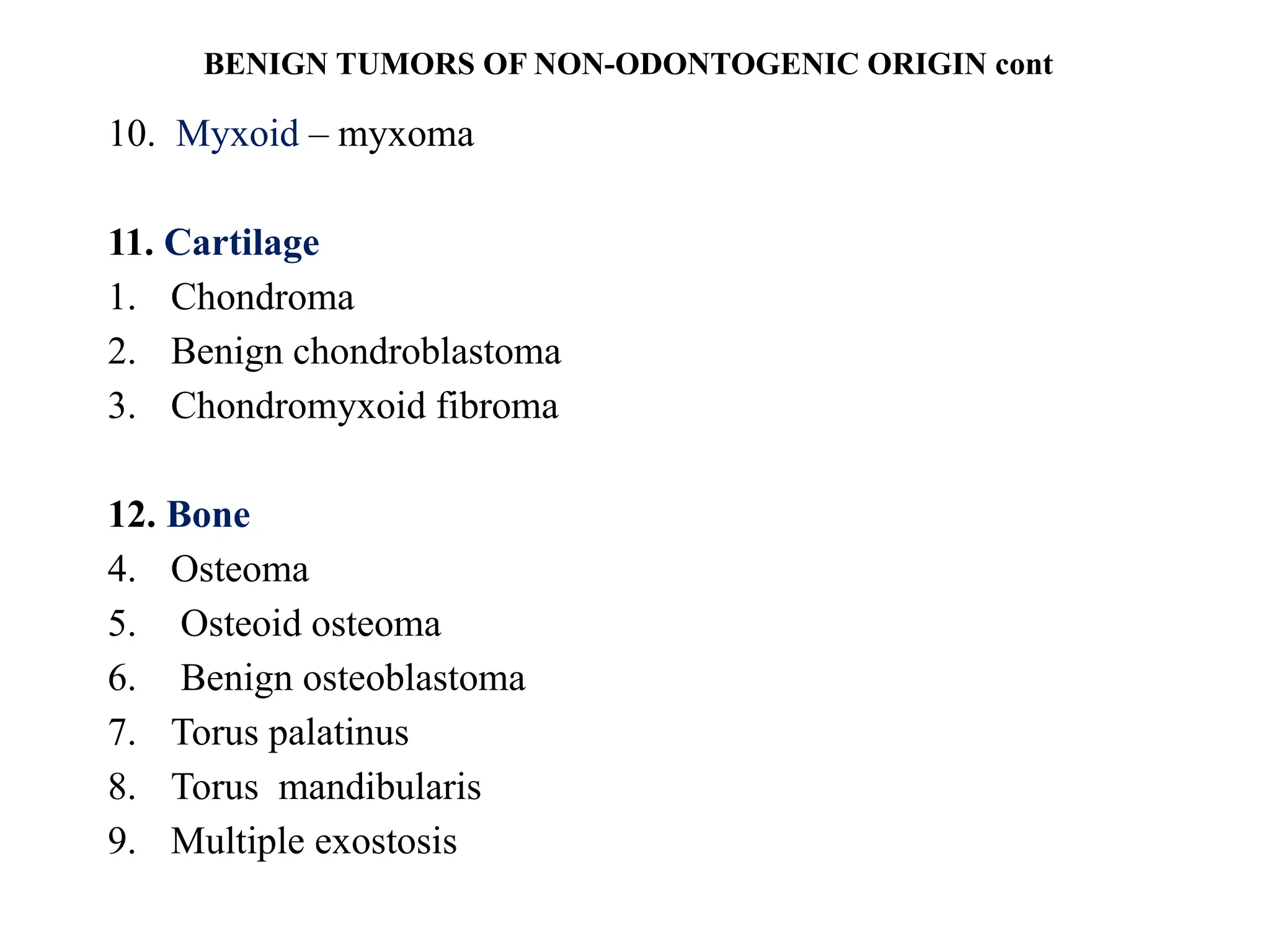
BENIGN: Osteoma, osteoblastoma, osteoid osteoma, ossifying
fibroma, chondroma, chondroblastoma, chondromyxoid fibroma, desmoplastic
fibroma, hemangioma, schwannoma, neurofibroma.
MALIGNANT: Osteosarcoma, Ewing's sarcoma, chondrosarcoma,
fibrosarcoma, angiosarcoma, leiomyosarcoma,
liposarcoma, undifferentiated pleomorphic sarcoma.
Non-Odontogenic Tumors Classification (WHO)
10.
3. GIANT CELLLESIONS AND BONE CYSTS
This group includes various lesions and cysts characterized by the
presence of giant cells.
Central giant cell granuloma.
Peripheral giant cell granuloma.
Cherubism.
Aneurysmal bone cyst.
Simple bone cyst.
4. OTHER LESIONS
This category encompasses other non-odontogenic lesions,
including a subset of fibrous dysplastic conditions.
Fibrous dysplasia
Non-Odontogenic Tumors Classification (WHO) cont
11.
CLASSIFICATION
• BENIGN TUMORSOF ODONTOGENIC ORIGIN
• Odontogenic epithelium tissue origin
Ameloblastoma
Squamous odontogenic tumor
Calcifying epithelial odontogenic tumor
Adenomatoid odontogenic tumor
• Odontogenic epithelium with odontogenic ectomesenchyme
with or without hard tissue formation
Ameloblastic fibroma
Ameloblastic fibro-odontoma
Ameloblastic fibrodentinoma
Odontoameloblastoma
12.
Calcifying odontogeniccyst
Complex odontoma
Compound odontoma
• Odontogenic ectomesenchyme with or without included
odontogenic epithelium
Odontogenic fibroma
Myxoma
Cementoblastoma(benign cementoblastoma, true
cementoblastoma)
SQUAMOUS PAPILLOMA
• Fourthmost common
• HPV 6 and 11
• Not infective
• CLINICAL FEATURES
• Exophytic growth – numerous finger like projections- roughened,
verrucous or cauliflower like surface
• Well circumscibed pedunculated tumor, occassionally sessile, white but
sometimes pink
22.
• Sites- tongue,lips, buccal mucosa, gingiva, palate
• painless , few mm
• Any age
• DIFFERENTIAL DIAGNOSIS
1. verruca vulgaris
2. Cowden’s Syndrome
• H/P- long thin finger like projections
• central connective tissue core
• Acanthosis
• chronic inflammatory cells
• Treatment : surgical excision including the base of the mucosa , recurrence rare
23.
KERATOACANTHOMA
• Also k/aSelf-healing Carcinoma, Molluscum
Pseudocarcinomatosum, Molluscum Sebaceum,
Verrucoma
• Common low grade malignancy - pilosebaceous glands
• A variant of invasive squamous cell carcinoma
24.
• ETIOLOGY
Unclear,
• Sunlight,
•Chemical Carcinogens(pitch And Tar),
• Trauma,
• Human Papilloma Virus,
• Genetic Factors,
• Immunocompromised Status,
• Chromosomal Aberrations Such As Gain On 8q, 1p, And 9q
With Deletions On 3p, 9p, 19p, And 19q
25.
• CLINICAL FEATURES
•Age: all ages, incidence increases with age
• Sex: twice in men
• Complexion: less common in dark –skinned people
• Site - sun- exposed areas.
• Face, neck, and dorsum of the upper extremities.
• Intraoral lesions are uncommon
26.
• Solitary -begin as firm, round , skin colored or reddish
papules - dome shaped nodules - central crateriform
ulceration or keratin plugging - 4- 8 weeks - next 4-8
weeks.-> regression with expulsion of the keratin core
• Elevated umbilicated or crateriform one with a depressed
central core or plug .
• Less than 1 to 1.5 cm
• Recurrence rare
27.
• D/D -Actinic Keratosis, Molluscum Contagiosum, Muir- Torre
Syndrome, Squamous Cell Carcinoma, And Verrucous Cell Carcinoma
• H/F:
• Hyperplastic squamous epithelium
• Surface covered by a thickened layer of parakeratin or orthokeratin with
central plugging.
• No cellular atypia but sometimes dysplasia.
• TREATMENT :
• Surgical excision. Excellent prognosis.
• Follow up for primary skin cancers esp SCC/ BCC
28.
ORAL NEVI
• AlsoK/A Oral Melanocytic Neus, Nevo cellular Nevus, mole,
Mucosal Melanocytic Nevi
• Benign proliferations of the nevus cells in either epithelium or
connective tissue
• Classified as congenital or acquired(Buchner and Hansen).
• On basis histologic location- junctional nevi, compound nevus ,
and intradermal nevus(common mole)
• Blue nevus is the second most common type found in the oral
cavity after intramucosal nevi.
29.
• CLINICAL FEATURES:
•Small (greater than 1 cm and usually 3 to 5 cm) or
• Garment nevi(greater than 10 cm)
• Congenital nevi - 1 to 2.5% neonates- change from flat pale tan
macules to elevated, verrucous hairy lesion.
• 15% occur in head and neck
• Acquired nevi are common.
• 8th month of life
• Increase in number with age with peak in 3rd
decade
30.
• Intradermal mostcommon type
• Spindle cell and/or epitheliod cell nevus- clinically benign and
histologically malignant.
• Blue nevus- dermal melanocytes
• Rarely undergo malignant transformation.
• Feet, hand, on the face.
• Majority present at birth and remain unchanged.
• Smooth ,exhibits hair growing from its surface, and varies in color from
brown to blue or bluish black
31.
• ORAL MANIFESTATIONS
•Mostly in younger than 40 years
• More in white, in all races
• More common in women
• Hard palate > buccal mucosa, vermilian border of the lip , labial
mucosa, gingiva
• Asymptomatic,
• Color varies from brown to black or blue. 15% amelanotic
• Well circumscibed , round or oval, rarely raised.
32.
• D/D
1. Melanoticmacule,
2. Amalgam tattoo,
3. Physiologic pigmentation,
4. Smoker’s melanosis,
5. Melanoma,
6. Vascular lesions
• TREATMENT : Removal of pigmented moles if they
suddenly increase in size, deepen in color or ulcerate
33.
ORAL FIBROMA
• CLINICALFEATURES:
• Any age, 3rd
, 4th
, 5th
decade
• Any site, more on buccal mucosa, gingiva,
tongue, lips and palate.
• Appear as elevated nodule of normal color
with a smooth surface and a sessile or
pedunculated base.
• Superficial ulcerations or hyperkeratosis
Also k/a irritational fibroma
Most common connective tissue tumor
Reactive focal fibrous hyperplasia due to trauma or local irritation
34.
• D/D-
1. Giantcell fibroma,
2. Neurofibroma,
3. Peripheral giant cell granuloma,
4. Mucocele
• Benign and malignant salivary gland tumors
• H/P
• Bundles of interlacing collagenous fibres interspersed with varying
numbers of fibroblasts and small blood vessels.
• Inflammation - vasodilatation, edema, inflammatory cell infiltrate
• True fibroma also similar to irritational fibroma
• TREATMENT:
• Conservative surgical excision.
• Seldom reoccur
35.
PERIPHERAL OSSIFYING FIBROMA
PeripheralOdontogenic Fibroma, Peripheral Cementifying Fibroma,
Peripheral Fibroma With Calcification
• CLINICAL FEATURES:
• Any age
• Children and young adults
• Female> Males
• Maxilla = mandible
• Anterior to molar area
36.
• Interdental papillaemost
• Well demarcated focal mass
• Sessile or pedunculated
• Same color or slightly red
• Surface intact or ulcerated
• RADIOGRAPHIC
FEATURES:
• no changes, rarely superficial
erosion of bone
37.
• H/F:
• Stratifiedsquamous epithelium
• Plump proliferating fibroblasts
• Cellular mass of connective tissue
• Vascularity – not prominent feature
• Calcifications
• TREATMENT: surgical excision
• Recurrence may occur
38.
PERIPHERAL GIANT CELLGRANULOMA
Peripheral Giant Cell Epulis, Peripheral Giant Cell Reparative
Granuloma
• Reactive lesion
• CLINICAL FEATURES:
• Any age, young children to elderly
• Females twice
• Site: Gingiva or alveolar process, anterior to the molars
• Asymptomatic
39.
• Rapid growth
•Pedunculated or sessile
• 0.5- 1.5 cm
• Dark red, vascular and hemorrhagic, ulcerated surface
• H/F:
• Non- encapsulated mass
• Ovoid or spindle cells
• Multinucleated giant cells Delicate reticular and fibrillar connective tissue
• Numerous capillaries
• Foci of hemmorrhage- hemosiderin pigment
• Spicules of osteoid or bone
40.
• R/F:
• Mayor may not involve bone
• Peripheral cuffing of the bone
• TREATMENT: conservative surgical excision
• Excellent prognosis
41.
CENTRAL GIANT CELLGRANULOMA
• Uncommon, benign proliferative lesion
• Etiology unknown
• CLINICAL FEATURES:
• All age group, young>, below 30 years
• Females twice
• Mandible>
• Anterior segment>
42.
• Nonaggressive andaggressive
• Non – aggressive -> slow growing,
no root resorption or cortical
perforation, new bone formation
• Aggressive -> fast growing, root
resorption and cortical perforation,
pain
• R/F:
• Destructive lesion
• Multilocular or unilocular
• Soap bubble appearance
43.
• H/F:
• Looseconnective tissue stroma
• Proliferating fibroblasts and small capillaries
• Multinucleated giant cells
• Foci of extravasated blood, hemosiderin pigment
• Foci of new trabeculae of osteoid or bone
• NOTE: in the presence of bilateral or multifocal lesion
hyperparathyroidism or cherubism should be investigated
• TREATMENT: curettage or surgical excision
• Recurrence rare
• Radiotherapy contraindicated
44.
Adipose tissue
LIPOMA
• Relativelyrare tumor
• Benign , slow growing neoplasm of mature fat cells
• Clinical features:
• Adults with no gender predilection
• Size: mostly less than 3 cm, can increase upto 5- 6cm
• Site: tongue, floor of mouth, buccal mucosa, gingiva,
mucobuccal folds
• Classified as superficial and encapsulated, diffuse form
45.
• Solitary- singleor a lobulated, sessile or pedunculated,
painless lesion, yellow color, well encapsulated , freely
movable, soft to palpation
• Diffuse: slight elevation due to location in deeper tissues
• Multiple lipomas of head and neck region
1. Gardner’s syndrome,
2. Neurofibromatosis,
3. Multiple familial lipomatosis,
4. Proteus syndrome
46.
• H/F
• Predominantlymature adipocytes, mixed with collagenic
streaks well demarcated from surrounding connective tissue
• Infiltrating lipoma- striated muscles
• Fibrolipoma, Angiolipoma, Myxoid Lipoma Or
Myxolipoma, Spindle Cell Lipoma, Osteolipoma,
Myelolipoma, Adenolipoma
• Treatment : surgical excision. Prognosis good.
47.
VERRUCIFORM XANTHOMA
• AlsoK/A Verrucous Xanthoma, Inflammatory Papillary
Hyperplasia With Foam Cell Response
• Uncommon lesion, unknown etiology, unknown nature
• Clinical features
• Age: 2- 89 years, mean 40-50 years
• Sex: no predilection
• Site: alveolar ridge, gingiva, buccal mucosa, palate, floor of
mouth, lip and lower mucobuccal fold
48.
• Solitary lesion,
• Normal or reddish but sometimes pale or “hyperkeratotic”
• Rough , pebbly surface,
• Pedunculated or sessile,
• Asymptomatic,
• Verrucous/papillary/ lichenoid oral lesion
• Size: 2mm – 1.5 cm
49.
• H/F:
• Mayappear verrucous, papillary, or lichenoid pattern
• Presence of large foam cells in the connective tissue papillae
between the epithelial pegs confined to the papillae and do not
extend into the dermis beneath the pegs.
• Slight inflammation
• Treatment: surgical excision. Recurrence rare
50.
ORAL HEMANGIOMAAND VASCULAR
MALFORMATION
VasformativeTumors- Hemagiomas And Vascular
Malformations
Vascular malformations are divided into
• venous,
• capillary,
• arteriovenous and
• lymphatic malformations
Hemangiomas can be classified as
• capillary hemangioma(strawberry hemangioma),
• cavernous hemangioma(juvenile),
• mixed hemangioma(parotid hemangioma)
51.
HEMANGIOMAS VASCULAR
MALFORMATIONS
Not presentat birth Present at birth
Age Peak- Second decade Broad range
Race More common in whites More common in whites
Sex More common in females Equal gender
Origin Rapid endothelial cell proliferation
in early infancy
Anomalous development
of vascular plexuses and
have a normal endothelial
cell growth
Manifestat
ions
First month of life, exhibit rapid
growth and slowly involute to
nonexistent
Vascular malformations
are stable and do not
regress
52.
• Hemangiomas a-tumorlike malformations
• seemingly disorganized masses of endothelium-lined
vessels - filled with blood and connected to the main
blood vascular system
• Hemangiomas – 10 -20 % incompletely involve
Associated with syndromes
1. Rendu- Osler Weber Syndrome,
2. Sturge Weber Syndrome, Kasabach Merritt Syndrome,
3. Maffucci Syndrome,
4. Von Hippel Lindau Syndrome,
5. Klippel Trenaunay Syndrome
53.
• Clinical features:
•Flat or raised
• Usually deep red or bluish red ,
• Seldom well circumscribed ,
• Readily compressible and reducible
• Sites:lip, tongue, buccal mucosa, and palate
• Tramatized ulcerated and secondary infection
• Intramuscuar hemangioma: rare, masseter
• Central hemangiomas: mandible> maxilla
54.
• Radiographic features:
•Honeycomb pattern, well demarcated
• Sun burst appearance
• D/D- ameloblastoma, giant cell lesion
• H/F:
• Many small capillaries lined by a single layer
of endothelial layer
• Supported by a connective tissue stroma of
varying
• Cavernous form: large dilated blood sinuses
with thin walls lined by endothelial cells
55.
• Treatment: undergospontaneous regression - early age
• Surgery, radiation therapy
• Sclerosing agents like sodium morrhuate or psylliate
• Carbondioxide snow
• Cryotherapy
• Compression
• Prognosis excellent
56.
LYMPHANGIOMA
• Benign hamartomatoushyperplasia of lymphatic vessels
• Three fourth - in the head and neck region
• Watson and McCarthy classified as
1. Simple lymphangioma
2. Cavernous lymphangioma
3. Cellular or hyperthrophic lymphangioma
4. Diffuse systemic lymphangioma
5. Cystic lymphangioma or hygroma
57.
• Clinical features:
•Majority present at birth
• Equal sex distribution
• The most common head and neck location is lateral neck
• Intraorally: tongue > palate, buccal mucosa, gingiva, lips
• Anterior dorsum of tongue most commonly involved,
• Irregular nodularity of the surface with grey and pink
projections
58.
• Superficial lesions:papillary lesions of
same color as surrounding mucosa or of
a slightly redder hue
• Deeper lesions: diffuse nodules or
masses without any significant change
in texture or color
• An unsual form of lymphangiom in
neonates-: lymphangioma of the
alveolar ridge in neonates
• Occasional – central lymphangioma
59.
• H/F:
• Multiple,interwining lymph vessels in a loose fibrovascular
stroma
• Cavernous type most common – numerous dilated
lymphatics, single layer of endothelial cells with flattened
plump nuclei and containing lymph
• No encapsulation
• Treatment and prognosis:
• Surgical excision
• Noncapsulated and infiltrating nature
• Complete removal not possible without excessive removal of
surrounding normal structures.
60.
CHONDROMA
• A benigncentral tumor of mature cartilage
• Clinical features:
• Develops at any age
• No gender predilection
• Arise as painless, slowly progressive swelling of the jaw -
loosening of teeth
• Site: anterior maxilla, posterior to cuspid in mandible or
coronoid or condylar processes, nasal septum
61.
• Radiographic features:
•Irregular radiolucent or mottled area
in the bone
• Root resorption
• H/F: Mass of hyaline cartilage with
areas of calcification
• Treatment: surgical excision
• Radioresistant
• Undergo malignant transformation
62.
OSTEOMA
• Cancellous orcompact bone proliferation,
• Endosteal or periosteal
• Clinical features:
• Not a common oral lesion
• Age: any age, more common in young
• Slow growing, seldom painful
• Periosteal- circumscribed swelling on the jaw - obvious asymmetry
63.
• Endosteal –slower to present clinical manifestations
• Multiple osteomas- Gardner’s syndrome
• Soft tissue osteoma- uncommon , tongue, firm nodule upto 2 cm
• Radiagraphic features:
• Central lesion – well circumscibed radiopaque mass which is
indistinguishable from scar bone
• D/D- chronic sclerosing osteomyelitis
64.
• H/f
• Extremelydense, compact bone or of coarse cancellous
bone
• Well circumscribed but not encapsulated
• Cartilage or myxomatous tissue may be found
• Treatment
• Surgical removal
• No recurrence
65.
OSTEOID OSTEOMA
• Youngadults
• Males >
• Femur most commonly involved
• Severe pain, unrelenting and sharp, worse at a night
• Classically relieved by aspirin
• Radiagraphic features: Small ovoid or round radiolucent
area surrounded by a rim of sclerotic border.
• Central radiolucency may show calcifications. Periosteal
reaction
66.
• H/F:
• compactosteoid tissue interpersed with vascular connective
tissue
• periosteal new bone formation
• Treatment: surgical removal
• Complete excision- no recur
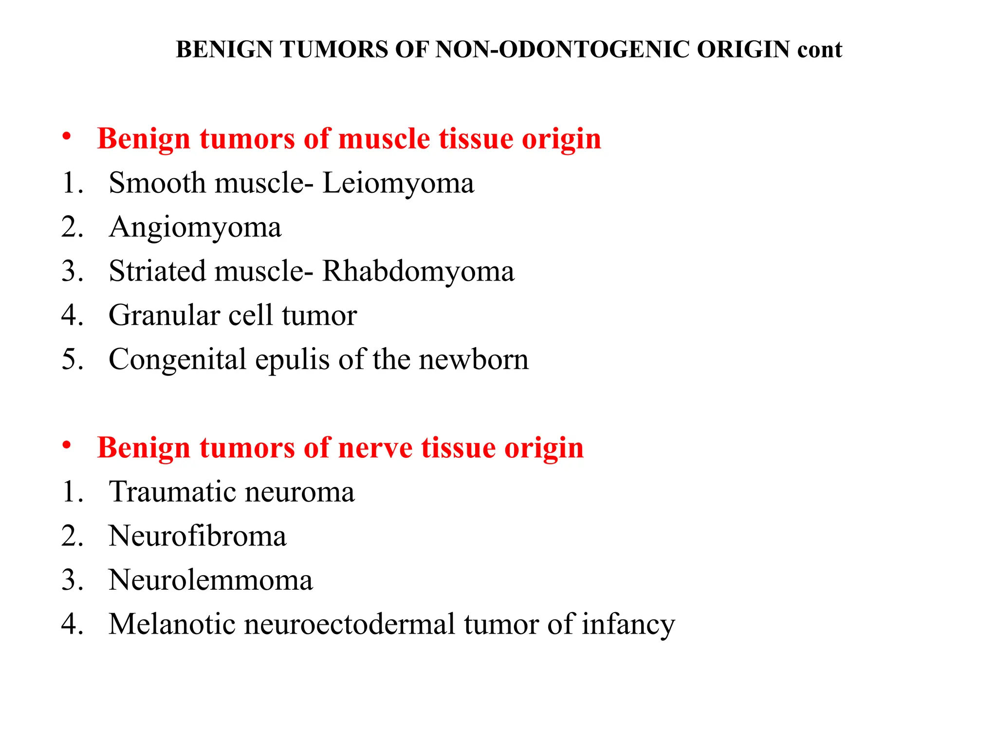
LEIOMYOMA
• Benign tumorof smooth muscles
• skin, subcutaneous tissues and the oral cavity
• Uncommon in oral cavity
• Clinical features:
• Posterior portion of the tongue, palate, cheeks, gingiva, lips
and salivary glands
• Middle decades of life
69.
• Slow growing
•Painless lesion
• Superficial
• Often pedunculated
• Normal mucosal color and texture
• Central leiomyoma- rare
70.
• H/F:
• Interlacingbundles of smooth muscle fibres interspersed
with fibrous connective tissue.
• The muscle nuclei are typically spindle shaped with blunt
ends and quite vesicular
• The bundles of fibres form whorls
• Treatment and prognosis:
• Conservative surgical excision .
• Do not recur
71.
RHABDOMYOMA
• Benign tumorof striated muscles
• Etiology unknown.
• Clonal balanced translocation in chromosome 15 and 17
• Clinical features:
• Adult rhabdomyoma- Middle age (16-82 years)
• Male > female
• Pharynx, oral cavity
72.
• Floor ofmouth > base of the tongue and soft
palate
• Fetal rhamdomyoma- newborns and young
children
• Male >
• Post and preauricular region, or face followed
by nasopharynx but not in mouth
• A nodule or submucosal mass
• Several cms in size
73.
• H/F
• Largeround cells - granular eosinophilic vacuolated cytoplasm
- show irregular striations
• Cytoplasm - rich in glycogen and glycoprotein
• Fibrous stroma, mitotic activity low
• Fetal rhabdomyoma- less mature ,somewhat pleomorphic,
polygonal muscle cells admixed with spindle cells
• More cellular & myxoid stroma
• Treatment: conservative surgical excision, recurrence
uncommon
74.
CONGENITAL EPULIS OFNEWBORN
• Also k/a Neuman’s tumor
• Benign , mostly single, rarely multiple
• Present at birth
• Theories of origin include- hamartomas, fibroblastic,
histiocytic, myogenic and neurogenic
• Clinical features:
• Present at birth
75.
• Maxillary >mandibular gingiva
• Pedunculated lesion - crest of the alveolar ridge or process
• Few mm to several cms
• H/F
• Sheets of large closely packed cells-fine, granular eosinophilic
cytoplasm
• Neither mitosis nor cross striations
• Numerous capillaries
• Treatment:
• Surgical excision. Recurrence rare
76.
TRAUMATIC NEUROMA
• Alsok/a amputation neuroma
• Not a true neoplasm
• Hyperplasia of nerve fibres and their supporting tissues
• Accidental or purposeful sectioning of a nerve or sequelae to
dental extraction
• Pathogenesis: Degeneration of the distal portion of the nerve
• Fragmentation and disintegration of the axis cylinders and myelin
sheath.
77.
• The neurilemmalsheath shrink - distal degenerating fibres
consist only of strands of connective tissue and neurilemma
• Repair begins with proliferation of axis cylinders, the cells
of neurilemmal sheath and endoneurium
• Reinnervation usually occurs unless meet scar tissue or
malaligned bone
• Nerve continues to grow into an unorganised bulbous or
nodular mass of nerve fibres and schwann cells
• Traumatic neuroma
78.
• Clinical features:
•Small nodule or swelling of the mucosa
• Slow growing
• Seldom greater than a cms
• Digital pressure may cause pain and along the course of nerve
• Typically near mental foramina, on the alveolar ridge in the
edentulous areas or on the lips or tongue
• Central lesion may also occur
79.
• H/F
• Massof irregular and often
interlacing neurofibrils (small
discrete bundles or spread diffusely)
• Schwann cells in connective tissue
stroma
• Treatment:
• Surgical excision along with a small
proximal portion of the involved
nerve
• Recurrence is not common
80.
NEUROFIBROMA
• Benign tumorof nerve tissue origin
• Solitary lesion or as a part of the generalized syndrome of
neurofibromatosis (Von Recklinghausen’s disease of the
skin)
• Origin- Perineural fibroblasts, gene mutations
• Inherited as an Autosomal Dominant Trait with a high
degree of penetrance
• Malignant transformation occurs subsequently in
neurofibromatosis
81.
• Clinical features:
•All races
• No sex predilection
• Discrete, nonulcerated nodules , same color as mucosa
• Buccal mucosa, palate, alveolar ridge, vestibule, and tongue
• Tongue involvement- macroglossia
• Central lesion > mandible, facial pain or paresthesia,
radiographically fusiform enlargement of mandibular canal
82.
• H/F:
• Mayor may not be well-circumscibed
• Spindle cells - thin, wavy nuclei intermingled with neurites in an
irregular pattern - delicate interwining connective tissue fibrils
• Cellular and myxoid pattern
• Melanocytes may be found , Mast cells common
• Treatment: Surgical excision for solitary lesion
• Recurrence may be noted. Multiple recurrences- malignant
transformation.
• Neurofibromatosis- functional and cosmetic, genetic counselling
and evaluation
83.
NEUROLEMMOMA
• Schwannoma, Neurilemmoma,Neurinoma, Lemmoma
• Derived neuroectodermal from schwann cells that produce
the myelin sheath surrounding the axons of peripheral nerves
• CLINICAL FEATURES:
• Any age, no sex predilection
• Slow growing
• Tongue> palate> floor of mouth, buccal mucosa, gingiva, lip,
vestibule, maxillary sinus
84.
• Central inmandible- destructive – pain and paresthesia
• Single, circumscribed nodule
• Varying size
expansion of the inferior alveolar nerve canal till the mental
foramen
85.
• H/F:
• Antonytype A and Antony type B
• Antony type A: elongated or spindle shaped nuclei
• Palisading pattern, parallel fashion – intercellular fibres
• Antony type B: no characteristic palisading pattern
• Disorderly arrangement of cells and fibres
• Verocay bodies- small hyaline structures
• Encapsulated- always
• TREATMENT: Surgical Excision, Radioresistant
• Recurrence uncommon
86.
Due to timeconstraints, the diagnostic
aspects—including radiological
interpretation, findings, and differential
diagnosis—have not been covered.
A detailed study is required for topics such as
fibro-osseous lesions, osteosarcoma,
osteochondroma, and osteoblastoma.
ACKNOWLEDGEMENT
Organizing team ofDept Of Oral Medicine
& Radiology VS Dental College and
Hospital in association with IAOMR
Karnataka State Branch, Bangalore.
My hod & colleagues of dept of oral
medicine & radiology j s s dental college.