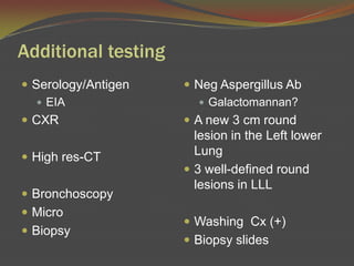
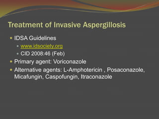
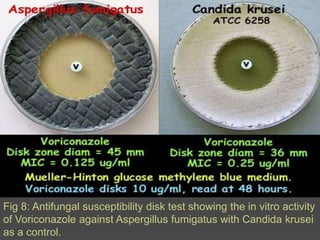
AspergillosisDiagnosis and Treatment
CaseA 50 year old WM with ESRD secondary to diabetes on HD whounderwent a cadaveric renal transplant.  Immunosuppressive regimen consisted of prednisone, Mycophenolatemofetil, and cyclosporine.  Patient developed thrombocytopenia and was taken off the Mycophenolatemofetil.  Developed catheter-related sepsis and bacteremia due to P. aeruginosa. The catheter was removed and he received a 2-week course of Meropenemand Ciprofloxacin then discharged.
1 month posttransplant he developed a nonproductive cough.  He was maintained on prednisone and cyclosporine and was given Ganciclovir for CMV prophylaxis.  He was again started on Mycophenolatemofetil.  2 months post-transplant he was readmitted to the hospital for possible rejection.
ROS was negative for cough chest pain, hemoptysis, SOB, fever and night sweats.  PE revealed that the patient was afebrile.  Clear lungs.  Heart sounds normal.Abdomen non tender.CBC: WBC 5.0, Hb10.5, Plt49Serum Cr 3.4 BUN 70What additional information or testing would you require?
Additional testingSerology/AntigenEIA CXRHigh res-CTBronchoscopyMicroBiopsyNegAspergillusAbGalactomannan?A new 3 cm round lesion in the Left lower Lung3 well-defined round lesions in LLLWashing Cx (+)Biopsy slides
Fig:1 Aspergilloma found at post-mortem in the lung of a child with leukemia.
Fig 2: Aspergilloma found at post-mortem in the lung of a child with leukemia.  Note fungus ball occupying cavity.
AspergillosisAspergillosis is a spectrum of diseases in humans and animals caused by members of the genus Aspergillus.  These include Mycotoxicosisdue to ingestion of contaminated foods Allergy and sequelae to the presence of conidia or transient growth of the organism in body orificesColonization without extension in preformed cavities and debilitated tissuesInvasive, inflammatory, granulomatous, necrotizing disease of lungs, and other organs Systemic and fatal disseminated disease.
The type of disease and severity depends upon the physiologic state of the host and the species of Aspergillus involved.Distribution:  World-wide.Etiological Agents:  Aspergillusfumigatus, A. flavus, A. niger, A. nidulans and A. terreus.
Invasive Aspergillosis DXHistopathologyacute angle branchingseptatednonpigmented hyphae, measuring 2-4 microns in widthculture yielding Aspergillus sp.
Fig 3: Grocott’smethenamine silver (GMS) stained tissue section of lung showing fungal balls of hyphae of Aspergillusfumigatus.
AspergillusFig 4: Grocott’smethenamine silver (GMS) stained tissue sections showing Aspergillusfumigatus in lung tissue, note conidial heads forming in an alveolus.
Fig 5: Microscopic morphology of Aspergillusniger showing large, globose, dark brown conidial heads, which become radiate, tending to split into several loose columns with age.  Conidiophores are smooth-walled, hyaline or turning dark towards the vesicle.  Conidial heads are biseriate with the phialides borne on brown, often septatemetulae. Conidia are globose to subglobose, dark brown to black and rough-walled.
Fig 6: Aspergillusterreus on Czapekdox agar showing typical suede-like cinnamon-buff to sand brown colonies.  Reverse yellow to deep dirty brown.
Fig 7: Conidial head of Aspergillusterreus.  Conidial heads are compact, columnar and biseriate.  Conidiophores are hyaline and smooth-walled.  Conidia are globose to ellipsoidal, hyaline to slightly yellow and smooth-walled.

Asperg

  • 1.
  • 2.
    CaseA 50 yearold WM with ESRD secondary to diabetes on HD whounderwent a cadaveric renal transplant. Immunosuppressive regimen consisted of prednisone, Mycophenolatemofetil, and cyclosporine. Patient developed thrombocytopenia and was taken off the Mycophenolatemofetil. Developed catheter-related sepsis and bacteremia due to P. aeruginosa. The catheter was removed and he received a 2-week course of Meropenemand Ciprofloxacin then discharged.
  • 3.
    1 month posttransplanthe developed a nonproductive cough. He was maintained on prednisone and cyclosporine and was given Ganciclovir for CMV prophylaxis. He was again started on Mycophenolatemofetil. 2 months post-transplant he was readmitted to the hospital for possible rejection.
  • 4.
    ROS was negativefor cough chest pain, hemoptysis, SOB, fever and night sweats. PE revealed that the patient was afebrile. Clear lungs. Heart sounds normal.Abdomen non tender.CBC: WBC 5.0, Hb10.5, Plt49Serum Cr 3.4 BUN 70What additional information or testing would you require?
  • 5.
    Additional testingSerology/AntigenEIA CXRHighres-CTBronchoscopyMicroBiopsyNegAspergillusAbGalactomannan?A new 3 cm round lesion in the Left lower Lung3 well-defined round lesions in LLLWashing Cx (+)Biopsy slides
  • 6.
    Fig:1 Aspergilloma foundat post-mortem in the lung of a child with leukemia.
  • 7.
    Fig 2: Aspergillomafound at post-mortem in the lung of a child with leukemia. Note fungus ball occupying cavity.
  • 8.
    AspergillosisAspergillosis is aspectrum of diseases in humans and animals caused by members of the genus Aspergillus. These include Mycotoxicosisdue to ingestion of contaminated foods Allergy and sequelae to the presence of conidia or transient growth of the organism in body orificesColonization without extension in preformed cavities and debilitated tissuesInvasive, inflammatory, granulomatous, necrotizing disease of lungs, and other organs Systemic and fatal disseminated disease.
  • 9.
    The type ofdisease and severity depends upon the physiologic state of the host and the species of Aspergillus involved.Distribution: World-wide.Etiological Agents: Aspergillusfumigatus, A. flavus, A. niger, A. nidulans and A. terreus.
  • 10.
    Invasive Aspergillosis DXHistopathologyacuteangle branchingseptatednonpigmented hyphae, measuring 2-4 microns in widthculture yielding Aspergillus sp.
  • 11.
    Fig 3: Grocott’smethenaminesilver (GMS) stained tissue section of lung showing fungal balls of hyphae of Aspergillusfumigatus.
  • 12.
    AspergillusFig 4: Grocott’smethenaminesilver (GMS) stained tissue sections showing Aspergillusfumigatus in lung tissue, note conidial heads forming in an alveolus.
  • 13.
    Fig 5: Microscopicmorphology of Aspergillusniger showing large, globose, dark brown conidial heads, which become radiate, tending to split into several loose columns with age. Conidiophores are smooth-walled, hyaline or turning dark towards the vesicle. Conidial heads are biseriate with the phialides borne on brown, often septatemetulae. Conidia are globose to subglobose, dark brown to black and rough-walled.
  • 14.
    Fig 6: Aspergillusterreuson Czapekdox agar showing typical suede-like cinnamon-buff to sand brown colonies. Reverse yellow to deep dirty brown.
  • 15.
    Fig 7: Conidialhead of Aspergillusterreus. Conidial heads are compact, columnar and biseriate. Conidiophores are hyaline and smooth-walled. Conidia are globose to ellipsoidal, hyaline to slightly yellow and smooth-walled.