Definition-
Pathway ofcell death
Induced by tightly regulated suicide program
Cells destined to die activate intrinsic enzymes
These intrinsic enzymes degrade the cell's own DNA , nuclear
& cytoplasmic proteins.
4.
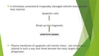
It eliminatesunwanted & irreparably damaged cellwith least possible
host reaction
Apoptotic cells
Break up into fragments
Plasma membrane of apoptotic cell remain intact , but structure is
altered in such a way that these become the tasty targets for
phagocytes.
APOPTOTIC BODIES
5.
Causes-
Physiological:
Serves to eliminatecells that are no longer needed
1. Destruction of cells during embryogenesis
2. Involvement of hormone dependent tissue upon hormone withdrawl
Endometrial cell breakdown during menstrual cycle
Ovarian follicular atresia in menopause
Regression of lactataing breast after weaning
Prostatic atrophy after castration
6.
3. Cell lossin proliferating cell population
Immature lymphocytes in bonemarrow & thymus & B-lymphocytes
in germinal centres that fail to express useful antigen receptors
Epithelial cells in intestinal crypts to maintain homeostasis
4. Elimination of potentially harmful self-reactive lymphocytes to prevent
reaction
against one's own tissue
5. Death of host cells that have served their useful purpose
Neutrophils in acute inflammatory response
Lymphocytes at end of an immune system.
7.
Pathological:
1. Radiation ,cytotoxic, anti-cancer drugs & hypoxia can damage DNA.
Repair followed by apoptosis
Elimination of cell is better alternative than risking mutation in
damaged DNA which may result in malignant transformation
2. Accumulation of misfolded proteins
Basis for degenerative diseases of CNS & other organs
Excessive accumulation of these proteins in the ER leads to condition
called ER stress, which results in Apoptotic cell death
8.
3. Pathologic atrophyin parenchymal organs after duct obstruction, seen
in pancreas, parotid gland
.
4. Cell death in certain infections either due to apoptosis induced
by microorganism or by host immune response
9.
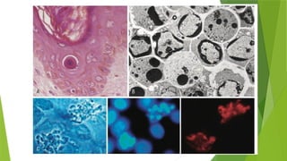
Morphological changes-
Cellshrinkage
Chromatin condensation
Formation of cytoplasmic blebs & Apoptotic bodies
Phagocytosis of Apoptotic cells , usually by macrophages
11.
Phases of Apoptosis-
Involvesactivation of enzymes called CASPASES
Initiation phase:
Some caspases are
catalytically activated in
this phase.
Execution phase:
Activated caspases trigger
the degradation of cellular
components.
Activation of caspases is dependent on Pro-apoptotic and Anti-
apoptotic proteins
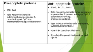
12.
Pro-apoptotic proteins Anti-apoptoticproteins
• BAK, BAX
• Role: Keep mitochondrial
outer membrane permeable &
cause leakage of CytC from
intermembranous space into cytosol
• BCL-2, BCL-XL, MCL-1
• Role: Keep mitochondrial outer membrane
impermeable & prevent leakage of CytC &
other death inducing
proteins into cytosol.
• Seen in Outer mitochondrial membrane,
Cytosol & ER membrane
• Have 4 BH domains called BH-4
• Stimulated by growth factors & survival
signals
13.
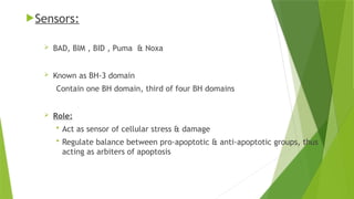
Sensors:
BAD, BIM, BID , Puma & Noxa
Known as BH-3 domain
Contain one BH domain, third of four BH domains
Role:
Act as sensor of cellular stress & damage
Regulate balance between pro-apoptotic & anti-apoptotic groups, thus
acting as arbiters of apoptosis
14.
Mechanisms:
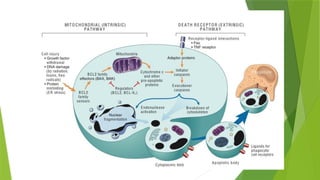
Intrinsic orMitochondrial pathway of Apoptosis
Extrinsic or Death Receptor-Initiated pathway of Apoptosis
15.
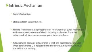
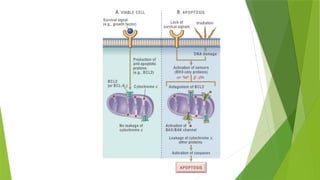
Intrinsic Mechanism
MajorMechanism
Stimulus from inside the cell
Results from increase permeability of mitochondrial outer membrane
with consequent release of death inducing molecules from the
mitochondrial intermembranous space into cytoplasm.
Mitochondria contains cytochrome C that are essential for life ,but
when cytochrome C is released into the cytoplasm it indicates that
the cell is not healthy
16.
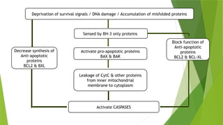
Sensed by BH-3only proteins
Activate pro-apoptotic proteins
BAX & BAK
Leakage of CytC & other proteins
from inner mitochondrial
membrane to cytoplasm
Decrease synthesis of
Anti-apoptotic
proteins
BCL2 & BXL
Deprivation of survival signals / DNA damage / Accumulation of misfolded proteins
Block function of
Anti-apoptotic
proteins
BCL2 & BCL-XL
Activate CASPASES
17.
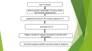
CytC in cytosol
Bindto protein called APAF-1 & forms wheel
like hexamer (Apoptosome)
Apoptosome bind to C-9 & Cleaves adjacent C-9
Activated caspases mediate execution phase of apoptotis
Activation of C-9
Triggers cascade of caspase activation & activate other
pro caspasas
18.
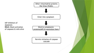
Other mitochodrial proteins
likeSmac/Diablo
Enter into cytoplasm
Bind to cytoplasmic
proteins(IAP) & neutralize them
Permits intitation of caspase
cascade
IAP Inhibitors of
Apoptosis
ROLE: Block activation
of caspases & cells alive
20.
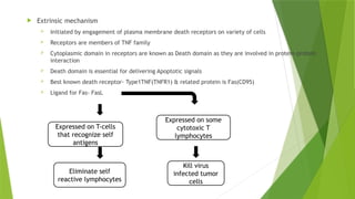
Extrinsic mechanism
Initiated by engagement of plasma membrane death receptors on variety of cells
Receptors are members of TNF family
Cytoplasmic domain in receptors are known as Death domain as they are involved in protein-protein
interaction
Death domain is essential for delivering Apoptotic signals
Best known death receptor- Type1TNF(TNFR1) & related protein is Fas(CD95)
Ligand for Fas- FasL
Expressed on T-cells
that recognize self
antigens
Eliminate self
reactive lymphocytes
Expressed on some
cytotoxic T
lymphocytes
Kill virus
infected tumor
cells
21.
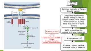
FasL bind
to FaS
3or more molecules of FaS are
brought together
Cytoplasmic death domain
form a binding site for an
adaptor protien which contain
death domain and is called
FADD (Fas-associated death
domain)
FADD binds to inactive
caspase 8,10 (procaspse-8,10)
Active caspase 8,10
Activated caspases mediate
execution phase of apoptotis
FLIP binds to ProC8
Some viruses use this FLIP
protein
to protect themselves
from FaS mediated Apoptosis
22.
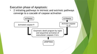
Execution phase ofApoptosis:
• 2 initiating pathways in intrinsic and extrinsic pathways
converge to a cascade of caspase activation
INTRINSIC EXTRINSIC
Activated caspase 9
Activate caspase
8,10
Enzymatic death program by rapid
and sequential activation of
executioner caspases 3 & 6.
APOPTOSIS
24.
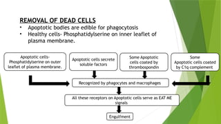
REMOVAL OF DEADCELLS
• Apoptotic bodies are edible for phagocytosis
• Healthy cells- Phosphatidylserine on inner leaflet of
plasma membrane.
Apoptotic cells-
Phosphatidylserine on outer
leaflet of plasma membrane.
Apoptotic cells secrete
soluble factors
Some Apoptotic
cells coated by
thrombospondin
Some
Apoptotic cells coated
by C1q complement
Recognized by phagocytes and macrophages
All these receptors on Apoptotic cells serve as EAT ME
signals
Engulfment
25.
• Apoptotic cellsand their fragments are cleared before
they undergo necrosis and release their cellular contents.
• This often occurs within minutes without leaving a
trace and inflammation is absent even in
extensive Apoptosis.
• This is the reason for least possible host reaction and
without any collateral tissue damage.
26.
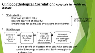
Clinicopathological Correlation: Apoptosisin health and
disease
1. GF deprivation :
Hormone sensitive cells
Neurons deprived of nerve GF
Lymphocytes not stimulated by antigens and cytokines
2. DNA Damage :
Apoptosis is triggered
by intrinsic pathway
Exposure of
cells to
radiation or
Chemo
DNA damage
(Genotoxic
stress)
Gene p53
arrests cell
cycle at G1
phase to allow
time for repair
If damage is
beyond repair,
then p53 triggers
APOPTOSIS
If p53 is absent or mutated, then cells with damaged DNA
survive & undergo mutation that leads to neoplastic
27.
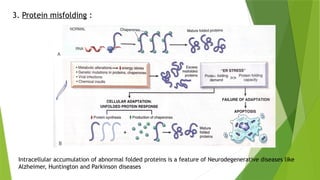
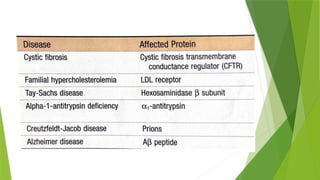
3. Protein misfolding:
Intracellular accumulation of abnormal folded proteins is a feature of Neurodegenerative diseases like
Alzheimer, Huntington and Parkinson diseases
29.
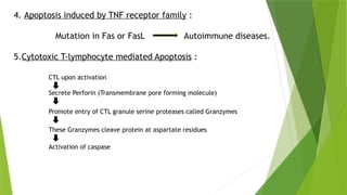
4. Apoptosis inducedby TNF receptor family :
Mutation in Fas or FasL Autoimmune diseases.
5.Cytotoxic T-lymphocyte mediated Apoptosis :
CTL upon activation
Secrete Perforin (Transmembrane pore forming molecule)
Promote entry of CTL granule serine proteases called Granzymes
These Granzymes cleave protein at aspartate residues
Activation of caspase
30.
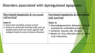
Disorders associated withdysregulated Apoptosis :
Decreased Apoptosis & Increased
cell survival
Seen in
Gene p53 mutation causes cancer
Failure to eliminate harmful cells such as
lymphocytes that can react against self-
antigens leads to auto immune diseases
Increased Apoptosis & Decreased
cell survival
Seen in
Neurodegenerative diseases ( Due to
mutation & misfolded proteins)
Ischemic injuries (MI, Stroke)
Death of virus infected cells in many
viral infections.
31.
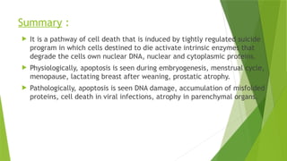
Summary :
Itis a pathway of cell death that is induced by tightly regulated suicide
program in which cells destined to die activate intrinsic enzymes that
degrade the cells own nuclear DNA, nuclear and cytoplasmic proteins.
Physiologically, apoptosis is seen during embryogenesis, menstrual cycle,
menopause, lactating breast after weaning, prostatic atrophy.
Pathologically, apoptosis is seen DNA damage, accumulation of misfolded
proteins, cell death in viral infections, atrophy in parenchymal organs.
32.
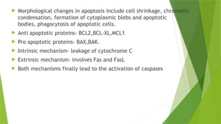
Morphological changesin apoptosis include cell shrinkage, chromatin
condensation, formation of cytoplasmic blebs and apoptotic
bodies, phagocytosis of apoptotic cells.
Anti apoptotic proteins- BCL2,BCL-XL,MCL1
Pro apoptotic proteins- BAX,BAK.
Intrinsic mechanism- leakage of cytochrome C
Extrinsic mechanism- involves Fas and FasL
Both mechanisms finally lead to the activation of caspases
33.
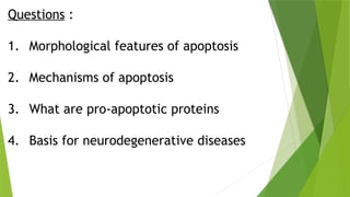
Questions :
1. Morphologicalfeatures of apoptosis
2. Mechanisms of apoptosis
3. What are pro-apoptotic proteins
4. Basis for neurodegenerative diseases
34.
References :
Robbins& Cotran pathologic basis of disease
Anderson's Pathology
Walter & Israel General pathology


![Hemangiblastoma CNS PPT.pptx [Autosaved].pptx](https://cdn.slidesharecdn.com/ss_thumbnails/hemangiblastomacnsppt-251005155615-644fd7bd-thumbnail.jpg?width=640&height=640&fit=bounds)


![PERI-PROSTHETIC FRACTURE NAIL-PLATE CONSTRUCT [NPC].pptx](https://cdn.slidesharecdn.com/ss_thumbnails/drarunkumardrmohamedashrafperiprostheticfrasturenail-plateconstructnpc-260209164459-7e9d15a1-thumbnail.jpg?width=640&height=640&fit=bounds)


