1. ORIGINAL ARTICLE
Office-based olfactory mucosa biopsies
Eric H. Holbrook, MD1,2
, Lina Rebeiz2
and James E. Schwob, MD, PhD2
Background: Requests from researchers for olfactory mu-
cosal biopsies are increasing as a result of advances in
the fields of neuroscience and stem cell biology. Published
studies report variable rates of success in obtaining true
olfactory tissue, o en below 50%. In cases where biopsies
are not obtained carefully and confirmed through histologi-
cal techniques, erroneous conclusions are made. A ention
to the epithelium alone without submucosal analysis may
add to the confusion. A consistent biopsy technique can
help rhinologists obtain higher yields of olfactory mucosa.
Confirmatory tissue staining analysis assures olfactory mu-
cosa has been obtained, thereby strengthening clinical cor-
relations and scientific conclusions.
Methods: Biopsies of the septum within the anterior ol-
factory cle were obtained under endoscopic guidance in
an office procedure room using topical local anesthetic (li-
docaine). A er mucosal incision, a small, cupped, biopsy
forceps was used to obtain specimens approximately 2 to
3 mm in size. Specimens were sectioned and analyzed with
immunohistochemistry for presence of olfactory epithe-
lium and/or olfactory fascicles.
Results: A total of 14 subjects were biopsied in this analysis.
Four subjects had biopsies in the operating room (OR). The
remaining 10 underwent biopsies in the clinic. All biopsies
obtained in the OR revealed evidence of olfactory mucosa.
Of the 10 clinic biopsies, 8 (80%) revealed evidence of ol-
factory mucosa. No complications were encountered.
Conclusion: High yields of olfactory mucosa can be ob-
tained safely in an office-based se ing. Technique, includ-
ing a ention to the area of biopsy, and confirmatory anal-
ysis are important in assuring presence of olfactory tissue.
C 2016 ARS-AAOA, LLC.
Key Words:
olfactory epithelium; olfactory fila; humans; immunohisto-
chemistry; S100; intermediate filaments; p75 neurotrophin
receptor; olfactory ensheathing cells
How to Cite this Article:
Holbrook EH, Rebeiz L, Schwob JE. Office-based olfactory
mucosa biopsies. Int Forum Allergy Rhinol. 2016;xx:1–8.
The olfactory epithelium (OE), including the olfactory
receptor neurons (ORNs) that maintain first-order
synapses with the olfactory bulb, is unique in its ability to
regenerate throughout life.1
The basal cells of the epithe-
lium have a major role in this capacity and respond with
upregulation of cell division after injury to the system.2,3
The olfactory mucosa of humans has been shown to be
very similar in to other mammals such as rodents in regard
1Department of Otolaryngology, Massachusetts Eye and Ear
Infirmary/Harvard Medical School, Boston, MA; 2Department of
Developmental, Molecular, and Chemical Biology, Tufts University
School of Medicine, Boston, MA
Correspondence to: Eric H. Holbrook, MD, 243 Charles Street, Boston,
MA 02114; e-mail: eric_holbrook@meei.harvard.edu
Additional Supporting Information may be found in the online version of this
article.
Funding sources for the study: NIH R01 DC010242; NIH NIDCD (JES).
Potential conflict of interest: None provided.
Presented at the Annual ARS Meeting on September 26, 2015, in Dallas, TX.
Received: 8 September 2015; Revised: 23 November 2015; Accepted:
3 December 2015
DOI: 10.1002/alr.21711
View this article online at wileyonlinelibrary.com.
to cellular consistency and immunohistochemical staining
characteristics.4
The regenerative capacity of the OE also
appears to be maintained in humans based on biopsy cul-
ture results5
and the continued presence of both mature
and immature olfactory sensory neurons (OSNs) regardless
of age with expression of cell division markers in the basal
cells.4
In humans, as opposed to rodents, it appears that the
regenerative process in response to insult may be less suc-
cessful in terms of function. Common causes for loss of ol-
faction have been consistently identified among smell and
taste centers and include those related to head injury, up-
per respiratory tract infections (URIs), chronic rhinosinusi-
tis, and age, while a large proportion have no identifiable
cause (idiopathic).6,7
However, we have a poor understand-
ing of the pathophysiology underlying these most common
causes. As the basic mechanisms of olfactory physiology
and regenerative processes are discovered in mouse mod-
els, comparisons with biopsies of human olfactory mucosa
will remain critical in identifying causality.
In addition, the basal cells of the OE—with their char-
acteristic features that satisfy the criteria of bona fide stem
1 International Forum of Allergy & Rhinology, Vol. 00, No. 0, xxxx 2016
2. Holbrook et al.
cells and their accessibility for biopsy—are of interest to
researches in the fields of neurodegenerative disorders and
neural regeneration. There has been a surge of interest in
culturing these basal stem cells for potential use in repair
of various forms of central nervous system disorders.8
In
addition, the olfactory ensheathing cells enveloping the ol-
factory axons and bundled together as fila olfactoria in the
lamina propria are thought to have special properties to
allow for enhanced axonal guidance and regeneration af-
ter injury to motor neurons of the spinal cord and facial
nerve.9–11
Obtaining biopsies of human olfactory mucosa is not
new,12–16
but the technique is often poorly described. Fre-
quently, published results from research laboratories pro-
vide conclusions depending on biopsies from “olfactory
areas” reported by a surgeon’s general anatomic descrip-
tion. In certain cases the biopsies are cultured without his-
tological confirmation that the tissue is indeed olfactory,
with conclusions made entirely on stained cells that have
been grown and passaged in various culture conditions.17
For purposes of OE analysis for olfactory disorders, the per-
vasive presence of respiratory epithelium in a biopsy could
be a result of sample selection error and result in erroneous
conclusions of respiratory metaplasia within an olfactory
region.13
For these reasons, consistency in the biopsy tech-
nique as well as care in sample evaluation is crucial for
advancing the field.
We describe an endoscopic biopsy technique for consis-
tently obtaining human olfactory mucosa in awake, non-
sedated subjects in a clinic setting using widely available
otolaryngology instruments. We also argue for the impor-
tance of confirming olfactory origin of the mucosa using
described methods that can be used regardless of epithelial
presence or respiratory status.
Subjects and methods
Subjects
Research subjects were recruited from patients visiting
the Sinus Center at Massachusetts Eye and Ear Infirmary
(MEEI) for smell disorders. Subjects with an identifiable
cause of smell loss either related to URI or head trauma
were enrolled. Olfactory function was assessed using a 40-
item, scratch-and-sniff, forced-choice, smell-identification
test (SIT; Sensonics, Inc., Haddon Heights, NJ). A group of
subjects undergoing septoplasty (1 subject with URI-related
anosmia) were also chosen for operating room (OR)-
based biopsies as a comparison. All subjects provided in-
formed consent for participation in this study, which was
approved by the Human Subjects Committee at MEEI.
OR-based olfactory biopsies
Biopsies performed in the OR were obtained under general
anesthesia after the septoplasty procedure using injections
of 1% lidocaine with 1:100,000 parts epinephrine. Three
specimens were obtained from 1 side only to reduce the risk
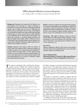
FIGURE 1. Cartoon representation of a sagittal section through the hu-
man nasal cavity showing the high-yield area of olfactory epithelium. The
peppered pattern demonstrates the area of the nasal septum most often
associated with olfactory epithelium based on averaging from multiple au-
topsy specimens and shows the positional relationship among easily iden-
tifiable nasal structures. Notice that the Olfactory Epithelium typically does
not reach the anterior edge of the middle turbinate in adults. Asterisk (*)
= cribriform plate; F = frontal sinus; MT = middle turbinate; S = sphenoid
sinus.
of smell loss. Specimens were obtained from the superior
septum within the olfactory cleft and adjacent to the middle
turbinate using a sickle knife and pediatric blakesley forceps
or cupped forceps. The area considered high-yield for OE
(Fig. 1) was defined using averaged areas calculated from
immunostaining for neurons over multiple whole-mount
specimens (data not shown).18
Clinic-based olfactory biopsies
Biopsies performed in the clinic were obtained from sub-
jects without the use of sedation in an upright sitting posi-
tion. The nasal cavity was sprayed with a mixture of lido-
caine (2%) and oxymetazoline (0.025%), and after several
minutes rigid endoscopy was performed using a 2.7-mm,
0-degree endoscope (Hopkins II; Karl Storz, El Segundo,
CA) to identify the side with the greatest olfactory cleft
space. In initial biopsies, a small cotton wisp soaked in the
lidocaine/oxymetazoline mixture was inserted into the ol-
factory cleft for further anesthetic (subjects HB5 and HB6);
however, concerns with exfoliation of the epithelium with
this technique resulted in a change in procedure. For the
remaining biopsies, further anesthetic was applied by plac-
ing the subject supine and hanging the head backward off
the edge of the chair with the vertex of the head parallel to
the floor (Fig. 2). A flexible angiocatheter was then used to
direct 0.5 to 1.0 mL of anesthetic solution into the superior
nasal cavity and olfactory cleft. The subjects remained in
this position for 2 to 3 minutes and then returned to the
upright sitting position. A sickle knife was then used to cre-
ate a 5-mm to 7-mm long, posterior-to-anterior, diagonal
incision along the septum approximately 5 to 8 mm below
the cribriform plate and greater than 5 mm posterior to
the anterior attachment of the middle turbinate (Support-
ing Video 1). Given the variability of OE anterior extent
and regression of OE area with age, attempts at accessing
the more posterior regions while still allowing for insertion
of the forceps with minimal discomfort to the subject were
made. The sickle knife was then used to carefully elevate
International Forum of Allergy & Rhinology, Vol. 00, No. 0, xxxx 2016 2
3. Office-based olfactory mucosa biopsies
FIGURE 2. Subject position used during topical anesthesia of the nose.
Illustration by Olivia Schwob.
a superior flap of mucosa away from the bone. A 3-mm
cupped biopsy forceps or pediatric blakesley forceps was
used to pinch off a portion of the superiorly raised flap. The
cupped biopsy forceps became the preferred instrument due
to its small size and ability to fit within the olfactory cleft.
Two biopsies were routinely obtained from each subject.
Minimal bleeding was encountered and controlled with a
lidocaine/oxymetazoline–soaked cotton wisp.
Tissue processing
Biopsy specimens were placed immediately in standard
10% neutral buffered formalin solution and placed on ice
for transport. The tissue was then rinsed in phosphate-
buffered saline (PBS) after 2 hours and then submerged
in 30% sucrose overnight. Orientation has been found to
be critical in the histological assessment of biopsy speci-
mens. To help identify the epithelial surface of the spec-
imens during processing, the area designated for biopsy
on the septum was initially inked with a standard surgical
marking pen early in the study (subjects HB3, HB4, and
HB5). This practice was then rejected for fear of causing
epithelial loss or decrease in cellular viability. Instead, a
dissecting microscope was used to identify the epithelial
and lamina propria surfaces. The biopsy was then placed
in OCT mounting medium (Miles Inc., Elkhart, IN), tak-
ing care to orient the biopsy in the cassette for sectioning
in the proper cross-sectional plane through the epithelium.
The tissue was snap frozen in liquid nitrogen, cryosectioned
at 10 µm (Leica CM3050S; Leica, Bannockburn, IL), and
mounted on Plus slides (Fisher Scientific, Pittsburg, PA) and
stored at −20°C for future use.
Immunohistochemistry
Slides were immersed in PBS for removal of OCT for
5 minutes and then placed in 3% hydrogen peroxide for
10 minutes to quench inherent peroxidase activity. Sec-
tions were then puddled with 0.01 M citric acid buffer
(pH 6.0) and placed in a commercial food steamer con-
taining water in its reservoir for 10 minutes for antigen
retrieval. Slides were rinsed in PBS and then incubated in
blocking solution (10% donkey serum + 5% nonfat dry
milk + 4% bovine serum albumin [BSA] + 0.1% Triton
X-100 in PBS) for 10 minutes. Incubation with primary
antibodies occurred overnight at 4°C in a humid cham-
ber. The dilutions and antigen specificity for each primary
antibody are listed in Table 1. Bound primary antibodies
were visualized by incubating with Alexa 488 or Alexa
594-conjugated secondary antibody (Invitrogen, Carlsbad,
CA) or through a biotinylated secondary antibody (Jackson
ImmunoResearch Laboratories, Inc., West Grove, PA) and
Alexa 488 or Alexa 594-conjugated strep-avidin system
(Invitrogen) incubating for 1 hour each at room temper-
ature. Immunofluorescent-stained sections were addition-
ally labeled for nuclei with 4 ,6-diamidino-2-phenylindole
(DAPI) and coverslipped with glycerol/n-propyl gallate
(NPG) mounting medium.
Photography
Sections were imaged with a Spot RT color digital cam-
era (Spot, Sterling Heights, MI) attached to a Nikon 800
E microscope (Nikon Instruments Inc., Melville, NY). Im-
age preparation, assembly, and analysis were performed
in Photoshop CS6 (Adobe Systems, San Jose, CA). In all
cases, only balance, contrast, brightness, and evenness of
illumination were altered.
Results
Subject profiles
A total of 14 subjects (7 females) had biopsies of the sep-
tal olfactory epithelium (Table 2). The average age was
53.4 years (range, 22–68 years). Seven subjects (4 female)
had smell loss related to URI, 3 subjects (1 female) had
smell loss related to head trauma, 2 subjects had no subjec-
tive loss and tested normal, and 2 subjects had no subjective
loss and tested hyposmic. Of the 7 URI-related smell loss
subjects, 6 were anosmic and 1 was hyposmic on testing.
All subjects with traumatic smell loss were anosmic. Four
of the subjects were biopsied in the OR (3 subjective nor-
mal and 1 URI-related anosmia). No complications relat-
ing to bleeding, infection, cerebrospinal fluid (CSF) leak, or
changes in smell function were experienced with any sub-
ject, as noted.19,20
Any reports of discomfort were related
to pressure during access to the olfactory cleft region, and
all subjects entering the protocol tolerated the procedure
without requests to abort.
Immunohistochemistry of the epithelium
In most cases (13/14 subjects) an epithelium was observed
in the biopsy specimen. To confirm biopsies as true OE,
an immunohistochemical staining analysis using 4 different
antibodies provided information on the presence of olfac-
tory neurons, the relative overall maturity of the neurons,
and the presence of respiratory epithelium. We used anti-
bodies against olfactory marker protein (OMP) to identify
mature OSNs; and to identify all OSNs of both mature and
immature age, we used antibodies against the protein gene
product 9.5 (PGP) or antibodies against beta-tubulin III
3 International Forum of Allergy & Rhinology, Vol. 00, No. 0, xxxx 2016
4. Holbrook et al.
TABLE 1. Primary antibody details
Antibody Species Concentration Company Catalog# Antigen
Staining
characteristic
Tuj1 Mouse 1:300 Covance, Emeryville, CA MMS-435P Rat brain beta-tubulin III All neurons
Beta-tubulin IV Mouse 1:1000 Sigma, St. Louis, MO T7941 C-terminal synthetic peptide
beta-tubulin IV
Respiratory cilia
hNF Mouse 1:50 Dako North America, Inc.,
Carpinteria, CA
2F11 Neurofilament from adult
human brain (70KD)
Trigeminal axons
OMP Goat 1:100 Santa Cruz Biotechnology,
Inc., Dallas, TX
SC-49070 Rodent olfactory marker
protein
Mature olfactory neurons
p75 (NTR) Mouse 1:50 Millipore, Billerica, MA MAB365 Protein isolated from plasma
membrane of PC-12 cells
Perineurium and
ensheathing cells
PGP9.5 Rabbit 1:1000 Cedarlane, Burlington, NC RA95101 Human brain protein gene
product 9.5
All neurons
S100 Rabbit 1:100 Dako North America, Inc.,
Carpinteria, CA
Z0311 S100 protein from cow
extracts
Neurons, ensheathing
cells, some gland tissue
TABLE 2. Subject demographics and biopsy results
Specimen Age (years) Gender Olfactory function PMH Location Marker Pledget Biopsy forceps Epithelium Fascicles
HB5 57 F Anosmia URI Clinic X X Blakesley Mix
HB6 24 F Normal — Clinic X Cup Absent Trigeminal
HB7 61 M Anosmia Trauma Clinic Cup Respiratory None
HB8 64 M Anosmia URI Clinic Cup Respiratory Empty
HB9 47 F Anosmia URI Clinic Cup Mix
HB10 62 F Anosmia URI Clinic Cup Mix
HB11 59 F Anosmia Trauma Clinic Cup Mix
HB12 60 M Anosmia Trauma Clinic Cup Olfactory
HB13 58 M Anosmia URI Clinic Cup Respiratory Empty
HB14 41 M Hyposmia URI Clinic Cup Respiratory Empty
F = female; M = male; PMH = past medical history; URI = upper respiratory infection; X = used during the biopsy.
(Tuj1). Respiratory cells were labeled at the ciliated surface
with antibodies against beta-tubulin IV (beta4).4
As shown
in Figure 3A, stained, adjacent sections of a biopsy from
a control subject (HB4) reveal the typical multicell layered
epithelium comprised of many PGP(+)/Tuj1(+) neurons
and absent beta4(+) respiratory cells. Many of the OSNs
are mature, with OMP(+) staining in the more apically
positioned neurons as expected.
In contrast, biopsies from subjects with olfactory loss
often revealed deviation from the normal pattern of stain-
ing. Stained sections from a subject with URI-related anos-
mia (HB10) demonstrates a more disorganized presence
of PGP(+)/Tuj1(+) neurons within the epithelium and less
distinct apically positioned mature OMP(+) cells (Fig. 3B).
In addition, there is evidence of respiratory cell meta-
plasia with patches of beta4(+) apical staining and in-
traepithelial neuromas, which in this case appears to be
comprised of mature and immature axons by double-
labeling.
The epithelium from a subject with trauma-related anos-
mia (HB12) has a more ordered appearance to the OE sim-
ilar to the control subject, but the OSNs are largely imma-
ture with infrequent OMP(+) mature cells (Fig. 3C). In this
case, respiratory epithelium is absent; but another intraep-
ithelial neuroma is present, although composed mostly of
Tuj1(+)/OMP(–) immature axons.
Immunohistochemistry of the submucosa
Loss of the epithelial layer may occur while obtaining the
biopsy or during tissue processing. In addition, respiratory
cell replacement is common during normal aging and in-
creases in various pathologic states.15,21–23
Biopsies may
also be mistakenly obtained outside the region of OE. For
this reason, an analysis of the epithelium alone may not be
International Forum of Allergy & Rhinology, Vol. 00, No. 0, xxxx 2016 4
5. Office-based olfactory mucosa biopsies
FIGURE 3. Immunohistochemistry demonstrates the presence of OE. Adjacent section from 3 separate biopsies are double labeled with antibodies against
PGP9.5 (PGP) and Beta4 or Tuj1 and OMP with DAPI nuclear staining in blue. (A) A biopsy from subject HB4 provides an example of normal OE with
multiple layers of PGP(+)/Tuj1(+) neurons. The epithelium is without any respiratory cells as confirmed with absent Beta4 staining. A healthy mix of immature
Tuj1(+)/OMP(–) and mature Tuj1(+)/OMP(+) OSNs are appreciated. (B) In comparison, a biopsy from subject HB10 with URI-related anosmia reveals a
disordered epithelium with less defined OSN cell bodies. OSN staining with PGP, Tuj1, and OMP is present confirming the epithelium as OE, but patches
of Beta4(+) respiratory cilia can also be seen. An asterisk shows a large intraepithelial neuroma. Notice how the neuroma and disorganized OSNs remain
compartmentalized between the row of supporting cell nuclei apically and the basal cell nuclei below. (C) A biopsy from subject HB12 with trauma-related
anosmia has an overall immature OE. The OSNs are confirmed with PGP(+)/Tuj1(+) labeling but mature OMP(+) cells are limited. Once again, an intraepithelial
neuroma is seen (asterisks), but in this example there is disruption of the supporting cell layer. Arrowheads = basal lamina; scale bar = 25 µm. Beta4 =
beta-tubulin IV; DAPI = 4 ,6-diamidino-2-phenylindole; OE = olfactory epithelium; OMP = olfactory marker protein; OSN = olfactory sensory neuron; PGP =
protein gene product; Tuj1 = beta-tubulin III; URI = upper respiratory infection.
sufficient to determine the biopsy is olfactory in origin. In
cases where OE was not identified in the biopsy, immuno-
histochemical characterization of the axon bundles within
the submucosa was performed. Similar antibodies used in
the epithelial analysis (Tuj1, OMP) identified axon bun-
dles in a control biopsy (HB4) with presence of mature
Tuj1(+)/OMP(+) OSN axons (Fig. 4A). In cases where
the epithelium is missing or has been identified as respi-
ratory, nerve fascicle-type structures can be found on rou-
tine hematoxylin and eosin (H&E) staining with elongated
nuclei of the olfactory ensheathing cells (OECs) and con-
nective tissue elements. These fascicles can be further iden-
tified as olfactory fascicles, empty olfactory fascicles, or
non-olfactory fascicles when stained with specific antibod-
ies (Fig. 4B). Antibodies against S100 (S100) label OECs
surrounding olfactory axons within the fila olfactoria.24–26
When OSN axons are present, double-labeling of fascicles
with Tuj1 and S100 is seen (Fig. 4B1). However, fascicles
empty of OSN axons remain S100(+) but lack Tuj1(+)
staining (Fig. 4B1, 4B2). The S100 staining in these fascicles
appear more dispersed with distinct rounded ensheathing
cell bodies with their associated DAPI(+) nucleus as op-
posed to the more evenly stained axon-full fascicles (com-
parison in Fig. 4B1). Antibodies against p75 neurotrophin
receptor (p75) label perineural fibroblasts.25
Staining with
p75 surrounds both intact and empty olfactory fascicles
(Fig. 4B1, 4B2), which help to confirm S100(+)/Tuj1(–)
structures as empty nerve fascicles as opposed to other sub-
mucosal structures (ie, glands).
In our analysis of the submucosa, small S100(+)/Tuj1(+)
bundles were frequently identified. With absence of
OMP(+) labeling, these small fascicles could be small im-
mature olfactory fascicles or trigeminal nerve branches. We
used antibodies to human neurofilament proteins (hNF)
known to label trigeminal nerve fibers.27
Adjacent sections
stained with hNF antibodies labeled these small Tuj1(+)
fascicles and supports their identification as trigeminal
(Fig. 4B).
Office-based biopsy efficiency
Immunohistochemical staining was performed on all the
biopsies to identify them as olfactory. Of our 10 biop-
sies performed in the clinic, 9 contained epithelium for im-
munohistochemical analysis (Table 2). Of these 9 samples,
5 contained either a mix of OE and respiratory epithelium
or OE alone. When only respiratory epithelium was en-
countered (4/10) or in the absence of epithelium, further
staining of the submucosa was performed. Evidence for
empty olfactory fascicles was present in 3 samples (3/5).
Interestingly, the 1 normal subject biopsied in the clinic
(HB6) had no epithelium present and only trigeminal nerve
branches. When taken together, an evaluation of both the
5 International Forum of Allergy & Rhinology, Vol. 00, No. 0, xxxx 2016
6. Holbrook et al.
FIGURE 4. Immunohistochemistry helps to confirm olfactory nerve fascicles. (A) Adjacent sections through a biopsy from subject HB4 reveals fila olfactoria
that contains abundant mature Tuj1(+)/OMP(+) axons and is reflective of the health of the overlying OE. Arrowhead = basal lamina; scale bar = 100 µm.
(B) Sections through biopsies stained with H&E reveal structures resembling nerve fascicles. Immunohistochemical double labeling with antibodies against
S100 and Tuj1, p75 neurotrophic receptor (p75), or hNF further define the fascicles. (B1) H&E and immunofluorescent staining of sections through a biopsy
from subject HB1 shows a typical large olfactory fascicle (asterisk) stained with S100 and Tuj1. The absence of hNF staining confirms the axons as olfactory
and not trigeminal. Two smaller fascicles (double arrows) are S100(+) but lack Tuj1(+) axon staining. The p75(+) staining of perineural tissue surrounding the
S100(+) OECs confirms these structures as empty olfactory fascicles. A smaller trigeminal nerve fascicle (single arrow) stained with S100, Tuj1, and hNF is also
present. Scale bar = 50 µm. (B2) H&E and immunofluorescent staining of adjacent sections through a biopsy from a subject with URI-related hyposmia (HB14)
at higher power magnification demonstrates a group of 3 empty fascicles stained with S100 and absent of Tuj1(+) axons. Perineural staining with p75 confirms
the structures as nerve fascicles. Note the wider-spaced S100(+) OECs in the empty fascicles as opposed to the larger Tuj1(+) olfactory nerve fascicle in B1.
Again, a small trigeminal nerve branch is seen (arrow). Scale bar = 25 µm. H&E = hematoxylin and eosin; hNF = human neurofilament protein; OE = olfactory
epithelium; OEC = olfactory ensheathing cell; OMP = olfactory marker protein; Tuj1 = beta-tubulin III; URI = upper respiratory infection.
epithelium and submucosa in our population of office-
based biopsies revealed successful sampling from olfactory
regions in 80% of the subjects.
Discussion
Olfactory mucosal biopsies have been reported in the lit-
erature with variable rates of success, often less than
50%.5,28,29
Intraoperative sampling of olfactory tissue
would be expected to result in higher success given the
ability to access more confined regions and the creation of
working space during planned surgical procedures under
general anesthesia. However, requests for olfactory biop-
sies on subjects without planned nasal surgery are becom-
ing more frequent either for the study of olfactory loss, for
diseases with related olfactory pathology, or for harvesting
OE stem cells in normal subjects. In these cases, a technique
is needed with high yield and relatively low discomfort.
We present a technique of olfactory mucosal biopsy that
can be performed under endoscopic visualization using
routine clinic instruments in an office-based setting with
a success rate of 80%. Our samples were obtained from
anterior septal mucosa in a region known to contain ol-
factory neurons based on prior autopsy analysis. In this
manner, we avoided problems with patient discomfort and
restrictions in instrumentation typically encountered with
attempts to access more posterior regions, such as the su-
perior turbinate. Although procedure discomfort was not
a major focus of this study and specific measurements of
pain were not included, we found the technique to be well
tolerated by the subjects. Our technique also minimizes in-
troduction of anesthetic materials including injections and
cotton pledgets that may disrupt the histology and viabil-
ity of the cells. We did not receive reports of olfactory
loss from subjects after biopsies were performed, and fur-
ther investigations on this potential complication would be
International Forum of Allergy & Rhinology, Vol. 00, No. 0, xxxx 2016 6
7. Office-based olfactory mucosa biopsies
TABLE 3. Staining characteristics of nerve fascicles within
olfactory mucosa
S100 Tuj1/PGP OMP p75 hNF
Olfactory X X X X O
Immature olfactory X X O X O
Empty olfactory X O O X O
Trigeminal X X O X or O X
hNF = human neurofilament proteins; O = absent staining; OMP = olfactory
marker protein; PGP = protein gene product; Tuj1 = neuron-specific class III
beta-tubulin; X = staining present.
beneficial. However, previously published papers with hu-
man OE biopsies were also without complications of smell
loss.19,20
Our analysis of the success rate in obtaining olfactory
mucosal biopsies takes into account both the epithelium as
well as the submucosa. The presence of Tuj1(+)/PGP(+)
neurons within the epithelium, either mature OMP(+)
or immature OMP(–), confirms the mucosa as olfactory.
However, replacement of OE with respiratory epithelium
is known to occur in humans and appears to increase with
age.21,23
In addition, conditions resulting in smell loss such
as URI-related anosmia may result in increased respiratory
metaplasia.13,30,31
Therefore, finding only respiratory ep-
ithelium in a biopsy specimen does not preclude it from
being olfactory in nature. Conversely, one cannot assume
a biopsy containing only respiratory epithelium obtained
from an area thought to be olfactory reflects respiratory
metaplasia when in reality a sampling error has occurred.
This is not trivial given previously published reports of
neurospheres cultured from olfactory epithelium when the
source tissue was never confirmed as olfactory prior to cell
dissociation.17
This possibility becomes more problematic
as reports of nasal cell cultures from areas far outside those
known to be olfactory result in neurosphere formation,
perhaps due to the presence of neural crest-derived cells.32
Furthermore, the act of the biopsy or tissue processing can
result in the loss of epithelium for analysis. For these rea-
sons, a deeper analysis of the submucosa is needed for es-
tablishing olfactory origin of biopsies.
Immunohistochemistry using antibodies important for
epithelial analysis can help identify the presence of olfactory
nerve fascicles below the surface. Positive OMP staining of
fascicles confirms the presence of fila olfactoria and the
epithelial surface as olfactory in origin. In humans, the ol-
factory axon bundles travel directly to the cribriform plate
and do not span across regions known to be originally res-
piratory in nature. This is supported by our observations
and others of whole complete sheets of OE in fetal tissue
without intervening respiratory zones.21,33
Therefore, posi-
tive identification of OMP(+) axons within nerve olfactory
axon fascicles run deep to epithelium that was originally
olfactory.
Further characterization of the underlying fascicles is
helpful when OMP staining is absent (Table 3) and has
proven useful for other investigators interested in harvest-
ing OECs.34
Neuronal staining with antibodies Tuj1 and
PGP in the absence of OMP denotes the presence of ax-
ons that may reflect dominance of immature OSNs. Ad-
ditional positive staining of these fascicles with hNF iden-
tifies them as trigeminal in nature. In our experience, the
hNF(+) bundles are much smaller than the fila olfacto-
ria. Empty olfactory nerve fascicles, absent of axons and
with collagen infiltration, have been previously reported
and associated with olfactory disorders.13
In these cases,
staining with neuronal markers including OMP, Tuj1, and
PGP is lacking. Immunostaining with antibodies to S100
can identify the OECs remaining within the axon-deficient
fascicles. S100 is not specific to just OECs and the addi-
tional staining with antibodies against p75 neurotrophic
receptor will identify perineuronal fibroblasts surrounding
the S100-labeled empty fascicles. Identification of these fas-
cicles not only helps to confirm the olfactory nature of the
biopsy but also lends additional description to the overall
status of the OE.13
Confirming mucosal biopsy samples as olfactory is im-
portant in making statements regarding the origin of cells
grown in culture. Although we believe adjacent biopsies re-
duce sampling error in obtaining true olfactory mucosa, it is
possible that they may be taken from 2 different regions—
olfactory or respiratory. However, if 1 sample demonstrates
olfactory mucosa regardless of the overlying epithelium dis-
playing respiratory or olfactory cells, the second adjacent
sample will have a high likelihood of originating from the
same. To increase confirmation of samples as either olfac-
tory or respiratory, biopsy samples should be divided into
2 pieces with 1 portion dedicated to immunohistochemical
confirmation.
We recognize the low numbers of office-based biopsies
used in this report; however, our technique has been suc-
cessfully used previously for obtaining OE samples.35
The
overall purpose of this manuscript is twofold. First, it pro-
vides rhinologists with a reproducible technique for obtain-
ing olfactory mucosa biopsies in an office setting under local
anesthesia with minimal patient discomfort and with sim-
ple office instruments. Our limitations relate to the space
restrictions of the olfactory cleft, and we recognize better
results could be obtained with specially designed thinner-
cutting instruments. We recognize the value of preserving
as much of the mucosa as possible—especially the epithe-
lial surface. Therefore, our technique evolved to avoid any
insertion of anesthetic-soaked cotton wisps or pledgets for
concern of possible epithelial exfoliation. This certainly re-
duced our ability to reach more posterior anatomy, which
has been successfully biopsied by others.34
Second, this
manuscript stresses the importance of analyzing the tis-
sue below the epithelium with focus on the nerve fascicles
contained within this compartment, and we provide an im-
munohistochemical staining protocol helpful for character-
izing these structures.
7 International Forum of Allergy & Rhinology, Vol. 00, No. 0, xxxx 2016
8. Holbrook et al.
Conclusion
Olfactory biopsies can be reproducibly obtained with
high yields in an office-based setting using common clinic
instruments with low patient discomfort under local anes-
thetic. It is the responsibility of both the surgeon and the
research team to ensure the biopsy material being studied
is in fact olfactory in nature before conclusions are made.
This can be done with a high level of certainty using rou-
tine immunohistochemistry and a small battery of specific
antibodies.
References
1. Graziadei PP, Monti Graziadei GA. Neurogenesis and
neuron regeneration in the olfactory system of mam-
mals. I. Morphological aspects of differentiation and
structural organization of the olfactory sensory neu-
rons. J Neurocytol. 1979;8:1–18.
2. Huard JM, Youngentob SL, Goldstein BJ, Luskin MB,
Schwob JE. Adult olfactory epithelium contains mul-
tipotent progenitors that give rise to neurons and non-
neural cells. J Comp Neurol. 1998;400:469–486.
3. Schnittke N, Herrick DB, Lin B, et al. Transcription
factor p63 controls the reserve status but not the stem-
ness of horizontal basal cells in the olfactory epithe-
lium. Proc Natl Acad Sci U S A. 2015;112:E5068–
E5077.
4. Holbrook EH, Wu E, Curry WT, Lin DT, Schwob JE.
Immunohistochemical characterization of human ol-
factory tissue. Laryngoscope. 2011;121:1687–1701.
5. Feron F, Perry C, McGrath JJ, Mackay-Sim A. New
techniques for biopsy and culture of human olfactory
epithelial neurons. Arch Otolaryngol Head Neck Surg.
1998;124:861–866.
6. Deems DA, Doty RL, Settle RG, et al. Smell and taste
disorders, a study of 750 patients from the University
of Pennsylvania Smell and Taste Center. Arch Oto-
laryngol Head Neck Surg. 1991;117:519–528.
7. Landis BN, Konnerth CG, Hummel T. A study on
the frequency of olfactory dysfunction. Laryngoscope.
2004;114:1764–1769.
8. Duan D, Lu M. Olfactory mucosa: a rich source of cell
therapy for central nervous system repair. Rev Neu-
rosci. 2015;26:281–293.
9. Chen L, Huang H, Xi H, et al. A prospective random-
ized double-blind clinical trial using a combination
of olfactory ensheathing cells and Schwann cells for
the treatment of chronic complete spinal cord injuries.
Cell Transplant. 2014;23(Suppl 1):S35–S44.
10. Tabakow P, Raisman G, Fortuna W, et al. Functional
regeneration of supraspinal connections in a patient
with transected spinal cord following transplantation
of bulbar olfactory ensheathing cells with peripheral
nerve bridging. Cell Transplant. 2014;23:1631–1655.
11. Guntinas-Lichius O, Wewetzer K, Tomov TL, et al.
Transplantation of olfactory mucosa minimizes ax-
onal branching and promotes the recovery of vibris-
sae motor performance after facial nerve repair in rats.
J Neurosci. 2002;22:7121–7131.
12. Rawson NE, Gomez G, Cowart B, et al. Selectivity and
response characteristics of human olfactory neurons.
J Neurophysiol. 1997;77:1606–1613.
13. Holbrook EH, Leopold DA, Schwob JE. Abnormal-
ities of axon growth in human olfactory mucosa.
Laryngoscope. 2005;115:2144–2154.
14. Borgmann-Winter KE, Rawson NE, Wang HY, et al.
Human olfactory epithelial cells generated in vitro ex-
press diverse neuronal characteristics. Neuroscience.
2009;158:642–653.
15. Yee KK, Pribitkin EA, Cowart BJ, et al. Neuropathol-
ogy of the olfactory mucosa in chronic rhinosinusitis.
Am J Rhinol Allergy. 2010;24:110–120.
16. Feron F, Perry C, Girard SD, Mackay-Sim A. Isolation
of adult stem cells from the human olfactory mucosa.
Methods Mol Biol. 2013;1059:107–114.
17. Wrobel BB, Mazza JM, Evgrafov OV, Knowles JA.
Assessing the efficacy of endoscopic office olfactory
biopsy sites to produce neural progenitor cell cultures
for the study of neuropsychiatric disorders. Int Forum
Allergy Rhinol. 2013;3:133–138.
18. Holbrook EH, Schwob JE. An integrated im-
munohistochemical analysis of human whole-mount
and cryosectioned olfactory tissue. Chem Senses.
2008;33:S78.
19. Lanza DC, Deems DA, Doty RL, et al. The effect of
human olfactory biopsy on olfaction: a preliminary
report. Laryngoscope. 1994;104:837–840.
20. Feron F, Mackay-Sim A, Andrieu JL, Matthaei KI,
Holley A, Sicard G. Stress induces neurogenesis in
non-neuronal cell cultures of adult olfactory epithe-
lium. Neuroscience. 1999;88:571–583.
21. Nakashima T, Kimmelman CP, Snow JBJr. Structure
of human fetal and adult olfactory neuroepithelium.
Arch Otolaryngol. 1984;110:641–646.
22. Hasegawa S, Yamagishi M, Nakano Y. Micro-
scopic studies of human olfactory epithelia fol-
lowing traumatic anosmia. Arch Otorhinolaryngol.
1986;243:112–116.
23. Paik SI, Lehman MN, Seiden AM, Duncan HJ, Smith
DV. Human olfactory biopsy. The influence of age and
receptor distribution.Arch Otolaryngol Head Neck
Surg. 1992;118:731–738.
24. Vincent AJ, West AK, Chuah MI. Morphological and
functional plasticity of olfactory ensheathing cells.
J Neurocytol. 2005;34(1-2):65–80.
25. Smithson LJ, Kawaja MD. A comparative examina-
tion of biomarkers for olfactory ensheathing cells in
cats and guinea pigs. Brain Res. 2009;1284:41–53.
26. Rawji KS, Zhang SX, Tsai YY, Smithson LJ, Kawaja
MD. Olfactory ensheathing cells of hamsters, rab-
bits, monkeys, and mice express alpha-smooth muscle
actin. Brain Res. 2013;1521:31–50.
27. Bae JY, Kim JH, Cho YS, Mah W, Bae YC. Quan-
titative analysis of afferents expressing substance P,
calcitonin gene-related peptide, isolectin B4, neurofila-
ment 200, and Peripherin in the sensory root of the rat
trigeminal ganglion. J Comp Neurol. 2015;523:126–
138.
28. Winstead W, Marshall CT, Lu CL, Klueber KM,
Roisen FJ. Endoscopic biopsy of human olfactory ep-
ithelium as a source of progenitor cells. Am J Rhinol.
2005;19:83–90.
29. Rawson NE, Gomez G, Cowart BJ, Kriete A, Pribitkin
E, Restrepo D. Age-associated loss of selectivity in
human olfactory sensory neurons. Neurobiol Aging.
2012;33:1913–1919.
30. Jafek BW, Eller PM, Esses BA, Moran DT. Post-
traumatic anosmia. Ultrastructural correlates. Arch
Neurol. 1989;46:300–304.
31. Yamagishi M, Fujiwara M, Nakamura H. Olfactory
mucosal findings and clinical course in patients with
olfactory disorders following upper respiratory viral
infection. Rhinology. 1994;32:113–118.
32. Hauser S, Widera D, Qunneis F, et al. Isolation
of novel multipotent neural crest-derived stem cells
from adult human inferior turbinate. Stem Cells Dev.
2012;21:742–756.
33. Naessen R. The identification and topographical local-
isation of the olfactory epithelium in man and other
mammals. Acta Otolaryngol. 1970;70:51–57.
34. Kachramanoglou C, Law S, Andrews P, Li D, Choi D.
Culture of olfactory ensheathing cells for central nerve
repair: the limitations and potential of endoscopic ol-
factory mucosal biopsy. Neurosurgery. 2013;72:170–
178; discussion 178–179.
35. Pantazopoulos H, Boyer-Boiteau A, Holbrook EH,
et al. Proteoglycan abnormalities in olfactory epithe-
lium tissue from subjects diagnosed with schizophre-
nia. Schizophr Res. 2013;150(2-3):366–372.
International Forum of Allergy & Rhinology, Vol. 00, No. 0, xxxx 2016 8